
Sarcoidosis: Non-Caseating Granulomas in Every Organ
Key points
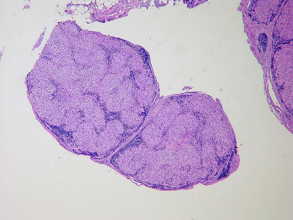
- Sarcoidosis: a multisystem inflammatory disease of unknown cause characterised by non-caseating (non-necrotising) epithelioid granulomas.
- Who it affects: typically adults aged 20 to 40, with a second peak in women over 50. More common and more severe in people of Black African and Caribbean heritage, and more prevalent in Northern Europe.
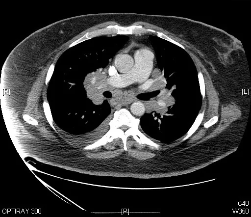
- The classic radiograph: bilateral hilar lymphadenopathy, which is symmetrical and usually painless. Around half of patients are asymptomatic at diagnosis.
- Lofgren syndrome: erythema nodosum, bilateral hilar lymphadenopathy, fever and arthralgia, typically of the ankles. An acute presentation with an excellent prognosis that usually needs no treatment.
- Diagnosis: compatible clinical and radiological features, histology showing non-caseating granulomas, and exclusion of other causes - above all tuberculosis, lymphoma and berylliosis.
- Serum ACE: raised in around 60% of patients but neither sensitive nor specific. It does not make or exclude the diagnosis.
- Hypercalcaemia: caused by granuloma macrophages expressing 1-alpha-hydroxylase, which converts vitamin D to its active form. It is an indication for corticosteroids.
- Treatment: most patients need none. Corticosteroids are reserved for progressive lung disease, hypercalcaemia, and cardiac, neurological, ocular or renal involvement.
Introduction
Sarcoidosis is a multisystem inflammatory disease of unknown cause, defined by the presence of non-caseating (non-necrotising) epithelioid granulomas in affected tissues. It can involve almost any organ, but the lungs and intrathoracic lymph nodes are affected in over 90% of patients.
The prevailing hypothesis is that an unidentified environmental antigen triggers an exaggerated Th1 immune response in a genetically susceptible individual. Macrophages and CD4 T lymphocytes aggregate into granulomas which, unlike those of tuberculosis, do not undergo central caseous necrosis. Occupational and environmental associations have been reported - including with insecticides, mouldy environments and, notably, in first responders to the World Trade Center site - but no single cause has been established.
Epidemiology
- Peak incidence at 20 to 40 years, with a second peak in women over 50
- More common and more severe in people of Black African and Caribbean heritage, who have higher rates of chronic and extrapulmonary disease
- Higher prevalence in Northern Europe, particularly Scandinavia
- Slightly more common in women, and less common in smokers - one of the few conditions where smoking appears protective, which is not a reason to recommend it
Clinical features
Presentation ranges from an entirely incidental radiographic finding to life-threatening cardiac or neurological disease. Around half of patients are asymptomatic at diagnosis, the abnormality having been found on a chest radiograph taken for another reason.
Constitutional
- Fatigue - extremely common and often the most disabling symptom of all, frequently persisting after the granulomatous disease has settled and consistently under-recognised
- Fever, night sweats and weight loss
- Arthralgia
Respiratory
- Dry cough, exertional breathlessness and vague chest discomfort
- Examination is frequently normal, even with extensive radiographic change - a striking and characteristic mismatch. Crackles appear only with established fibrosis.
- Clubbing is not a feature, which helps separate advanced sarcoidosis from other fibrotic lung diseases
- Endobronchial involvement may cause wheeze and airflow obstruction
Two named acute syndromes
Skin
- Erythema nodosum - tender, red, raised nodules on the shins. It is a reactive, non-granulomatous phenomenon rather than sarcoid tissue itself, and indicates acute disease with a good prognosis.
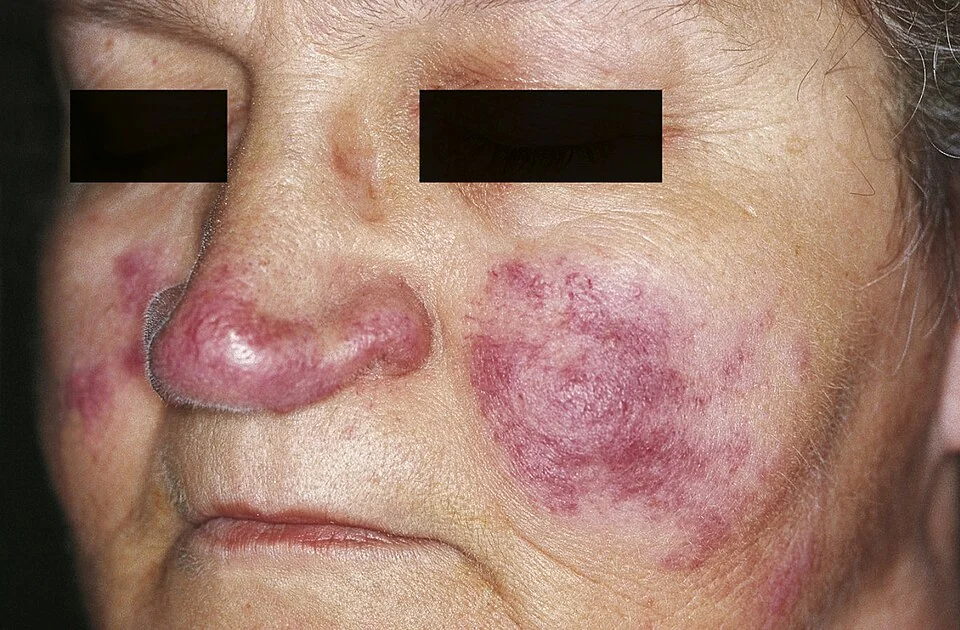
- Lupus pernio - indurated violaceous plaques on the nose, cheeks and ears. This is genuine granulomatous infiltration and is a marker of chronic, treatment-resistant disease, often with upper airway and bone involvement. Its prognostic implication is the opposite of erythema nodosum, which is worth fixing in mind.
- Scar sarcoidosis - granulomas infiltrating old scars and tattoos, which become raised, purple and indurated
- Maculopapular lesions, plaques and subcutaneous nodules
Eyes
- Anterior uveitis - the commonest ocular manifestation, presenting with a painful red eye, photophobia and blurred vision
- Posterior uveitis, optic neuritis and retinal vasculitis
- Keratoconjunctivitis sicca from lacrimal gland involvement
- Every patient with sarcoidosis needs an ophthalmology assessment, because significant eye disease may be asymptomatic and untreated inflammation can cause permanent visual loss
Cardiac
- Clinically apparent in around 5% but found at post-mortem in up to 25%, so it is substantially under-diagnosed
- Conduction disease - heart block of any degree, and a common cause of complete heart block in a young patient
- Arrhythmias - ventricular tachycardia in particular
- Cardiomyopathy and heart failure
- Sudden cardiac death - cardiac sarcoidosis is a leading cause of death from the disease, which is why any syncope, palpitations or ECG abnormality must be taken seriously
Neurological
- Affects around 5 to 10%
- Cranial nerve palsies - the facial nerve most commonly, and it may be bilateral
- Aseptic (lymphocytic) meningitis
- Hypothalamic and pituitary involvement - causing diabetes insipidus and hypopituitarism
- Space-occupying lesions, seizures, peripheral neuropathy and small fibre neuropathy
Other systems
- Hypercalcaemia and hypercalciuria - from unregulated 1-alpha-hydroxylase activity in granuloma macrophages, converting 25-hydroxyvitamin D to active 1,25-dihydroxyvitamin D. Causes nephrocalcinosis, renal stones and renal impairment.
- Renal - interstitial nephritis and, less often, glomerulonephritis
- Liver and spleen - hepatosplenomegaly with a cholestatic liver profile; usually asymptomatic
- Lymphadenopathy - peripheral as well as intrathoracic
- Bone and joints - dactylitis, punched-out cystic lesions in the phalanges, and an inflammatory arthritis
- Parotid and salivary gland enlargement
Investigations
Imaging and staging
The chest radiograph is staged using the Scadding classification, which correlates loosely with the likelihood of spontaneous remission.
| Stage | Chest radiograph | Spontaneous resolution |
|---|---|---|
| 0 | Normal | - |
| I | Bilateral hilar lymphadenopathy alone | Around 60-80% |
| II | Bilateral hilar lymphadenopathy with parenchymal infiltrates | Around 50-60% |
| III | Parenchymal infiltrates alone, without lymphadenopathy | Around 20-30% |
| IV | Pulmonary fibrosis with volume loss and honeycombing | Rare - irreversible |
- HRCT - perilymphatic nodules distributed along the bronchovascular bundles, fissures and subpleural surfaces, with upper and mid zone predominance, together with symmetrical hilar and mediastinal lymphadenopathy. Advanced disease shows traction bronchiectasis and fibrosis.
- Cardiac MRI or FDG-PET - for suspected cardiac sarcoidosis, showing late gadolinium enhancement or active inflammation
- MRI brain and spine with contrast - for suspected neurosarcoidosis
Blood and urine tests
- Serum calcium - hypercalcaemia in around 10%, and hypercalciuria in up to a third. Check a 24-hour urinary calcium.
- Serum ACE - raised in around 60%, but also raised in tuberculosis, lymphoma, hyperthyroidism, diabetes and liver disease. It is neither sensitive nor specific enough to diagnose or exclude sarcoidosis, and is used at most to follow disease activity in a patient in whom it was raised initially.
- FBC - lymphopenia is common, and anaemia or thrombocytopenia may occur
- ESR and CRP - often raised
- LFTs - a cholestatic pattern with a raised ALP suggests hepatic involvement
- U&Es and creatinine - for renal involvement
- Immunoglobulins - often polyclonally raised
- Vitamin D - and note that supplementation may precipitate hypercalcaemia and should be given cautiously with monitoring
Physiological and organ assessment
- Spirometry and transfer factor - typically a restrictive pattern with a reduced TLCO, though an obstructive pattern occurs with endobronchial disease. Serial measurement is the standard way of following pulmonary disease.
- ECG in every patient - looking for conduction abnormality. Extend to Holter monitoring, echocardiography and cardiac MRI if there are symptoms or ECG changes.
- Ophthalmology review in every patient
- Urinalysis and renal function
Tissue diagnosis
- Biopsy is required in most patients, showing non-caseating epithelioid granulomas. Endobronchial ultrasound-guided transbronchial needle aspiration (EBUS-TBNA) of mediastinal nodes is now the first-line approach, with a high yield and low complication rate.
- Biopsy the most accessible site - skin lesions, palpable lymph nodes or the lip (for minor salivary glands) may be much simpler than the chest
- Biopsy may be omitted where the picture is characteristic and the risk of an alternative diagnosis is low - classical Lofgren syndrome and asymptomatic stage I disease are the accepted examples
- Always request stains and cultures for mycobacteria and fungi on the specimen, since granulomas alone cannot distinguish sarcoidosis from infection
- Bronchoalveolar lavage - a raised CD4 to CD8 ratio (above 3.5) supports the diagnosis but is not specific, and lavage is principally useful for excluding infection
Management
Who does not need treatment
Most patients require no treatment at all. Asymptomatic stage I disease, and mild stage II disease with preserved lung function, are simply observed with periodic clinical review, spirometry and imaging, because the majority remit spontaneously. Treating everyone would expose a large number of people who were going to get better anyway to the harms of long-term corticosteroids.
Drug treatment
- Prednisolone - typically 0.5 mg/kg or 20 to 40 mg daily for 4 to 6 weeks, then tapered to a maintenance dose, with a total course usually of 6 to 24 months. Relapse on withdrawal is common.
- Bone protection - calcium and vitamin D supplementation is the usual accompaniment to steroids but must be given cautiously here because of the risk of hypercalcaemia; monitor calcium closely, and consider a bisphosphonate with careful assessment
- Methotrexate - the usual first-line steroid-sparing agent, added where steroids cannot be tapered or are poorly tolerated
- Azathioprine, mycophenolate and leflunomide - alternative steroid-sparing options
- Hydroxychloroquine - particularly useful for cutaneous disease and hypercalcaemia
- TNF-alpha inhibitors (infliximab, adalimumab) - for refractory disease, especially neurosarcoidosis, cardiac disease and lupus pernio
- Topical corticosteroids for isolated skin disease, and topical or intraocular steroids for uveitis under ophthalmology supervision
- NSAIDs for erythema nodosum and arthralgia in Lofgren syndrome, which usually needs nothing more
Organ-specific and supportive management
- Cardiac sarcoidosis - immunosuppression plus, frequently, an implantable cardioverter defibrillator, given the risk of sudden death. Manage jointly with cardiology.
- Hypercalcaemia - fluids, corticosteroids, and avoidance of excess vitamin D, calcium supplements and strong sunlight
- Fatigue - explain it, screen for depression, hypothyroidism, anaemia and sleep disturbance, and consider a graded exercise programme. It is often not steroid-responsive, which needs to be said clearly rather than treated with escalating doses.
- Pulmonary rehabilitation and oxygen in advanced fibrotic disease
- Lung transplantation for end-stage stage IV disease
- Monitoring - regular clinical review with spirometry, TLCO, calcium, renal and liver function, imaging as indicated, and continuing ophthalmology and cardiac surveillance
Complications
- Pulmonary fibrosis and respiratory failure - the endpoint in stage IV disease
- Pulmonary hypertension and cor pulmonale
- Aspergilloma - a fungal ball colonising a fibrotic cavity, which may cause massive haemoptysis
- Sudden cardiac death, complete heart block and ventricular arrhythmia
- Permanent visual loss from untreated uveitis or optic neuritis
- Chronic renal failure and nephrocalcinosis from persistent hypercalcaemia
- Diabetes insipidus and hypopituitarism from hypothalamic involvement
- Disfigurement from lupus pernio and cutaneous disease
- Corticosteroid toxicity - osteoporosis, diabetes, hypertension, weight gain, cataracts and adrenal suppression, which accumulates over the long courses often required
- Chronic fatigue and reduced quality of life, frequently persisting after the disease itself is quiescent
Red flags
Prognosis
The overall outlook is good. Around 60 to 70% of patients remit spontaneously, usually within 2 to 5 years, and the disease is fatal in only about 1 to 5% - most often from progressive pulmonary fibrosis, cardiac involvement or neurosarcoidosis.
| Good prognosis | Poor prognosis |
|---|---|
| Lofgren syndrome | Lupus pernio |
| Erythema nodosum | Chronic uveitis |
| Stage I disease | Stage III or IV disease |
| Acute onset | Insidious onset with symptoms for more than 6 months |
| Age under 40 | Age over 40 at onset |
| White European ethnicity | Black African or Caribbean heritage |
| Asymptomatic presentation | Cardiac or neurological involvement |
| Chronic hypercalcaemia and nephrocalcinosis | |
| Nasal mucosal involvement and cystic bone lesions | |
| Splenomegaly and extensive extrapulmonary disease |
Two practical points are worth carrying into clinic. First, a patient who remits may relapse, most often within the first few years and particularly after steroid withdrawal, so discharge should not be premature. Second, fatigue frequently outlasts the granulomatous disease and is the symptom patients most often say is dismissed - acknowledging it, excluding treatable contributors and offering realistic support does more for many patients than any adjustment of immunosuppression.
References
- Crouser ED, Maier LA, Wilson KC et al. Diagnosis and detection of sarcoidosis: an official ATS clinical practice guideline. 2020. Available here
- British Thoracic Society. Interstitial lung disease guidelines and resources. Available here
- NICE Clinical Knowledge Summaries. Sarcoidosis. Available here
- Baughman RP, Valeyre D, Korsten P et al. ERS clinical practice guidelines on treatment of sarcoidosis. European Respiratory Journal. 2021. Available here
- Scadding JG. Prognosis of intrathoracic sarcoidosis in England. BMJ. 1961. Available here
- BNF. Prednisolone, methotrexate and hydroxychloroquine - indications and dosing. Available here
- Yale Rosen, CC BY-SA 2.0, via Wikimedia Commons. Available here
- Sand M, Sand D, Thrandorf C, Paech V, Altmeyer P, Bechara FG. Cutaneous lesions of the nose. Head and Face Medicine. 2010. CC BY 2.0, via Wikimedia Commons. Available here
This article is written for revision and education. It is not clinical guidance and must not be used to make decisions about the care of a patient. Always check current NICE guidance and local protocols.