
Diabetes Insipidus
Key points
- Diabetes insipidus (DI): passage of large volumes of dilute urine due to impaired ADH (vasopressin) secretion - cranial DI - or renal resistance to ADH - nephrogenic DI.
- Cranial causes: idiopathic, pituitary or hypothalamic tumours, pituitary surgery or trauma, Sheehan syndrome, infiltrative disease, and familial forms.
- Nephrogenic causes: lithium is the classic cause; also hypercalcaemia, hypokalaemia, chronic kidney disease and the X-linked AVPR2 mutation.
- Presentation: polyuria, polydipsia and nocturia. Risk of severe dehydration and hypernatraemia if fluid access is restricted, e.g. post-operatively or with reduced consciousness.
- Diagnosis: high or high-normal plasma osmolality with inappropriately dilute urine is the key clue. Confirm with a water deprivation test followed by desmopressin, which distinguishes cranial from nephrogenic DI.
- Management: cranial DI - desmopressin. Nephrogenic DI - treat the cause (stop lithium if possible), thiazide diuretics, NSAIDs, and a low salt/protein diet.
- Key differential: primary (psychogenic) polydipsia, where urine concentrates on fluid deprivation alone without desmopressin, and poorly controlled diabetes mellitus, where glucose drives an osmotic diuresis.
- Danger of over-treatment: excess desmopressin causes dilutional hyponatraemia. DI can also occur transiently in pregnancy from placental vasopressinase.
Introduction
Diabetes insipidus is the passage of abnormally large volumes of dilute urine, caused either by impaired secretion of antidiuretic hormone (ADH, also called arginine vasopressin) from the posterior pituitary - cranial (central) DI - or by resistance of the renal collecting duct to ADH - nephrogenic DI.1
Normally, ADH released from the posterior pituitary acts on V2 receptors in the renal collecting duct, inserting aquaporin-2 water channels and allowing water reabsorption. Without effective ADH action, the collecting duct remains impermeable to water, and large volumes of dilute urine are lost regardless of the body's hydration state.
Aetiology
| Type | Causes |
|---|---|
| Cranial (central) | Idiopathic (up to half of cases); pituitary or hypothalamic tumours (craniopharyngioma, germinoma, pituitary adenoma); pituitary/hypothalamic surgery or trauma; traumatic brain injury; Sheehan syndrome; infiltrative disease - sarcoidosis, Langerhans cell histiocytosis, lymphocytic infundibulo-neurohypophysitis; infection - meningitis, encephalitis; familial - autosomal dominant AVP gene mutations; congenital malformations such as septo-optic dysplasia |
| Nephrogenic | Lithium - the classic and commonest acquired cause, affecting up to 40% of long-term users; hypercalcaemia; hypokalaemia; chronic kidney disease and tubulointerstitial disease; relief of chronic urinary tract obstruction; genetic - X-linked AVPR2 mutation (the majority of congenital cases, presenting in male infants) or autosomal aquaporin-2 (AQP2) mutation |
Diabetes insipidus in pregnancy is a distinct, usually transient entity: the placenta produces an enzyme, vasopressinase, that degrades circulating ADH. It typically resolves within weeks of delivery, and is managed with desmopressin, which is resistant to vasopressinase and safe in pregnancy.
Clinical features
Polyuria (often several litres a day, sometimes exceeding 10 litres in severe cases), polydipsia and nocturia are the cardinal features.5 Patients often describe an unusual craving for cold water. Urine is pale and dilute.
Differential diagnosis
The essential differential for polyuria and polydipsia is threefold, and distinguishing between them is the entire point of the investigation pathway.
| Condition | Mechanism | Key distinguishing feature |
|---|---|---|
| Cranial/nephrogenic DI | Failure of ADH secretion or action | Plasma osmolality high or high-normal; urine fails to concentrate on fluid deprivation |
| Primary (psychogenic) polydipsia | Excessive water intake driving physiological suppression of ADH | Plasma osmolality low-normal; urine does concentrate on fluid deprivation, without needing desmopressin |
| Diabetes mellitus | Osmotic diuresis from glycosuria | Glucose present in the urine; plasma glucose raised; a completely different mechanism, but can also present with polyuria and polydipsia |
Investigations
Initial tests
Paired plasma and urine osmolality is the key first step. The combination of a high or high-normal plasma osmolality with an inappropriately dilute urine osmolality (relative to the plasma) strongly suggests DI, and if plasma osmolality is already high with dilute urine, a formal water deprivation test may not even be needed. Also check U&Es (sodium, calcium, potassium), glucose (to exclude diabetes mellitus) and a drug history, particularly lithium.2
Water deprivation test
Performed under close supervision when initial osmolalities are equivocal. Fluids are withheld, and plasma and urine osmolality are measured hourly along with weight, stopping if more than 3% of body weight is lost. Desmopressin is then given at the end of the test, and the response distinguishes the cause.
| Diagnosis | Urine osmolality after fluid deprivation | Response to desmopressin |
|---|---|---|
| Normal | Concentrates appropriately (typically over 600 mosm/kg) | Little further change |
| Cranial DI | Remains inappropriately dilute | Urine concentrates significantly (classically by more than 50%) - ADH is deficient but the kidney can still respond |
| Nephrogenic DI | Remains inappropriately dilute | No significant concentration - the kidney cannot respond to ADH regardless of the source |
| Primary polydipsia | Concentrates on deprivation alone, without desmopressin (though the maximum achieved may be blunted by chronic medullary washout) | Little further change |
Imaging
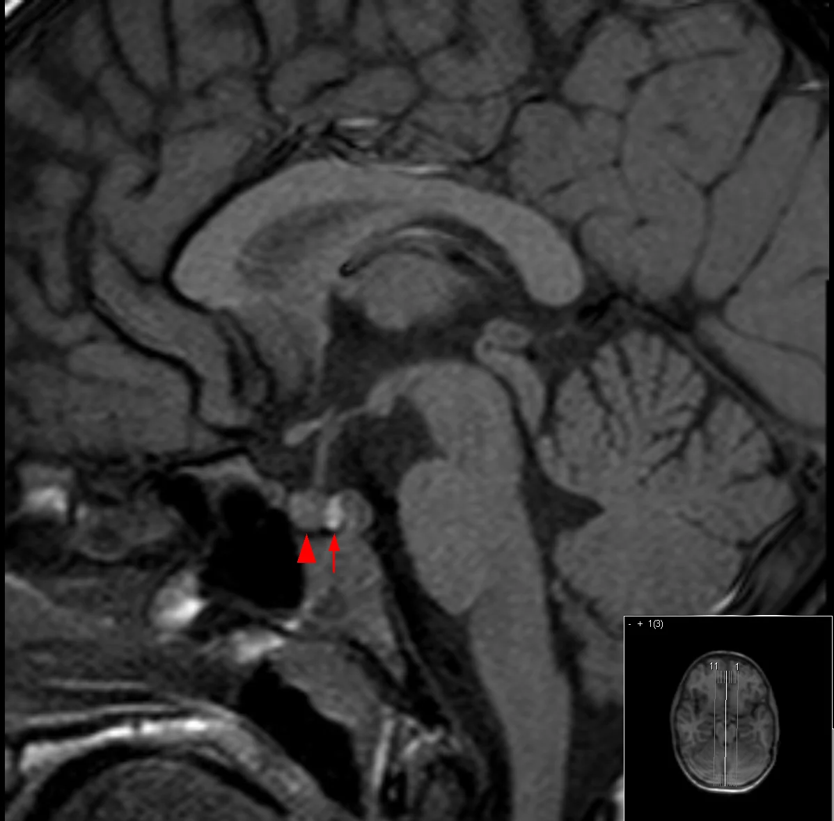
MRI pituitary is used for suspected cranial DI, looking for a structural cause and for loss of the normal posterior pituitary 'bright spot' on unenhanced T1-weighted imaging - a signal thought to reflect stored vasopressin, which is characteristically absent in cranial DI.3
Management
| Type | Management |
|---|---|
| Cranial DI | Desmopressin (DDAVP)4 - oral, intranasal or subcutaneous, titrated to symptoms, urine output and sodium. Also treat the underlying cause where identifiable (e.g. resect a tumour). |
| Nephrogenic DI | Treat/remove the cause - stop lithium if clinically possible (in discussion with psychiatry), correct hypercalcaemia or hypokalaemia. Thiazide diuretics paradoxically reduce urine volume by causing mild volume depletion, which increases proximal tubular water and sodium reabsorption. NSAIDs reduce renal prostaglandins, which normally antagonise ADH, and can further reduce urine volume. A low-salt, low-protein diet reduces the osmotic load that must be excreted. |
Complications
- Severe dehydration and hypernatraemia if fluid access is lost, which can cause confusion, seizures and death
- Dilutional hyponatraemia from over-treatment with desmopressin
- Hydronephrosis and a dilated, poorly contractile bladder from chronically high urine volumes in longstanding untreated disease
- Sleep disruption from severe nocturia
- Complications of the underlying cause - tumour, TBI, infiltrative disease
Red flags
Prognosis
With adequate desmopressin replacement, patients with cranial DI can expect normal fluid balance, normal sodium and an essentially normal life, provided they have reliable access to water and medication. Nephrogenic DI caused by lithium may improve after stopping the drug, though recovery can be incomplete after long-term use, and DI from irreversible structural causes (e.g. destructive tumour, severe TBI) is usually permanent.
Pregnancy-related DI from placental vasopressinase resolves within weeks of delivery. The main determinant of long-term outcome is reliable access to water and medication, and patient education about sick day management and the risks of both under- and over-treatment with desmopressin.
References
- NICE Clinical Knowledge Summaries. Diabetes insipidus. Available here
- Society for Endocrinology. Clinical practice guidance - disorders of water balance. Available here
- Di Iorgi N, Napoli F, Allegri AE et al. Diabetes insipidus - diagnosis and management. Hormone Research in Paediatrics. 2012. Available here
- BNF. Desmopressin acetate. Available here
- NHS. Diabetes insipidus. Available here
- Hellerhoff, CC BY-SA 3.0, via Wikimedia Commons. Available here
This article is written for revision and education. It is not clinical guidance and must not be used to make decisions about the care of a patient. Always check current NICE guidance and local protocols.