
Pulmonary Hypertension
Key points
- Definition: a mean pulmonary artery pressure above 20 mmHg at rest, measured by right heart catheterisation.
- The classification matters: the five WHO groups have entirely different treatments. Giving pulmonary vasodilators to the wrong group causes harm.
- Group 1: pulmonary arterial hypertension - idiopathic, heritable, drug-induced, or associated with connective tissue disease, HIV, portal hypertension or congenital heart disease.
- Group 2: pulmonary hypertension due to left heart disease. The commonest group by far, and treated by treating the left heart.
- Group 3: due to lung disease or hypoxia - COPD, interstitial lung disease, obstructive sleep apnoea. Treat the lung disease and give long-term oxygen.
- Presentation: insidious exertional breathlessness with a normal chest examination, later exertional syncope, chest pain and signs of right heart failure.
- Screening test: transthoracic echocardiography estimates pulmonary artery systolic pressure from the tricuspid regurgitant jet, but right heart catheterisation is required to confirm the diagnosis and group.
- Group 4: chronic thromboembolic pulmonary hypertension - the one potentially curable form, by pulmonary endarterectomy. Screen with a V/Q scan, not CTPA.
Introduction
Pulmonary hypertension is defined haemodynamically as a mean pulmonary artery pressure above 20 mmHg at rest, measured directly by right heart catheterisation. The threshold was lowered from 25 mmHg in the 2022 ESC/ERS guidelines, reflecting evidence that pressures above 20 mmHg carry prognostic significance.1
The pulmonary circulation is normally a low-pressure, high-compliance system, accommodating the entire cardiac output at roughly one sixth of systemic pressure. The right ventricle is a thin-walled, compliant chamber built for volume rather than pressure, and it tolerates a pressure load poorly. Almost all the morbidity and mortality of pulmonary hypertension results from progressive right ventricular failure.
Classification
| Group | Name | Typical causes | Treatment principle |
|---|---|---|---|
| 1 | Pulmonary arterial hypertension | Idiopathic; heritable (BMPR2 mutations); drug- and toxin-induced; associated with connective tissue disease (especially systemic sclerosis), HIV, portal hypertension, congenital heart disease and schistosomiasis | Targeted pulmonary vasodilator therapy in a specialist centre |
| 2 | Due to left heart disease | Heart failure with reduced or preserved ejection fraction, valvular disease (especially mitral) | Treat the left heart disease. Pulmonary vasodilators are contraindicated. |
| 3 | Due to lung disease or hypoxia | COPD, interstitial lung disease, obstructive sleep apnoea, obesity hypoventilation, chronic altitude exposure | Treat the lung disease, and give long-term oxygen therapy if hypoxic |
| 4 | Chronic thromboembolic pulmonary hypertension (CTEPH) and other pulmonary artery obstruction | Organised thrombus after pulmonary embolism; also tumour and arteritis | Lifelong anticoagulation and assessment for pulmonary endarterectomy - potentially curative |
| 5 | Unclear or multifactorial mechanisms | Sarcoidosis, haematological disorders (chronic haemolytic anaemia, myeloproliferative disease), metabolic disorders, chronic renal failure, fibrosing mediastinitis | Treat the underlying condition |
Group 2 is by far the commonest cause in clinical practice, since left heart disease is common and pulmonary hypertension is a frequent consequence of chronically raised left atrial pressure. Group 1 is rare - idiopathic pulmonary arterial hypertension affects around 5-15 per million - but it is the group with specific licensed treatment and dedicated commissioned services, which is why it receives disproportionate attention in teaching.
Pathophysiology
In pulmonary arterial hypertension, the small pulmonary arterioles undergo progressive remodelling: intimal proliferation and fibrosis, medial hypertrophy, adventitial thickening, in situ thrombosis, and the formation of plexiform lesions. The mechanism involves an imbalance between vasoconstrictor and proliferative mediators (endothelin-1, thromboxane) and vasodilator and antiproliferative mediators (nitric oxide, prostacyclin) - and these three pathways are precisely what the targeted drugs address.
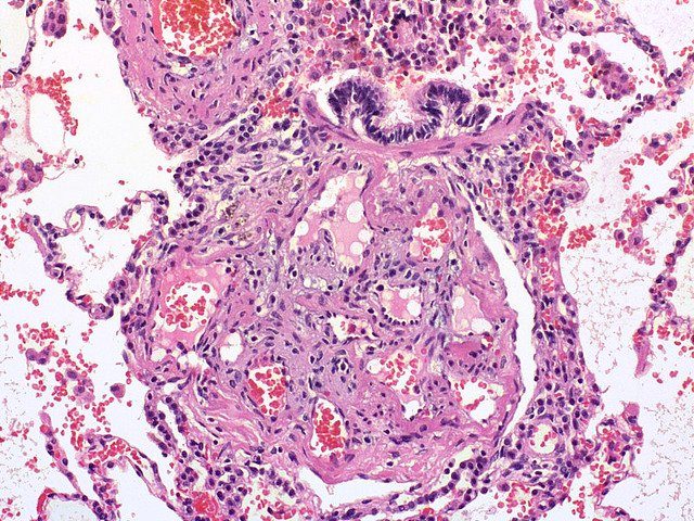
The consequence in every group is the same: increased right ventricular afterload. The right ventricle initially hypertrophies and maintains output, but it is poorly adapted to pressure work. Eventually it dilates, the tricuspid annulus stretches causing functional tricuspid regurgitation, and right ventricular output falls.
A dilated right ventricle also bows the interventricular septum leftwards, impairing left ventricular filling - a form of ventricular interdependence. This is why patients with severe pulmonary hypertension have a low cardiac output despite normal left ventricular contractility, and why they tolerate hypovolaemia, vasodilatation and anaesthesia so poorly.
Clinical features
Symptoms are insidious and non-specific, which is why the median delay from symptom onset to diagnosis remains around two years. Patients are commonly misdiagnosed with asthma, deconditioning, obesity or anxiety before the correct diagnosis is reached.
Symptoms
- Progressive exertional breathlessness - the cardinal and usually the first symptom, characteristically with a normal respiratory examination and a normal chest X-ray, which is what should prompt suspicion
- Fatigue and reduced exercise tolerance
- Exertional chest pain - from right ventricular ischaemia, as a hypertrophied right ventricle outgrows its blood supply
- Exertional presyncope and syncope - an ominous sign indicating the right ventricle cannot increase output to meet demand. Syncope on exertion is a red flag in any context and mandates urgent assessment.
- Palpitations - atrial arrhythmia is common and poorly tolerated
- Ankle swelling and abdominal distension - from right heart failure and ascites
- Hoarseness (Ortner syndrome) - compression of the left recurrent laryngeal nerve by a dilated pulmonary artery
- Haemoptysis - uncommon but recognised
Examination
- A loud pulmonary component of the second heart sound (P2) - often the earliest sign, and one worth listening for specifically
- A right ventricular heave at the left sternal edge, from right ventricular hypertrophy
- Raised JVP with prominent a waves - from forceful right atrial contraction against a hypertrophied right ventricle. Once tricuspid regurgitation develops, giant v waves appear.
- A pansystolic murmur at the left sternal edge, louder on inspiration - functional tricuspid regurgitation
- An early diastolic murmur (Graham Steell murmur) - pulmonary regurgitation from a dilated pulmonary annulus
- A right ventricular third or fourth heart sound
- Peripheral oedema, hepatomegaly with a pulsatile liver, and ascites - established right heart failure
- Central cyanosis - in advanced disease or where there is a right-to-left shunt
- Signs of a cause - sclerodactyly, telangiectasia and Raynaud's phenomenon in systemic sclerosis; clubbing and crackles in interstitial lung disease; a barrel chest in COPD; stigmata of chronic liver disease
Investigations
The diagnostic pathway has two objectives: confirm that pulmonary hypertension is present, and determine which group it belongs to. The second is the harder and more important task.
Initial investigations
- ECG - right axis deviation, right ventricular hypertrophy with a dominant R wave in V1, right atrial enlargement (P pulmonale) with peaked P waves over 2.5 mm in lead II, right bundle branch block, and a right ventricular strain pattern with T inversion in V1-V4. A normal ECG makes severe pulmonary hypertension less likely but does not exclude it.
- Chest X-ray - enlarged proximal pulmonary arteries with peripheral pruning (reduced peripheral vascular markings), right ventricular enlargement filling the retrosternal space, and evidence of underlying lung disease
- Bloods - FBC, U&Es, LFTs, BNP or NT-proBNP (which correlates with severity and prognosis), thyroid function, autoimmune screen (ANA, anti-centromere, anti-Scl-70 for systemic sclerosis), HIV testing, and a thrombophilia screen where CTEPH is suspected
- Arterial blood gas and pulse oximetry
- Pulmonary function tests with transfer factor - to identify group 3 disease. An isolated reduced transfer factor with otherwise preserved lung volumes is characteristic of pulmonary vascular disease.
- Overnight oximetry or sleep study - for obstructive sleep apnoea
Echocardiography
The principal screening test. Pulmonary artery systolic pressure is estimated from the peak velocity of the tricuspid regurgitant jet, added to an estimate of right atrial pressure from inferior vena cava size and collapsibility.
- Estimated pulmonary artery systolic pressure - reported as a low, intermediate or high probability of pulmonary hypertension
- Right ventricular size and function - dilatation, hypertrophy and reduced systolic function (measured as TAPSE)
- Right atrial dilatation and inferior vena cava dilatation without inspiratory collapse
- Septal flattening with a D-shaped left ventricle in the short axis view
- Assessment of the left heart - crucially, to identify group 2 disease, by looking for left ventricular systolic or diastolic dysfunction, left atrial enlargement and mitral or aortic valve disease
- A bubble study - to detect an intracardiac shunt
Excluding chronic thromboembolic disease
Right heart catheterisation
Required to confirm the diagnosis in anyone being considered for targeted therapy, and performed in a specialist centre. It measures:
- Mean pulmonary artery pressure - above 20 mmHg confirms pulmonary hypertension
- Pulmonary artery wedge pressure - 15 mmHg or below indicates pre-capillary disease; above 15 mmHg indicates post-capillary (group 2) disease
- Pulmonary vascular resistance - above 2 Wood units indicates a pulmonary vascular component
- Cardiac output and right atrial pressure - both important prognostic markers
- Vasoreactivity testing with inhaled nitric oxide - a positive response identifies the small minority of patients with idiopathic PAH who respond to high-dose calcium channel blockers
Cardiac MRI is increasingly used to assess right ventricular volumes and function, and the six-minute walk test and WHO functional class are used to grade severity and monitor response to treatment.
Management
Management is dictated by the group. All patients with suspected group 1 or group 4 disease should be referred to one of the designated national pulmonary hypertension centres, since targeted therapy is commissioned only through these services.
General measures for all groups
- Diuretics - for right heart failure and fluid overload, with careful monitoring since the right ventricle is preload-dependent and over-diuresis reduces cardiac output
- Long-term oxygen therapy - where there is chronic hypoxaemia, particularly in group 3
- Supervised exercise rehabilitation - improves exercise capacity and quality of life
- Vaccination - influenza, pneumococcal and COVID-19
- Avoid pregnancy - maternal mortality in pulmonary arterial hypertension remains of the order of 20-30%, and effective contraception must be discussed. Oestrogen-containing methods are generally avoided.
- Avoid high altitude and unpressurised flight
- Psychological support and palliative care input where appropriate
- Careful perioperative management - these patients tolerate anaesthesia poorly, and non-cardiac surgery should be discussed with the specialist centre
Group 1: pulmonary arterial hypertension
Three pathways are targeted, and modern practice uses upfront combination therapy in most patients rather than sequential escalation.
| Pathway | Drug class | Examples | Notes |
|---|---|---|---|
| Endothelin | Endothelin receptor antagonists | Bosentan, ambrisentan, macitentan | Hepatotoxic (bosentan especially) - requires monthly liver function monitoring. Teratogenic - pregnancy prevention programme required. |
| Nitric oxide | Phosphodiesterase-5 inhibitors; guanylate cyclase stimulators | Sildenafil, tadalafil; riociguat | Never combine riociguat with a PDE5 inhibitor - profound hypotension. Riociguat is also licensed for CTEPH. |
| Prostacyclin | Prostacyclin analogues and receptor agonists | Epoprostenol (IV), iloprost (inhaled), treprostinil, selexipag | Intravenous epoprostenol is used in the most severe disease; it has a very short half-life, so interruption of the infusion is life-threatening |
| Calcium channel blockade | High-dose nifedipine, diltiazem, amlodipine | - | Only for the small minority (under 10%) with a positive vasoreactivity test. Giving these to non-responders causes harm. |
Anticoagulation is no longer routine in idiopathic PAH, its role having become uncertain. Atrial septostomy and lung or heart-lung transplantation are options in advanced disease refractory to medical therapy.
Group 2: left heart disease
Treat the left heart disease. Optimise heart failure therapy, control blood pressure and rate, and intervene on valve disease. Pulmonary hypertension frequently improves once left atrial pressure falls. Targeted pulmonary vasodilators are not indicated and may cause pulmonary oedema.
Group 3: lung disease and hypoxia
Treat the lung disease and correct hypoxia with long-term oxygen therapy, which is the only intervention shown to improve survival in cor pulmonale complicating COPD. Treat obstructive sleep apnoea with CPAP. Targeted vasodilators are generally avoided because they worsen ventilation-perfusion matching, though they may be considered in a specialist centre where pulmonary hypertension is severe and out of proportion to the lung disease.
Group 4: chronic thromboembolic disease
- Lifelong anticoagulation in all patients
- Pulmonary endarterectomy - the treatment of choice where the disease is surgically accessible, and it is potentially curative, frequently normalising pulmonary pressures. In the UK this is performed at a single national centre.
- Balloon pulmonary angioplasty - for distal disease not amenable to surgery
- Riociguat - licensed for inoperable or persistent CTEPH after endarterectomy
- Every patient with pulmonary hypertension should be screened for CTEPH, because this is the group where a diagnosis changes everything
Complications
- Progressive right ventricular failure - the principal cause of death
- Arrhythmia - atrial flutter and fibrillation are common and poorly tolerated, since the stiff right ventricle depends on atrial contribution to filling. Restoring sinus rhythm is often a priority.
- Hepatic congestion and cardiac cirrhosis, with ascites and secondary renal impairment (cardiorenal syndrome)
- Syncope and sudden cardiac death
- Haemoptysis and pulmonary artery dissection or rupture - rare but catastrophic
- Ortner syndrome - hoarseness from recurrent laryngeal nerve compression
- Complications of therapy - hepatotoxicity from endothelin receptor antagonists, systemic hypotension, and line infection or interruption with intravenous prostacyclin
- High-risk pregnancy and perioperative mortality
Red flags
Prognosis
Prognosis depends on the group, the severity at diagnosis, and above all on right ventricular function, which is a better predictor of survival than pulmonary artery pressure itself.
Idiopathic pulmonary arterial hypertension had a median survival of under three years before targeted therapy became available. Modern combination therapy has improved this substantially, with contemporary registry data showing five-year survival of around 60-70%, though it remains a life-limiting condition. Risk stratification tools combining WHO functional class, six-minute walk distance, NT-proBNP, right atrial pressure and cardiac index are used to guide escalation of therapy.
Group 2 and group 3 pulmonary hypertension carry the prognosis of the underlying disease, but the development of pulmonary hypertension is a marker of more advanced disease and worse outcome in both heart failure and COPD.
Group 4 (CTEPH) is the exception and the reason it must be actively sought. Untreated it progresses relentlessly, but pulmonary endarterectomy in operable disease can be curative, with the majority of patients returning to near-normal exercise capacity and long-term survival approaching that of the general population. Around 1-3% of patients develop CTEPH after an acute pulmonary embolism, and persistent breathlessness after a PE should never be dismissed.
Adverse prognostic markers across groups include WHO functional class III or IV, a six-minute walk distance under 165 metres, a markedly raised NT-proBNP, a raised right atrial pressure, a low cardiac index, and the presence of a pericardial effusion on echocardiography.
References
- Humbert M, Kovacs G, Hoeper MM et al. 2022 ESC/ERS Guidelines for the diagnosis and treatment of pulmonary hypertension. European Heart Journal. 2022. Available here
- NHS England. Specialised services for pulmonary hypertension. Available here
- NICE TA272. Riociguat for treating chronic thromboembolic pulmonary hypertension. See also NICE guidance on bosentan and sildenafil. Available here
- NICE NG115. Chronic obstructive pulmonary disease in over 16s. 2018, updated 2019. Available here
- NICE NG158. Venous thromboembolic diseases: diagnosis, management and thrombophilia testing. 2020, updated 2023. Available here
- Pulmonary Hypertension Association UK. Information for healthcare professionals. Available here
- Regitz-Zagrosek V, Roos-Hesselink JW, Bauersachs J et al. 2018 ESC Guidelines for the management of cardiovascular diseases during pregnancy. European Heart Journal. 2018. Available here
- BNF. Bosentan - indications, cautions and monitoring. Available here
This article is written for revision and education. It is not clinical guidance and must not be used to make decisions about the care of a patient. Always check current NICE guidance and local protocols.