
Erythema Nodosum
Key points
- Erythema nodosum: a delayed-type hypersensitivity reaction causing septal panniculitis - inflammation of the fat septae in the subcutaneous tissue - presenting as tender, erythematous nodules, classically on the shins.
- Evolution: individual nodules evolve through the colour changes of a resolving bruise over 2-6 weeks, without ulcerating or scarring - a key distinguishing feature from other panniculitides.
- It is a sign, not a diagnosis: erythema nodosum is a reaction pattern with a long list of possible triggers; finding the underlying cause is the point of the work-up, not confirming the rash itself.
- Common causes: up to half of cases are idiopathic; identifiable causes include streptococcal infection (especially in children), sarcoidosis, tuberculosis, inflammatory bowel disease, pregnancy, and drugs including the combined oral contraceptive and sulfonamides.
- Lofgren syndrome: the combination of erythema nodosum, bilateral hilar lymphadenopathy and polyarthralgia, representing an acute presentation of sarcoidosis with an excellent prognosis.
- Diagnosis: clinical, from the characteristic appearance and evolution; investigations are directed at identifying the underlying cause rather than confirming the rash.
- Management: treat the underlying cause, with rest, leg elevation and NSAIDs for symptomatic relief; potassium iodide or colchicine are options for persistent, idiopathic disease.
- Prognosis: usually self-limiting within 3-6 weeks without scarring; recurrence and long-term outlook depend on the underlying cause rather than the rash itself.
Introduction
Erythema nodosum is the commonest form of panniculitis - inflammation of subcutaneous fat - and presents with characteristic tender, red nodules, almost always on the shins. It occurs most often in young adults, is around five times more common in women, and has its peak incidence between the ages of 20 and 45.1
The most important thing to understand about erythema nodosum is that it is a reaction pattern rather than a disease in its own right - the skin's non-specific response to a wide range of underlying triggers. The clinical task is not simply to recognise the rash, which is usually straightforward, but to identify why it has occurred, since the list of possible causes ranges from a self-limiting streptococcal sore throat to tuberculosis or sarcoidosis.
Pathophysiology
Erythema nodosum is thought to be a delayed-type (type IV) hypersensitivity reaction, with immune complexes depositing in and around the septae of subcutaneous fat lobules, provoking a septal panniculitis - inflammation concentrated in the fibrous septae between fat lobules rather than within the lobules themselves, which is the histological hallmark that distinguishes it from lobular panniculitides.2
The shins are the classic site partly because of the relatively poor collateral circulation and lower ambient temperature of that region, which is thought to predispose the local vasculature and connective tissue to this type of inflammatory reaction. As the inflammatory infiltrate resolves, extravasated red cells within the fat are broken down in the same sequence as a bruise, which is why individual lesions visibly progress through red, purple, green and yellow before fading - a distinctive and diagnostically useful feature.
Causes
Roughly 30-50% of cases have no identifiable cause and are labelled idiopathic. Among the remainder, infection, systemic inflammatory disease, pregnancy and drugs account for most cases.3
| Category | Examples |
|---|---|
| Infection | Streptococcal pharyngitis (especially in children), tuberculosis, Yersinia enterocolitis, Mycoplasma pneumoniae, deep fungal infections (histoplasmosis, coccidioidomycosis in endemic areas) |
| Systemic inflammatory disease | Sarcoidosis (including Lofgren syndrome), inflammatory bowel disease (Crohn's disease more than ulcerative colitis), Behcet disease |
| Drugs | Combined oral contraceptive pill, sulfonamides, penicillins, NSAIDs |
| Other | Pregnancy, malignancy (rarely, particularly lymphoma), and idiopathic (no cause found in a substantial proportion) |
Clinical features
Erythema nodosum presents with tender, warm, erythematous nodules, typically 2-6 cm across, most often bilateral and symmetrical over the shins (the pretibial surface), though the forearms, thighs and trunk can occasionally be affected.
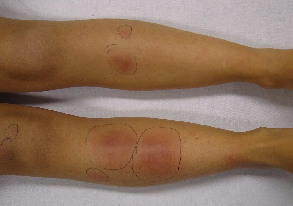
Nodules are ill-defined rather than sharply marginated, and unlike most other panniculitides, they never ulcerate or scar - a useful discriminating feature. Over 2-6 weeks, individual lesions flatten and fade through the colour sequence of a resolving bruise (red to purple-blue to green-yellow) before disappearing completely.
Systemic symptoms frequently accompany the rash and can precede it by days: fever, malaise, and arthralgia or frank arthritis, most often affecting the ankles and knees. These systemic features, alongside any symptoms pointing to a specific cause (sore throat, cough, diarrhoea, weight loss), are as important to elicit as the rash itself.
Clinical examination
- Distribution and evolution - bilateral pretibial nodules at varying stages of the bruise-like colour sequence is highly suggestive
- Absence of ulceration - important to note explicitly, since ulceration points towards an alternative panniculitis
- Lymphadenopathy - particularly cervical or generalised, which might point towards an infective or lymphomatous cause
- Joint examination - swelling or tenderness, especially ankles and knees
- Chest examination - for signs relevant to tuberculosis or sarcoidosis, though findings are often normal even with significant intrathoracic disease
- Abdominal examination and a brief systems review - for symptoms suggesting inflammatory bowel disease
Differential diagnosis
- Erythema induratum (nodular vasculitis) - a lobular panniculitis, typically on the calves rather than the shins, that characteristically does ulcerate; historically associated with tuberculosis4
- Superficial thrombophlebitis - a tender, cord-like, linear lesion following a vein rather than discrete round nodules
- Cellulitis - typically unilateral, with more diffuse spreading erythema and systemic signs of infection, rather than discrete symmetrical nodules
- Insect bite reactions - usually more acute in onset and less symmetrically distributed
- Polyarteritis nodosa - can cause tender subcutaneous nodules along the course of arteries, usually with other systemic vasculitic features
- Panniculitis of other causes (pancreatic panniculitis, lupus panniculitis) - distinguished by the clinical context and, where needed, biopsy
Investigations
The diagnosis of erythema nodosum itself is clinical. Investigation is directed entirely at finding the underlying cause.
- FBC, ESR/CRP - raised inflammatory markers are typical and support an active inflammatory process, though non-specific
- Throat swab and anti-streptolysin O (ASO) titre - if a preceding streptococcal infection is suspected, especially in children
- Chest X-ray - looking for bilateral hilar lymphadenopathy (sarcoidosis, Lofgren syndrome) or changes suggesting tuberculosis
- Interferon-gamma release assay or tuberculin skin test - if tuberculosis is a realistic possibility from the history or chest X-ray
- Stool studies and faecal calprotectin, or referral for colonoscopy - if bowel symptoms suggest inflammatory bowel disease
- Skin biopsy - reserved for atypical presentations, since it is rarely needed when the clinical picture is classical, but distinguishes septal from lobular panniculitis when the diagnosis is in doubt
Management
Management has two parallel strands: treating any identified underlying cause, and controlling the symptoms of the rash itself, which is usually self-limiting regardless.
- Treat the underlying cause where one is found - for example antibiotics for streptococcal infection, standard anti-tuberculous therapy for tuberculosis, or optimising control of underlying inflammatory bowel disease
- Rest and leg elevation - reduces oedema and discomfort in the affected legs
- NSAIDs - first-line for symptomatic relief of pain and inflammation, used with the usual cautions8
- Stop any potentially causative drug (combined oral contraceptive, sulfonamide, penicillin) where a drug cause is suspected
- Potassium iodide - a recognised, effective second-line option for persistent idiopathic disease not settling with the above
- Colchicine or a short course of oral corticosteroids - considered for severe or persistent disease once infection, particularly tuberculosis, has been excluded, since corticosteroids can worsen an unrecognised infective cause
Complications
- Chronic or recurrent erythema nodosum - a minority of patients, particularly with an ongoing underlying cause such as inflammatory bowel disease, have repeated episodes
- Progression of the underlying disease if the trigger is not identified and treated - the rash itself resolves regardless, so failing to investigate risks missing a condition such as tuberculosis or inflammatory bowel disease that needs its own treatment
- Persistent joint symptoms - arthralgia can occasionally outlast the skin lesions by weeks
- Post-inflammatory bruising/pigmentation at the site of resolved nodules, though true scarring does not occur
Red flags
Prognosis
Erythema nodosum itself is a self-limiting condition, with individual lesions resolving over 2-6 weeks without scarring, even without specific treatment for the rash. The long-term outlook is determined entirely by the underlying cause: idiopathic disease and streptococcal-associated disease in children generally have an excellent prognosis with no recurrence, while erythema nodosum associated with inflammatory bowel disease often recurs in parallel with flares of the underlying condition.6
Lofgren syndrome, despite its dramatic radiological appearance, resolves spontaneously in the great majority of patients within 6-24 months and rarely progresses to chronic sarcoidosis, which is worth explaining clearly to patients who understandably find a chest X-ray showing lymphadenopathy alarming.7
References
- DermNet NZ. Erythema nodosum. Available here
- Requena L, Sanchez Yus E. Erythema nodosum. Seminars in Cutaneous Medicine and Surgery. 2007. Available here
- Schwartz RA, Nervi SJ. Erythema nodosum: a sign of systemic disease. American Family Physician. 2007. Available here
- Gilchrist H, Patterson JW. Erythema nodosum and erythema induratum (nodular vasculitis): diagnosis and management. Dermatologic Therapy. 2010. Available here
- Mana J, Gomez-Vaquero C, Montero A et al. Lofgren's syndrome revisited: a study of 186 patients. American Journal of Medicine. 1999. Available here
- NICE Clinical Knowledge Summaries. Crohn's disease. Available here
- Iannuzzi MC, Rybicki BA, Teirstein AS. Sarcoidosis. New England Journal of Medicine. 2007. Available here
- BNF. Non-steroidal anti-inflammatory drugs - treatment summary. Available here
This article is written for revision and education. It is not clinical guidance and must not be used to make decisions about the care of a patient. Always check current NICE guidance and local protocols.