
Respiratory Failure: Type 1 and Type 2, and What to Do About Each
Key points
- Respiratory failure: failure of gas exchange, defined on an arterial blood gas as a PaO2 below 8 kPa breathing room air.
- Type 1: PaO2 below 8 kPa with a normal or low PaCO2. A failure of oxygenation, most often from ventilation-perfusion mismatch.
- Type 2: PaO2 below 8 kPa with a PaCO2 above 6 kPa. A failure of ventilation - the patient is not moving enough air, whatever the reason.
- Five mechanisms of hypoxaemia: V/Q mismatch, shunt, hypoventilation, diffusion impairment and a low inspired oxygen concentration.
- Shunt: the one form of hypoxaemia that does not correct with supplemental oxygen, because blood bypasses ventilated alveoli entirely.
- Acute or chronic: bicarbonate answers it. Acute type 2 failure has a low pH with a normal bicarbonate; chronic has a normal pH with a high bicarbonate; acute-on-chronic has a low pH with a high bicarbonate.
- Hypercapnia signs: headache, drowsiness, confusion, a bounding pulse, warm peripheries and a flapping tremor. Drowsiness in a breathless patient is an ominous sign.
- Treatment: treat the cause, give oxygen to the correct target, and add non-invasive ventilation for a persisting respiratory acidosis. Agree a ceiling of care early.
Introduction
Respiratory failure is the inability of the respiratory system to maintain adequate gas exchange. It is defined on an arterial blood gas, not clinically, as a PaO2 below 8 kPa (60 mmHg) breathing room air, and it is divided by the PaCO2.
| Type 1 | Type 2 | |
|---|---|---|
| PaO2 | Below 8 kPa | Below 8 kPa |
| PaCO2 | Normal or low | Above 6 kPa |
| The problem | Oxygenation - gas exchange at the alveolus | Ventilation - not enough air moving in and out |
| Typical mechanism | V/Q mismatch or shunt | Hypoventilation |
| Typical causes | Pneumonia, pulmonary oedema, PE, asthma, ARDS | COPD, opiates, neuromuscular disease, chest wall disease |
| Oxygen target | 94-98% | 88-92% if at risk of CO2 retention |
| Ventilatory support | CPAP or high-flow nasal oxygen | NIV (BiPAP) |
The distinction is worth making carefully, because it changes both the oxygen target and the mode of ventilatory support - and because type 2 failure means the patient is failing to ventilate, which is a more immediately dangerous situation than isolated hypoxaemia.
A useful additional grouping used in critical care describes type 3 (perioperative, from atelectasis) and type 4 (shock, where respiratory muscles cannot be adequately perfused), but for most purposes the type 1 and type 2 division is what matters.
Mechanisms of hypoxaemia
There are only five, and identifying which is operating explains both the blood gas and the response to oxygen.
| Mechanism | What is happening | Response to oxygen | Examples |
|---|---|---|---|
| V/Q mismatch | Alveoli are ventilated but underperfused, or perfused but underventilated. By far the commonest mechanism. | Corrects | Pneumonia, COPD, asthma, pulmonary embolism, atelectasis |
| Shunt | Blood passes from the right to the left side of the circulation without contacting ventilated alveoli | Does not correct - the shunted blood never meets the oxygen | Consolidation, ARDS, pulmonary oedema, right-to-left cardiac shunt, arteriovenous malformation |
| Hypoventilation | Insufficient alveolar ventilation, so alveolar oxygen falls as alveolar CO2 rises | Corrects, but the PaCO2 keeps rising unless ventilation is supported | Opiates, sedatives, neuromuscular disease, chest wall disease, severe COPD |
| Diffusion impairment | Thickened alveolar-capillary membrane slows equilibration | Corrects | Interstitial lung disease, emphysema, pulmonary oedema |
| Low inspired oxygen | Reduced FiO2 or barometric pressure | Corrects | Altitude, smoke or gas inhalation |
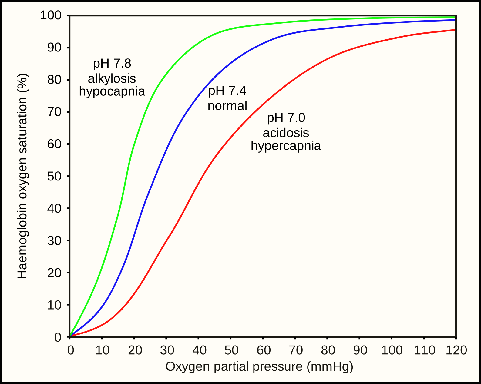
Two consequences of that curve are worth carrying to the bedside. First, a patient can lose a great deal of respiratory reserve before the saturation moves at all, so a normal SpO2 is not reassurance in a breathless patient - look at the respiratory rate. Second, once saturations start to fall the patient is already on the steep part of the curve and will deteriorate quickly. The curve shifts right (offloading oxygen to tissues) with acidosis, hypercapnia, pyrexia and raised 2,3-DPG, and left with alkalosis, hypocapnia, hypothermia, carboxyhaemoglobin and fetal haemoglobin.
Causes
Type 1 respiratory failure
- Pneumonia and other infection
- Pulmonary oedema - cardiogenic and non-cardiogenic
- Pulmonary embolism
- Acute asthma - type 1 initially, progressing to type 2 as the patient tires
- Pneumothorax
- Acute respiratory distress syndrome
- Interstitial lung disease and bronchiectasis
- Pulmonary haemorrhage, atelectasis and lobar collapse
Type 2 respiratory failure
It helps to work anatomically from the brain outwards, because that is the order in which the causes are missed.
| Level | Causes |
|---|---|
| Reduced central drive | Opioids, benzodiazepines and other sedatives, anaesthetic agents, alcohol, brainstem stroke, raised intracranial pressure, encephalitis, central hypoventilation syndromes |
| Spinal cord and nerve | High cervical cord injury, Guillain-Barre syndrome, motor neurone disease, poliomyelitis, phrenic nerve palsy, critical illness polyneuropathy |
| Neuromuscular junction and muscle | Myasthenia gravis, Lambert-Eaton syndrome, botulism, muscular dystrophies, myopathies, hypokalaemia and hypophosphataemia |
| Chest wall and pleura | Kyphoscoliosis, ankylosing spondylitis, flail chest, obesity hypoventilation syndrome, large pleural effusions, circumferential burns |
| Airways and lung | COPD - by far the commonest cause, severe asthma in the exhausted phase, obstructive sleep apnoea, cystic fibrosis, advanced bronchiectasis, upper airway obstruction |
Clinical features
Signs of hypoxaemia
- Breathlessness and tachypnoea - the respiratory rate is the most sensitive single sign and the most often unrecorded
- Restlessness, agitation and confusion - hypoxic patients are frequently mistaken for being difficult or intoxicated
- Central cyanosis - a bluish tinge to the lips and tongue, appearing when deoxygenated haemoglobin exceeds about 50 g/L. It is therefore absent in anaemia even when severely hypoxic, and appears early in polycythaemia.
- Tachycardia and arrhythmias, with bradycardia as a pre-terminal sign
- Use of accessory muscles, intercostal recession and tracheal tug
- In chronic hypoxaemia: secondary polycythaemia, pulmonary hypertension and cor pulmonale, and finger clubbing depending on the cause
Signs of hypercapnia
- Headache - from cerebral vasodilatation, characteristically worse in the morning in chronic retention
- Drowsiness, confusion and, eventually, coma - CO2 narcosis
- Flapping tremor (asterixis) - a coarse irregular flap of the outstretched hands
- Bounding pulse and warm peripheries - from peripheral vasodilatation
- Papilloedema - in severe chronic hypercapnia
- Myoclonic jerks and seizures in extreme cases
Investigations
Arterial blood gas
The ABG defines respiratory failure, classifies it, and tells you whether it is acute or chronic. Record the inspired oxygen concentration with every gas, since the numbers are meaningless without it.
| pH | PaCO2 | Bicarbonate | Base excess | |
|---|---|---|---|---|
| Acute | Low (under 7.35) | High | Normal | Normal |
| Chronic | Normal | High | High | Positive |
| Acute-on-chronic | Low | High | High | Positive |
The logic is that renal compensation takes 2 to 3 days. A patient whose CO2 rose this morning has not had time to retain bicarbonate, so their pH falls. A patient who has been hypercapnic for years has a high bicarbonate and a pH pulled back towards normal. The combination of a low pH with a high bicarbonate therefore identifies a chronic retainer who has acutely decompensated - the group who benefit most from non-invasive ventilation.
Other investigations
- Chest X-ray - for pneumonia, oedema, pneumothorax, effusion and collapse. A clear film in a hypoxic patient should prompt thought about pulmonary embolism, shunt, or a non-pulmonary cause.
- ECG - for arrhythmia, ischaemia and right heart strain
- Bloods - FBC (anaemia, polycythaemia, infection), U&Es, CRP, and bicarbonate which reveals chronic retention even without a gas
- Bedside spirometry with serial FVC - essential in suspected neuromuscular failure
- CT pulmonary angiogram - where pulmonary embolism is suspected
- Echocardiogram - for cardiac failure, pulmonary hypertension or intracardiac shunt
- Toxicology and a trial of naloxone where opioid toxicity is possible
- Sleep study and daytime blood gas - for suspected obesity hypoventilation or OSA
Management
Immediate approach
- ABCDE assessment, with continuous monitoring and early senior involvement
- Give oxygen to the correct target - 94-98% in most patients, 88-92% in anyone at risk of hypercapnic respiratory failure until a blood gas is available
- Take an arterial blood gas, recording the FiO2
- Treat the underlying cause - this is what actually resolves the failure: antibiotics for pneumonia, bronchodilators and steroids for COPD or asthma, diuretics for pulmonary oedema, anticoagulation for PE, a chest drain for pneumothorax, naloxone for opioid toxicity
- Reassess after 30 to 60 minutes with a repeat gas, and escalate if the pH is falling or the PaCO2 rising
Ventilatory support
| Modality | What it does | Main indications |
|---|---|---|
| Controlled oxygen | Raises inspired oxygen concentration | All hypoxaemic patients, at the appropriate target |
| High-flow nasal oxygen | Delivers heated humidified gas at high flow with a small positive pressure and washout of dead space | Type 1 failure not responding to standard oxygen, and as a comfortable alternative to CPAP |
| CPAP | Constant positive pressure that recruits collapsed alveoli and reduces the work of breathing. Does not augment ventilation. | Type 1 failure - cardiogenic pulmonary oedema, atelectasis, and obstructive sleep apnoea |
| NIV (BiPAP) | Separate inspiratory and expiratory pressures, so it augments tidal volume and clears CO2 | Type 2 failure with respiratory acidosis - typically COPD with pH 7.25-7.35 after optimal medical therapy, and also chest wall and neuromuscular disease and obesity hypoventilation |
| Invasive ventilation | Full control of ventilation with a protected airway | Failure of NIV, exhaustion, reduced conscious level, an unprotected airway, or a condition expected to resolve |
Chronic respiratory failure
- Long-term oxygen therapy - for chronic hypoxaemia meeting the criteria, used for at least 15 hours a day. It is the only treatment shown to improve survival in COPD.
- Home NIV - for chronic hypercapnic failure in COPD, obesity hypoventilation, chest wall disease and neuromuscular disease. It improves survival and reduces admissions in selected patients.
- Treat the underlying disease and its contributors - weight loss, smoking cessation, pulmonary rehabilitation, vaccination and nutritional support
- Cough assist and secretion management in neuromuscular disease
- Advance care planning, including preferences about ventilation and hospital admission
- Palliative management of breathlessness - opioids and benzodiazepines are effective and appropriate, and fear of respiratory depression should not prevent their proper use at the end of life
Complications
- Cardiac arrest - hypoxia is a reversible cause and one of the four Hs
- Hypoxic brain injury and cognitive impairment
- Arrhythmias - atrial fibrillation is common, and hypoxia lowers the threshold for ventricular arrhythmia
- Pulmonary hypertension and cor pulmonale from chronic hypoxic vasoconstriction
- Secondary polycythaemia with hyperviscosity and thrombotic risk
- CO2 narcosis, seizures and coma
- Acute kidney injury and multi-organ failure
- Complications of ventilation - ventilator-associated pneumonia, barotrauma and pneumothorax, tracheal injury, and the consequences of prolonged sedation and immobility
- Pressure ulceration from NIV masks, aspiration, and gastric distension
- ICU-acquired weakness and post-intensive care syndrome
Red flags
Prognosis
Prognosis is determined almost entirely by the underlying cause and its reversibility, not by the blood gas numbers themselves. Type 1 failure from pneumonia in a previously well patient usually resolves completely; type 2 failure in advanced COPD or progressive neuromuscular disease reflects a trajectory that ventilatory support can modify but not reverse.
- Acute hypercapnic failure in COPD treated with NIV - in-hospital mortality of around 10 to 20%, considerably better than before NIV became standard, but each admission marks a step down and around a third are readmitted within 3 months
- Guillain-Barre syndrome requiring ventilation - the great majority recover fully, though ventilation may be needed for weeks
- ARDS - mortality of around 35 to 45%, mainly from the underlying illness and multi-organ failure
- Chronic respiratory failure on home NIV - survival and admission rates improve substantially in selected groups, particularly obesity hypoventilation and chest wall disease
Two things consistently improve outcomes and are within the reach of any clinician: recognising deterioration early, which usually means acting on a respiratory rate and a repeat blood gas rather than waiting for saturations to fall, and having the escalation conversation in advance. Patients whose ceiling of care has been thought about, discussed and recorded receive treatment that matches their wishes, and their families are spared decisions made under pressure in the middle of the night.
References
- British Thoracic Society / ICS. Guideline for the ventilatory management of acute hypercapnic respiratory failure in adults. 2016. Available here
- British Thoracic Society. Guideline for oxygen use in adults in healthcare and emergency settings. 2017. Available here
- NICE NG115. Chronic obstructive pulmonary disease in over 16s: diagnosis and management. 2018, updated 2019. Available here
- NICE NG202. Obstructive sleep apnoea/hypopnoea syndrome and obesity hypoventilation syndrome in over 16s. 2021. Available here
- MHRA. Pulse oximeters: use in people with darker skin tone. Available here
- Resuscitation Council UK. Adult advanced life support guidelines: reversible causes. Available here
- British Thoracic Society. Guideline for home oxygen use in adults. 2015. Available here
- Peter Southwood, CC0, via Wikimedia Commons. Available here
This article is written for revision and education. It is not clinical guidance and must not be used to make decisions about the care of a patient. Always check current NICE guidance and local protocols.