
Prematurity: Complications and Care of the Preterm Infant
Key points
- Definition: birth before 37+0 weeks of gestation, subdivided into late preterm (34-36+6), moderate (32-33+6), very preterm (28-31+6) and extremely preterm (under 28 weeks).
- How common: around 7-8% of UK births. Prematurity is the leading cause of neonatal death and of death in children under 5 worldwide.
- Strongest risk factor: a previous preterm birth. Multiple pregnancy, a short cervix, previous cervical surgery and infection are the other major contributors.
- Antenatal corticosteroids: given between 24+0 and 33+6 weeks, they reduce respiratory distress syndrome, intraventricular haemorrhage, necrotising enterocolitis and neonatal death.
- Magnesium sulfate: given for fetal neuroprotection below 30 weeks, it reduces the risk of cerebral palsy.
- The central problem: every organ system is immature at once. Surfactant deficiency, a fragile germinal matrix, an immature gut and poor thermoregulation are the four that dominate the first days.
- Corrected age: used for assessing growth and development until 2 years. Immunisations, by contrast, are given at chronological age and are never postponed.
- Outcome: survival rises steeply with each additional week - from roughly a third at 23 weeks to over 95% beyond 28 weeks - and neurodevelopmental impairment follows the same gradient.
Introduction
Preterm birth is delivery before 37 completed weeks of gestation. It affects around 7-8% of UK births and accounts for the majority of neonatal intensive care activity, most neonatal deaths, and a substantial share of childhood disability.
The reason preterm infants are so vulnerable is straightforward: the last trimester is when the fetus lays down surfactant, brown fat, immunoglobulin, iron stores and bone mineral, and when the lungs, gut, brain and retina complete their structural development. A baby delivered before that has to do all of it ex utero, in an environment for which none of those systems is ready.
Two things follow for exam purposes. First, the complications of prematurity are best learned system by system rather than as a list, because each one traces back to a specific immaturity. Second, several of the most effective interventions happen before delivery - antenatal steroids, magnesium sulfate and in-utero transfer - which is why obstetric and neonatal management cannot be separated.1
Classification and causes
| By gestation | Definition | By birth weight | Definition |
|---|---|---|---|
| Late preterm | 34+0 to 36+6 weeks | Low birth weight (LBW) | Under 2,500 g |
| Moderate preterm | 32+0 to 33+6 weeks | Very low birth weight (VLBW) | Under 1,500 g |
| Very preterm | 28+0 to 31+6 weeks | Extremely low birth weight (ELBW) | Under 1,000 g |
| Extremely preterm | Under 28+0 weeks |
How preterm birth happens
- Spontaneous preterm labour - roughly half of cases, with intact membranes
- Preterm prelabour rupture of membranes (PPROM) - around a quarter
- Medically indicated (iatrogenic) delivery - around a quarter, where continuing the pregnancy is more dangerous than delivering: pre-eclampsia, fetal growth restriction, abnormal fetal monitoring, placenta praevia or abruption
Risk factors
- Previous preterm birth - the single strongest predictor, and the basis for prophylactic progesterone or cervical cerclage in a subsequent pregnancy
- Multiple pregnancy - around 60% of twins deliver preterm
- Short cervix on transvaginal ultrasound, and previous cervical surgery such as LLETZ or cone biopsy
- Uterine anomalies and fibroids
- Infection - bacterial vaginosis, urinary tract infection, and chorioamnionitis
- Smoking, alcohol and substance misuse
- Maternal age under 18 or over 40, low pre-pregnancy BMI, and a short interval since the last pregnancy
- Socioeconomic deprivation and Black ethnicity, both of which carry a substantially raised risk in UK data
- Assisted conception, polyhydramnios and antepartum haemorrhage
Antenatal and delivery room management
The interventions with the largest effect on outcome are delivered before the baby is born.1
- Antenatal corticosteroids - a course of betamethasone or dexamethasone offered between 24+0 and 33+6 weeks where preterm birth is anticipated within 7 days, and considered from 22+0 and up to 35+6. They accelerate type II pneumocyte maturation and surfactant production, and reduce respiratory distress syndrome, intraventricular haemorrhage, necrotising enterocolitis and neonatal mortality.
- Magnesium sulfate for fetal neuroprotection - offered below 30 weeks and considered between 30+0 and 33+6, reducing the risk of cerebral palsy
- Tocolysis - nifedipine, with atosiban as an alternative, used for up to 48 hours purely to create time for steroids to work and for in-utero transfer. It does not improve neonatal outcome by itself.
- In-utero transfer to a unit with an appropriate level of neonatal intensive care, which produces better outcomes than transferring the baby after delivery
- Antibiotics - erythromycin for PPROM, and intrapartum benzylpenicillin for group B streptococcal prophylaxis
- Deferred cord clamping for at least 60 seconds where the baby is stable, which increases circulating blood volume and reduces the need for transfusion and the risk of intraventricular haemorrhage
In the delivery room the priorities are thermal care and lung recruitment: the baby is placed in a polyethylene bag or wrap without drying under a radiant heater, a hat is applied, saturations are monitored, and respiratory support is given with early CPAP rather than routine intubation where possible.
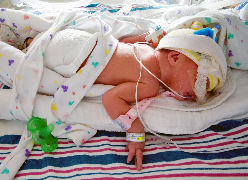
Complications by system
Respiratory
- Respiratory distress syndrome (RDS) - surfactant deficiency. Type II pneumocytes begin producing surfactant at around 24 weeks but adequate amounts are not present until 34-36 weeks. Without it, alveolar surface tension is unopposed, alveoli collapse at end-expiration, and compliance falls. The baby presents within minutes to hours with tachypnoea, grunting, nasal flaring, recession and cyanosis; the chest radiograph shows a diffuse ground-glass appearance with air bronchograms and reduced lung volumes.
- Treatment of RDS: early CPAP, with exogenous surfactant given via a fine catheter (less invasive surfactant administration, LISA) or by brief intubation, and mechanical ventilation where these fail2
- Apnoea of prematurity - immature central respiratory drive, treated with caffeine citrate, which also reduces the incidence of bronchopulmonary dysplasia and cerebral palsy
- Bronchopulmonary dysplasia - chronic lung disease resulting from the combination of immature lungs, oxygen toxicity and ventilator-induced injury
- Air leak - pneumothorax and pulmonary interstitial emphysema, and pulmonary haemorrhage
| Diagnosis | Typical picture |
|---|---|
| Respiratory distress syndrome | Preterm, onset within minutes to hours and worsening over 48 hours, ground-glass lungs with air bronchograms, responds to surfactant |
| Transient tachypnoea of the newborn | Term or late preterm, often after caesarean section, tachypnoea from delayed clearance of lung fluid, fluid in the horizontal fissure, resolves within 24-48 hours |
| Congenital pneumonia or early-onset sepsis | Risk factors such as prolonged rupture of membranes or maternal group B streptococcus, temperature instability, patchy or asymmetrical changes, raised inflammatory markers |
| Meconium aspiration syndrome | Term or post-term, meconium-stained liquor, patchy infiltrates with hyperinflation, often with pulmonary hypertension |
| Pneumothorax | Sudden deterioration, asymmetrical chest movement, reduced air entry, transillumination positive |
| Congenital heart disease | Cyanosis without marked respiratory distress, failure to improve with oxygen, abnormal pre- and post-ductal saturations |
| Congenital diaphragmatic hernia | Scaphoid abdomen, displaced apex beat, bowel loops in the chest on radiograph |
Cardiovascular
- Patent ductus arteriosus - the duct normally closes as oxygen tension rises and circulating prostaglandins fall, but in preterm infants it frequently stays open, producing a left-to-right shunt, a continuous machinery murmur, bounding pulses, a wide pulse pressure and difficulty weaning from respiratory support. Treated conservatively where possible, or with ibuprofen, paracetamol or indometacin to inhibit prostaglandin synthesis, and by surgical ligation or transcatheter device closure if that fails.
- Hypotension from immature vasomotor control and myocardial function
Neurological
- Intraventricular haemorrhage - bleeding from the germinal matrix, a highly vascular, fragile structure adjacent to the lateral ventricles that involutes by around 32 weeks. Risk is greatest in the first 72 hours and is increased by fluctuating blood pressure, hypercapnia, pneumothorax and handling. Graded I to IV, where grade III involves ventricular dilatation and grade IV extends into the parenchyma, and detected by cranial ultrasound.
- Post-haemorrhagic hydrocephalus, which may require a ventricular reservoir or shunt
- Periventricular leukomalacia - ischaemic white matter injury in the watershed zone, and the lesion most strongly associated with later spastic diplegic cerebral palsy
- Retinopathy of prematurity - abnormal retinal neovascularisation, driven by fluctuating oxygen exposure. Screening is offered to babies born under 32 weeks or weighing under 1,501 g, with treatment by laser or intravitreal anti-VEGF where disease is severe.5
Gastrointestinal and nutritional
- Necrotising enterocolitis - the major gastrointestinal emergency of prematurity, covered separately
- Feed intolerance and delayed establishment of enteral feeding, requiring parenteral nutrition and the central venous access that brings its own infection risk8
- Gastro-oesophageal reflux, and an uncoordinated suck-swallow-breathe reflex before about 34 weeks so that feeding is initially by nasogastric tube
- Inguinal hernia, which is markedly more common in preterm boys and carries a high risk of incarceration
- Metabolic bone disease of prematurity - inadequate calcium and phosphate accretion, since most is deposited in the third trimester
Metabolic, haematological and immune
- Hypoglycaemia - minimal glycogen and fat stores with high metabolic demand
- Hypocalcaemia and electrolyte disturbance, including hypernatraemic dehydration from transepidermal water loss
- Anaemia of prematurity - low iron stores, a short red cell lifespan, poor erythropoietin response and repeated blood sampling
- Jaundice at lower bilirubin thresholds, with a greater risk of kernicterus
- Vitamin K deficiency bleeding, prevented by intramuscular vitamin K at birth
- Sepsis - early-onset from group B streptococcus and E. coli, and late-onset predominantly from coagulase-negative staphylococci associated with indwelling lines. Immunoglobulin is transferred across the placenta mainly after 32 weeks, so preterm infants start with very little.6
Thermoregulation
Preterm infants lose heat rapidly because of a high surface area to volume ratio, thin skin with little keratin, minimal subcutaneous fat and almost no brown adipose tissue for non-shivering thermogenesis. Hypothermia increases oxygen consumption, worsens acidosis and independently increases mortality, which is why incubator care, high ambient humidity and a plastic wrap at delivery are treatments rather than comfort measures.
Ongoing neonatal care
- Nutrition - maternal expressed breast milk is strongly preferred, with pasteurised donor milk as the next choice, because formula substantially increases the risk of necrotising enterocolitis. Human milk fortifier is added for very preterm infants, along with supplemental iron and vitamins.
- Respiratory support weaned stepwise from ventilation to CPAP or high-flow to low-flow oxygen, with caffeine continued until apnoeas have resolved
- Infection control - meticulous hand hygiene and line care, since late-onset sepsis is largely nosocomial
- Minimal handling and clustered care, with developmentally supportive positioning, light and noise reduction
- Kangaroo (skin-to-skin) care, which improves thermal stability, breastfeeding rates and parental bonding
- Screening: cranial ultrasound, retinopathy screening, newborn hearing screening, and the newborn blood spot
- Parental involvement throughout, with honest and repeated communication - parents of preterm infants have high rates of anxiety, depression and post-traumatic stress
Outcomes
Survival and disability both follow a steep gradient with gestational age, and each additional week in utero matters.
| Gestation | Approximate survival | Neurodevelopmental impairment among survivors |
|---|---|---|
| 22 weeks | Low, and highly dependent on active management and antenatal steroids | High |
| 23 weeks | Around a third | Moderate or severe impairment in a substantial minority |
| 24 weeks | Around 60% | Around a third with moderate or severe impairment |
| 25 weeks | Around 75% | Falling steadily with each week |
| 26-27 weeks | Over 80% | Most survivors free of severe impairment |
| 28-31 weeks | Over 95% | Small excess of learning and behavioural difficulty |
| 32-36 weeks | Approaching that of term infants | Mild excess of school difficulties, particularly in late preterm children |
The EPICure studies followed cohorts of extremely preterm babies in England and showed both improving survival over time and a persisting burden of impairment among survivors - a combination that is central to counselling parents honestly.4
Longer-term problems
- Cerebral palsy, most often spastic diplegia following periventricular leukomalacia
- Learning difficulties and specific cognitive deficits, particularly in executive function, attention and mathematics
- ADHD and autism spectrum disorder, both more frequent than in term-born children
- Chronic lung disease with an increased frequency of wheeze, respiratory infection and admission in the first years
- Hearing impairment and visual problems, including myopia, strabismus and the sequelae of retinopathy
- Growth - many very preterm infants show catch-up growth by 2-3 years, but some remain small
- Adult cardiometabolic risk - preterm birth is associated with later hypertension, insulin resistance and reduced exercise capacity
Structured follow-up is therefore part of the treatment. Very preterm infants are seen in a neonatal follow-up clinic with a formal developmental assessment at around 2 years corrected age, with input from physiotherapy, occupational therapy, speech and language therapy, dietetics and community paediatrics as needed - and with early referral for additional educational support where difficulties emerge.
References
- NICE NG25. Preterm labour and birth. 2015, updated 2022. Available here
- NICE NG124. Specialist neonatal respiratory care for babies born preterm. 2019. Available here
- British Association of Perinatal Medicine. Perinatal management of extreme preterm birth before 27 weeks of gestation. 2019. Available here
- Costeloe KL, Hennessy EM, Haider S et al. Short term outcomes after extreme preterm birth in England: comparison of two birth cohorts in 1995 and 2006 (the EPICure studies). BMJ. 2012. Available here
- Royal College of Ophthalmologists and RCPCH. UK screening of retinopathy of prematurity guideline. Available here
- NICE NG195. Neonatal infection: antibiotics for prevention and treatment. 2021. Available here
- UKHSA. Immunisation schedule: the Green Book, chapter 11. Available here
- NICE NG154. Neonatal parenteral nutrition. 2020. Available here
This article is written for revision and education. It is not clinical guidance and must not be used to make decisions about the care of a patient. Always check current NICE guidance and local protocols.