
Bronchiolitis: Diagnosis and Management
Key points
- Definition: an acute viral lower respiratory tract infection of infants causing inflammation and obstruction of the bronchioles, with cough, tachypnoea, and wheeze or fine crackles.
- Who gets it: children under 2, with a peak between 3 and 6 months. Around a third of infants have bronchiolitis in their first year and 2-3% are admitted.
- Cause: respiratory syncytial virus in roughly 80% of cases, with a sharply seasonal epidemic between November and March.
- Classic history: a coryzal prodrome of 1-3 days, then a persistent cough with breathlessness and poor feeding, worst on days 3-5.
- Diagnosis: clinical. Chest radiographs are not routine and cause harm by prompting antibiotics for a wrongly labelled pneumonia.
- Management: supportive only - oxygen, nasogastric or intravenous fluids, and airway support if needed.
- What does not work: salbutamol, ipratropium, nebulised adrenaline, hypertonic saline, montelukast, corticosteroids and antibiotics. NICE recommends against all of them.
- Prevention: maternal RSV vaccination in pregnancy, and passive immunisation with a monoclonal antibody for the highest-risk infants.
Introduction
Bronchiolitis is an acute viral infection of the small airways in infants. It is the commonest cause of hospital admission in the first year of life in the UK, and every winter it fills paediatric wards from late November through to March.1
It is worth being clear about why it behaves so differently from a chest infection in an older child. The bronchioles of an infant are only a few millimetres across, and airway resistance rises with the fourth power of the radius. A degree of mucosal oedema and mucus plugging that an adult would not notice at all can halve the calibre of an infant bronchiole and increase resistance sixteen-fold. This is why a virus that gives an adult a bad cold gives a 6-week-old respiratory failure.
Bronchiolitis is also one of the clearest examples in paediatrics of a condition where the evidence tells you to do less. Nearly every therapeutic instinct - a bronchodilator for the wheeze, steroids for the inflammation, antibiotics for the fever - has been tested in large trials and found not to help. NICE guideline NG9 recommends against all of them, and the exam question is usually testing whether you know that.1
Aetiology and pathophysiology
Respiratory syncytial virus (RSV) accounts for around 80% of cases. The remainder are caused by human metapneumovirus, rhinovirus, parainfluenza, adenovirus, influenza, coronaviruses including SARS-CoV-2, and occasionally Mycoplasma pneumoniae. Co-infection with two viruses is common and is associated with more severe disease.
The virus is spread by respiratory droplets and by contaminated hands and surfaces, where RSV survives for several hours. It replicates in the nasopharyngeal epithelium and spreads over 1-3 days to the lower airway.
In the bronchioles, infection produces necrosis of the ciliated epithelium, submucosal oedema and a peribronchiolar lymphocytic infiltrate. Sloughed epithelium and mucus form plugs, and because the infant airway has little collateral ventilation these plugs cause both partial and complete obstruction:
- Partial obstruction acts as a ball valve - air enters on inspiration but is trapped on expiration, causing hyperinflation and the characteristic wheeze
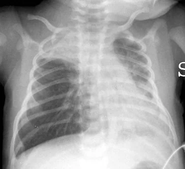
- Complete obstruction causes distal absorption of gas and patchy atelectasis, seen as segmental collapse on a radiograph if one is taken
- The resulting ventilation-perfusion mismatch produces hypoxaemia, which is why oxygen saturation is a more useful measure than any single examination sign
- Increased work of breathing eventually causes fatigue, carbon dioxide retention and respiratory failure in the smallest infants
- Apnoea occurs particularly in infants under 6 weeks and in those born preterm, and can be the presenting feature before any respiratory signs at all
Infection does not confer lasting immunity, so reinfection with RSV throughout life is normal - in adults and older children it simply produces a cold. It is the airway calibre, not the virus, that makes bronchiolitis a disease of infancy.
Risk factors
Risk factors for acquiring bronchiolitis
- Age under 12 months, and especially under 6 months
- Older siblings, or attendance at nursery
- Winter birth, placing the infant at their most vulnerable during the epidemic
- Exposure to tobacco smoke in the household
- Not having been breastfed
Risk factors for severe disease
These are the ones that matter clinically, because they lower the threshold for admission and identify the infants who may qualify for passive immunisation.1
- Age under 3 months, and particularly under 6 weeks, where apnoea is a real risk
- Prematurity, especially birth before 32 weeks
- Chronic lung disease of prematurity (bronchopulmonary dysplasia)
- Haemodynamically significant congenital heart disease
- Neuromuscular disease, which impairs the ability to clear secretions and to sustain the work of breathing
- Immunodeficiency, congenital or acquired
- Down syndrome and other conditions associated with airway hypotonia
- Cystic fibrosis and other chronic respiratory conditions
Clinical features
The history is stereotyped enough that it is often diagnostic on its own. A coryzal prodrome of 1-3 days is followed by a persistent dry cough, increasing breathlessness, and difficulty feeding. Fever, when present, is usually low grade - a temperature above 39°C should prompt you to reconsider pneumonia or another bacterial focus.
Symptoms peak between days 3 and 5 and then improve. Cough resolves within 3 weeks in 90% of infants. Knowing the natural history is essential: an infant presenting on day 2 may well get worse before they get better, and that shapes both the admission decision and the advice given to parents.
Examination findings
- Tachypnoea, often the earliest and most reliable sign
- Wheeze, fine end-inspiratory crackles, or both - NICE accepts either. Crackles are more typical of bronchiolitis and wheeze more typical of viral-induced wheeze, but the overlap is considerable.
- Increased work of breathing: subcostal and intercostal recession, nasal flaring, tracheal tug, head bobbing, and grunting in severe disease
- Hyperinflation: a barrel-shaped chest with a liver edge displaced downwards by flattened diaphragms, which can be mistaken for hepatomegaly
- Reduced feeding - a useful and quantifiable proxy for severity. Ask what proportion of the usual volume the infant is taking.
- Apnoea, particularly in infants under 6 weeks or born preterm
- Dehydration: reduced wet nappies, dry mucous membranes, sunken fontanelle
Assessment of severity and referral
NICE divides the decision into immediate emergency referral and consideration of referral.1
- Consider referral if the respiratory rate is above 60, if oral intake has fallen to 50-75% of the usual volume, or if there is clinical dehydration
- Have a lower threshold in any infant with the risk factors for severe disease listed above, and where social circumstances or distance from hospital would make deterioration at home dangerous
Differential diagnosis
| Diagnosis | Features pointing towards it |
|---|---|
| Viral-induced wheeze | Over 12 months, recurrent episodes, wheeze without crackles, responds to salbutamol |
| Bacterial pneumonia | High fever above 39°C, focal crackles or bronchial breathing, focal consolidation, persistently unwell |
| Heart failure from congenital heart disease | Murmur, hepatomegaly, poor weight gain, sweating on feeds, symptoms preceding the viral illness |
| Whooping cough | Paroxysmal cough with whoop or post-tussive vomiting, well between paroxysms, marked lymphocytosis |
| Inhaled foreign body | Sudden onset in a well child, unilateral signs, often a choking episode witnessed |
| Cystic fibrosis | Faltering growth, loose fatty stools, recurrent chest infections, positive newborn screening or family history |
| Gastro-oesophageal reflux with aspiration | Symptoms clearly related to feeds, back-arching, recurrent rather than seasonal |
Investigations
Bronchiolitis is a clinical diagnosis and most infants need no investigation at all.
- Pulse oximetry - the one measurement that should be made in every infant
- Chest radiograph - not routine. NICE specifically warns that radiographic changes in bronchiolitis mimic consolidation, and that imaging leads to unnecessary antibiotics. Reserve it for diagnostic uncertainty, an atypical course, or a child requiring intensive care.1
- Nasopharyngeal aspirate or throat swab for viral PCR - does not change the management of the individual infant, but is used for cohorting on the ward and for surveillance
- Capillary or venous blood gas - only where respiratory failure is suspected, in a child with worsening distress, or before escalating airway support. A rising carbon dioxide is a late and ominous sign.
- Blood tests and cultures - not routine. Secondary bacterial infection is uncommon, and a raised white cell count in bronchiolitis is not evidence of it.
- Urine culture - worth considering in a febrile infant, since a concurrent urinary tract infection occurs in a small proportion
Management
Treatment is entirely supportive. The aim is to maintain oxygenation and hydration while the infant clears the virus.
Supportive care in hospital
- Oxygen - supplemental oxygen for saturations persistently below 90% in infants over 6 weeks, or below 92% in younger infants and those with underlying conditions
- Fluids and feeding - if the infant cannot maintain oral intake, give fluids by nasogastric or orogastric tube first. Intravenous isotonic fluid is reserved for those who cannot tolerate enteral feeds or who have impending respiratory failure.
- Minimal handling - clustering care and disturbing the infant as little as possible genuinely reduces oxygen requirement
- Upper airway suction - not routine. NICE advises suction only where there is apnoea, or where secretions in the upper airway are causing respiratory distress or feeding difficulty.
- Nasal high-flow humidified oxygen - used where standard oxygen therapy is failing, to reduce the work of breathing
- CPAP - for impending respiratory failure, with escalation to intubation and ventilation in a small minority
- Isolation or cohorting and strict hand hygiene, since RSV spreads rapidly on a paediatric ward
Treatments that do not work
NICE recommends against the following in bronchiolitis. Being able to list them, and say why, is what an examiner is looking for:1
| Treatment | Why not |
|---|---|
| Salbutamol | The obstruction is oedema, mucus and epithelial debris, not smooth muscle constriction, and infant airway smooth muscle is poorly developed. Trials show no benefit and it can worsen ventilation-perfusion mismatch.4 |
| Ipratropium bromide | No demonstrated benefit on symptoms, admission or length of stay |
| Nebulised adrenaline | Produces transient improvement at most, with no effect on admission rate or length of stay |
| Corticosteroids, oral, inhaled or nebulised | No effect on admission, length of stay or duration of symptoms, in contrast to their role in croup and asthma |
| Nebulised hypertonic saline | Earlier promise has not been borne out in larger UK trials |
| Antibiotics | The illness is viral. Secondary bacterial infection is uncommon and antibiotics do not shorten the course or prevent it. |
| Montelukast | No benefit in acute bronchiolitis or in preventing subsequent wheeze |
| Chest physiotherapy | No benefit in uncomplicated bronchiolitis; reserved for infants with comorbidity impairing secretion clearance, such as neuromuscular disease |
Discharge and safety netting
An infant can be discharged when they are clinically stable, taking adequate oral fluid - conventionally at least 75% of their usual intake - and have maintained oxygen saturations above 90% in air for 4 hours including a period of sleep, since saturations dip during sleep and a daytime reading can be falsely reassuring.
- Explain the expected course: worst on days 3-5, cough for up to 3 weeks
- Advise on feeding little and often, and recognising dehydration by counting wet nappies
- Advise a smoke-free home, and explain that this reduces both severity now and wheeze later
- Give clear instructions to seek urgent help for worsening work of breathing, colour change, apnoea, or a fall in feeding to less than half the usual amount
- Warn that saline nose drops before feeds may help, but that over-the-counter cough medicines have no role
Prevention
Prevention has changed substantially in the UK in the last few years and is now an examinable topic in its own right.
- Maternal RSV vaccination - since September 2024 a single dose of the RSV vaccine has been offered to pregnant women from 28 weeks of gestation, giving passive protection to the infant through transplacental antibody during their first RSV season.3
- Passive immunisation of high-risk infants - a monoclonal antibody against the RSV F protein is offered to infants at highest risk, such as those with chronic lung disease of prematurity, haemodynamically significant congenital heart disease or severe combined immunodeficiency. Palivizumab required monthly intramuscular injections through the season; the longer-acting nirsevimab needs only a single dose and has replaced it in an increasing number of pathways.3,7
- Infection control - hand hygiene, cohorting of RSV-positive infants, and keeping unwell visitors away from newborns
- Avoiding tobacco smoke exposure and promoting breastfeeding, both of which reduce severity
Complications
- Apnoea - the reason infants under 6 weeks are admitted even when they look reasonably well
- Respiratory failure requiring CPAP or ventilation, in roughly 2-5% of admitted infants
- Dehydration from poor feeding combined with increased insensible losses
- Hyponatraemia from inappropriate ADH secretion, aggravated by hypotonic intravenous fluids - which is why isotonic fluid is used
- Secondary bacterial infection, uncommon but more likely in infants who are ventilated
- Persistent wheeze - around half of infants admitted with bronchiolitis wheeze again in the following year
- Bronchiolitis obliterans - rare, most often after adenovirus, causing permanent small airway fibrosis
The association between severe RSV bronchiolitis in infancy and later asthma is well established but the direction of causation is not settled: severe bronchiolitis may damage the developing airway, or it may simply identify infants whose airways were always going to be susceptible. Either way, parents should be told that further wheezy episodes are likely and do not mean the bronchiolitis was mismanaged.
Prognosis
The outlook for a previously well infant is excellent. Most are managed entirely at home, most admissions last 2-3 days, and mortality in the UK is very low - deaths occur almost exclusively in infants with significant comorbidity such as severe congenital heart disease, chronic lung disease of prematurity or immunodeficiency.2
The practical message for both parents and exam candidates is that bronchiolitis is a disease you support a child through rather than treat. The clinical skill lies in identifying the infant who is on the wrong trajectory - the one under 6 weeks with apnoea, the ex-premature infant whose feeding has stopped, the child with rising carbon dioxide - and in resisting the pull towards drugs that have all been tested and all been found wanting.
References
- NICE NG9. Bronchiolitis in children: diagnosis and management. 2015, updated 2021. Available here
- NICE Clinical Knowledge Summaries. Bronchiolitis. Available here
- UKHSA. Respiratory syncytial virus (RSV): the Green Book, chapter 27a. Available here
- Ralston SL, Lieberthal AS, Meissner HC et al. Clinical practice guideline: the diagnosis, management, and prevention of bronchiolitis. Pediatrics. 2014. Available here
- Cunningham S, Rodriguez A, Adams T et al. Oxygen saturation targets in infants with bronchiolitis (BIDS): a double-blind, randomised, equivalence trial. The Lancet. 2015. Available here
- Franklin D, Babl FE, Schlapbach LJ et al. A randomized trial of high-flow oxygen therapy in infants with bronchiolitis. New England Journal of Medicine. 2018. Available here
- BNF for Children. Palivizumab. Available here
This article is written for revision and education. It is not clinical guidance and must not be used to make decisions about the care of a patient. Always check current NICE guidance and local protocols.