
Necrotising Enterocolitis
Key points
- Definition: acute inflammatory necrosis of the bowel wall in a neonate, most often affecting the terminal ileum and proximal colon.
- Who gets it: 5-10% of very low birth weight infants. Risk rises steeply as gestation and birth weight fall, and onset is typically in the second or third week of life.
- Mechanism: an immature gut with an exaggerated inflammatory response, abnormal bacterial colonisation, and enteral feed as substrate - with ischaemia contributing in term infants.
- Early signs: feed intolerance with bilious aspirates, abdominal distension and bloody stools, alongside non-specific deterioration - apnoea, temperature instability and lethargy.
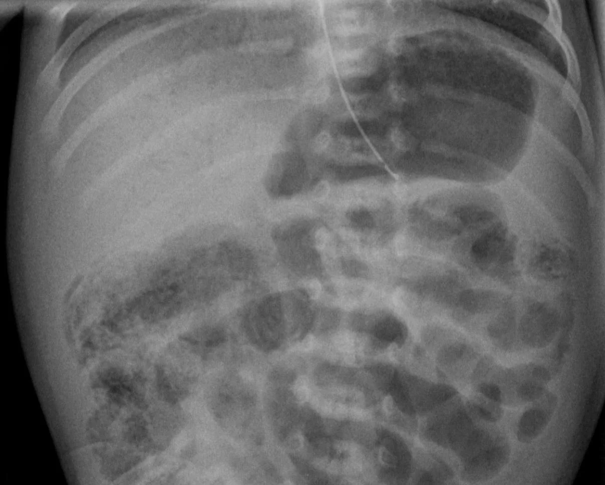
- The diagnostic sign: pneumatosis intestinalis - gas within the bowel wall on abdominal radiograph. Portal venous gas is more ominous and pneumoperitoneum means perforation.
- Medical management: nil by mouth, nasogastric tube on free drainage, parenteral nutrition, broad-spectrum antibiotics for 7-10 days, and serial radiographs.
- Surgery: pneumoperitoneum is an absolute indication. Otherwise operate for deterioration despite maximal medical therapy, a fixed loop, or an abdominal mass.
- Prevention: maternal or donor breast milk rather than formula, a standardised cautious feeding regimen, probiotics, and avoiding prolonged empirical antibiotics and acid suppression.
Introduction
Necrotising enterocolitis (NEC) is an acute inflammatory disease of the neonatal intestine that progresses from mucosal injury to transmural necrosis and, if unchecked, to perforation and peritonitis. It is the commonest gastrointestinal emergency in the newborn and the commonest cause of neonatal surgery.
Around 90% of cases occur in preterm infants, affecting 5-10% of those born weighing under 1,500 g, and incidence rises steeply with decreasing gestation. The remaining 10% occur in term infants, almost always in the setting of congenital heart disease, perinatal asphyxia, polycythaemia or growth restriction with abnormal umbilical artery Doppler - that is, situations in which the gut has been hypoperfused.6
What makes NEC clinically difficult is that the early presentation is non-specific: a preterm baby who has more aspirates than yesterday and one apnoea overnight looks much like a baby with sepsis, or with nothing at all. The disease can then progress from mild feed intolerance to perforation within hours. Careful serial examination of the abdomen, and a low threshold for stopping feeds and taking a film, are the practical skills this topic is really about.
Pathophysiology
NEC is not a single-cause disease. The prevailing model is a convergence of four factors in a gut that is not ready for extrauterine life.
- Intestinal immaturity. The preterm gut has poor motility, an immature mucosal barrier with reduced mucus and tight junction integrity, low immunoglobulin A secretion, and an exaggerated innate immune response - notably heightened toll-like receptor 4 signalling, which converts an ordinary bacterial encounter into a destructive inflammatory cascade.
- Abnormal bacterial colonisation. Neonatal unit flora, prolonged empirical antibiotics and acid suppression with H2 receptor antagonists or proton pump inhibitors all produce a dysbiotic, low-diversity microbiome dominated by Gram-negative organisms.
- Enteral feeding. Feed provides the substrate for bacterial proliferation and fermentation. Formula feeding increases risk several-fold compared with breast milk, and rapid advancement of feed volumes is a modifiable risk factor.4
- Ischaemia and hypoperfusion. Historically considered the primary event, and still the dominant mechanism in term infants with congenital heart disease or asphyxia, though in the preterm infant it is now regarded as contributory rather than initiating.
The result is mucosal injury, bacterial invasion of the bowel wall, and gas production by fermenting organisms within it - which is what produces the radiological hallmark of pneumatosis intestinalis. Inflammation and coagulative necrosis extend outwards through the wall; gas may track into mesenteric and portal veins, and full-thickness necrosis leads to perforation. The terminal ileum and proximal colon are most often affected, presumably because they combine the highest bacterial load with a relatively vulnerable blood supply.
Risk factors
- Prematurity and low birth weight - by far the dominant risk factors
- Formula feeding rather than maternal expressed or donor breast milk
- Rapid advancement of enteral feeds
- Prolonged empirical antibiotic exposure in the first week of life, and acid suppression with an H2 blocker or proton pump inhibitor
- Patent ductus arteriosus, and the drugs used to treat it, through effects on mesenteric perfusion
- Umbilical arterial or venous catheters, and exchange transfusion
- Intrauterine growth restriction with absent or reversed end-diastolic flow on umbilical artery Doppler
- Congenital heart disease, particularly duct-dependent systemic circulation and hypoplastic left heart syndrome - the main risk factor in a term infant
- Perinatal asphyxia, polycythaemia and hypotension
- Anaemia and red cell transfusion, an association that remains debated but influences transfusion and feeding practice on many units
Clinical features
Presentation is typically in the second or third week of life, and later in more immature infants - a 24-week baby may present at 5 or 6 weeks. Term infants tend to present within the first few days.
Gastrointestinal features
- Feed intolerance with increasing gastric residuals, and particularly bilious aspirates
- Abdominal distension, often the first thing a nurse notices, with an increasing abdominal girth
- Abdominal tenderness, guarding, and visible or palpable bowel loops
- Abdominal wall erythema or discolouration, indicating peritoneal involvement
- Blood in the stool - fresh, altered or occult
- Bilious vomiting
- A palpable mass in the right lower quadrant in more advanced disease
Systemic features
- Apnoea and bradycardia, or a new or increased oxygen requirement
- Temperature instability and lethargy
- Poor perfusion, mottling and hypotension
- Hypoglycaemia or hyperglycaemia
- Metabolic acidosis on blood gas
- In advanced disease, shock, disseminated intravascular coagulation and multi-organ failure
Investigations
Abdominal radiograph
The supine abdominal film is the key investigation, supplemented by a left lateral decubitus or cross-table lateral view when perforation is suspected, since free gas is easier to see over the liver with the baby on their left side.
| Sign | What it means |
|---|---|
| Dilated bowel loops and ileus | Non-specific and often the earliest change |
| Bowel wall thickening and loss of the normal gas pattern | Mucosal oedema |
| Pneumatosis intestinalis | Gas within the bowel wall, seen as a curvilinear or bubbly lucency following the bowel contour. Diagnostic of NEC. |
| Portal venous gas | Branching lucencies over the liver. Indicates more extensive disease and a worse prognosis. |
| Persistent fixed dilated loop | The same loop unchanged over serial films, suggesting an infarcted segment |
| Pneumoperitoneum | Free intraperitoneal gas - perforation, and an absolute indication for surgery. Signs include Rigler's sign (gas on both sides of the bowel wall), the football sign of a large central lucency, and outlining of the falciform ligament. |
| Ascites and a gasless abdomen | Ominous, suggesting extensive necrosis with fluid |
Other investigations
- Abdominal ultrasound - increasingly used alongside plain films. It detects free fluid, bowel wall thickness and perfusion, pneumatosis and portal venous gas, and can show necrotic bowel before the radiograph changes.
- FBC - thrombocytopenia is often the earliest laboratory abnormality and tracks disease severity; neutropenia carries a worse prognosis than neutrophilia
- CRP - useful as a trend, and a falling CRP is one of the criteria used when deciding to restart feeds
- Blood gas - metabolic acidosis reflecting tissue hypoperfusion
- U&Es and glucose - hyponatraemia is common from third-space losses
- Coagulation screen - for disseminated intravascular coagulation
- Blood culture before antibiotics, and stool culture where relevant
Differential diagnosis
- Neonatal sepsis - shares all the systemic features and frequently coexists, which is why blood cultures and antibiotics are part of NEC management anyway2
- Spontaneous intestinal perforation - typically earlier, in the first week, in an extremely low birth weight infant, with a bluish abdomen and free gas but without pneumatosis, and with a better prognosis than NEC
- Malrotation with volvulus - bilious vomiting in a term infant, an upper gastrointestinal contrast study is diagnostic, and this is a surgical emergency in its own right
- Hirschsprung's disease with enterocolitis - delayed passage of meconium, explosive stool on rectal examination, abdominal distension
- Cow's milk protein allergy - blood in the stool in a well, thriving, usually term infant
- Anal fissure or swallowed maternal blood - common benign causes of blood in the stool
- Ileus from any cause, including electrolyte disturbance and opiates
- Necrotising enterocolitis of the term infant secondary to congenital heart disease, which should prompt an echocardiogram
Management
Medical management
- Stop enteral feeds immediately and keep the infant nil by mouth
- Nasogastric tube on free drainage to decompress the bowel
- Intravenous fluids and parenteral nutrition through central access, with careful attention to third-space losses3
- Broad-spectrum intravenous antibiotics after blood cultures, covering Gram-negative and anaerobic organisms - a typical UK regimen is a penicillin with gentamicin and metronidazole, though local policy governs the exact combination8
- Cardiorespiratory support - many infants need intubation and inotropes as the illness progresses
- Correct thrombocytopenia, coagulopathy and anaemia with blood products as required
- Analgesia, which is often under-provided in a very tender abdomen
- Early surgical involvement, even when the initial plan is medical
- Serial abdominal radiographs, typically 6-hourly while the infant is unstable, looking for perforation and for a fixed loop
- Serial abdominal examination and girth measurement by the same clinician where possible
Bowel rest and antibiotics are usually continued for 7-10 days in definite NEC. Feeds are reintroduced cautiously once the abdomen is soft and non-tender, the infant is systemically well, the CRP is falling and the radiographs have normalised - starting with small volumes of maternal or donor breast milk.
Surgical management
- Absolute indication: pneumoperitoneum
- Relative indications: clinical deterioration despite maximal medical therapy, a persistent fixed dilated loop, a palpable abdominal mass, abdominal wall erythema or a positive paracentesis
- Laparotomy with resection of necrotic bowel and formation of a stoma is the usual approach, with a plan for later reversal
- Primary peritoneal drainage under local anaesthetic is used in extremely low birth weight or profoundly unstable infants, either as definitive treatment or to stabilise before laparotomy
- NEC totalis - necrosis of virtually the whole small and large bowel found at laparotomy - is not survivable, and the discussion becomes one of palliative care
Prevention
- Breast milk - maternal expressed milk first, pasteurised donor milk second. The Cochrane review of formula versus donor breast milk in preterm infants found roughly a doubling of NEC risk with formula, making this the single most effective preventive measure available.4
- Standardised feeding guidelines with cautious, protocolised advancement of volumes - the standardisation itself appears to reduce incidence
- Probiotics - a Cochrane review of very preterm and very low birth weight infants found that combination probiotics reduce severe NEC and all-cause mortality, and many UK neonatal units now use them routinely5
- Antenatal corticosteroids, which reduce NEC as well as respiratory distress syndrome1
- Avoid prolonged empirical antibiotics where cultures are negative, and avoid H2 blockers and proton pump inhibitors unless there is a compelling indication
- Careful management of a patent ductus arteriosus, and of feeding around transfusions in units where that practice is followed
- Meticulous infection control, since NEC sometimes occurs in clusters on a unit
Complications and prognosis
Complications
- Perforation, peritonitis and septic shock in the acute phase
- Disseminated intravascular coagulation and multi-organ failure
- Intestinal stricture - occurs in up to a third of infants treated medically, most often in the colon, presenting weeks later with obstruction or failure to tolerate feeds. A contrast study before stoma closure is standard.
- Short bowel syndrome after extensive resection, with malabsorption, long-term parenteral nutrition dependence and its complications
- Parenteral nutrition-associated cholestasis and liver disease
- Enterocutaneous fistula, adhesive obstruction and stoma complications
- Recurrent NEC, in a small proportion
- Growth failure and neurodevelopmental impairment - infants who require surgery for NEC have significantly worse cognitive and motor outcomes than gestation-matched peers
Prognosis
Overall mortality is in the region of 20-30%, and rises to around 50% in infants who require surgery and in those of extremely low birth weight. Death is usually from overwhelming sepsis and multi-organ failure, or from the consequences of extensive bowel loss.
Survivors of medically managed NEC generally do well, though they need watching for stricture over the following weeks and months. Survivors of surgical NEC carry a substantial long-term burden: prolonged hospital stay, dependence on parenteral nutrition, repeated operations, and a measurable excess of neurodevelopmental impairment that persists into school age.6
The gap between those two groups is the reason prevention and early recognition matter so much. Very little in neonatal medicine has as favourable a ratio of effort to benefit as ensuring a preterm baby receives their mother's milk, feeds are advanced by protocol, and the abdomen is examined properly every day.
References
- NICE NG25. Preterm labour and birth. 2015, updated 2022. Available here
- NICE NG195. Neonatal infection: antibiotics for prevention and treatment. 2021. Available here
- NICE NG154. Neonatal parenteral nutrition. 2020. Available here
- Quigley M, Embleton ND, McGuire W. Formula versus donor breast milk for feeding preterm or low birth weight infants. Cochrane Database of Systematic Reviews. 2019. Available here
- Sharif S, Meader N, Oddie SJ, Rojas-Reyes MX, McGuire W. Probiotics to prevent necrotising enterocolitis in very preterm or very low birth weight infants. Cochrane Database of Systematic Reviews. 2023. Available here
- Neu J, Walker WA. Necrotizing enterocolitis. New England Journal of Medicine. 2011. Available here
- Bell MJ, Ternberg JL, Feigin RD et al. Neonatal necrotizing enterocolitis: therapeutic decisions based upon clinical staging. Annals of Surgery. 1978. Available here
- BNF for Children. Metronidazole. Available here
This article is written for revision and education. It is not clinical guidance and must not be used to make decisions about the care of a patient. Always check current NICE guidance and local protocols.