
Chorioamnionitis
Key points
- Definition: infection or inflammation of the chorion, amnion, amniotic fluid and decidua, usually from organisms ascending from the vagina.
- Frequency: complicates 1-5% of term births but up to 40-70% of preterm births associated with prelabour rupture of membranes.
- Clinical criteria: maternal fever of 38.0 °C or above plus maternal tachycardia, fetal tachycardia, uterine tenderness, offensive liquor or leucocytosis.
- Organisms: typically polymicrobial: Ureaplasma and Mycoplasma species, group B streptococcus, Escherichia coli and anaerobes.
- Investigations: blood cultures and vaginal swabs before antibiotics, but never delay treatment to obtain them; CRP and white cell count are unreliable in labour.
- Treatment: immediate broad-spectrum intravenous antibiotics within one hour, plus expedited birth at any gestation.
- Tocolysis: is absolutely contraindicated; the uterus must be emptied, not quietened.
- Consequences: maternal sepsis, uterine atony and postpartum haemorrhage; neonatal sepsis, and a fetal inflammatory response linked to cerebral palsy and chronic lung disease.
Introduction
Chorioamnionitis is infection or inflammation of the fetal membranes, amniotic fluid, placenta and decidua. It complicates 1-5% of term deliveries but is far commoner at earlier gestations, being present in perhaps 40-70% of preterm births associated with preterm prelabour rupture of membranes. In many of those cases the infection is the cause of the preterm labour rather than a consequence of it.
It matters for two separate reasons. The mother is at risk of sepsis, which remains a leading cause of maternal death, and the fetus is at risk both of direct infection and of a systemic inflammatory response that damages the developing brain and lung even when the baby is never bacteraemic. Treatment therefore has to address both, and it has to be immediate: chorioamnionitis is one of the few obstetric diagnoses where the correct action is antibiotics and delivery, regardless of gestation.
Terminology has shifted. Because many cases show inflammation without demonstrable organisms, the term intra-amniotic inflammation or infection, sometimes abbreviated to Triple I, is increasingly used. In UK practice the older term remains in general use and the management is unaffected.
Pathogenesis and microbiology
The overwhelming majority of cases arise from organisms ascending from the vagina and cervix into the uterine cavity. Rupture of the membranes removes the principal mechanical and antimicrobial barrier, but ascent can also occur through intact membranes, which is why chorioamnionitis is found in preterm labour with intact membranes.
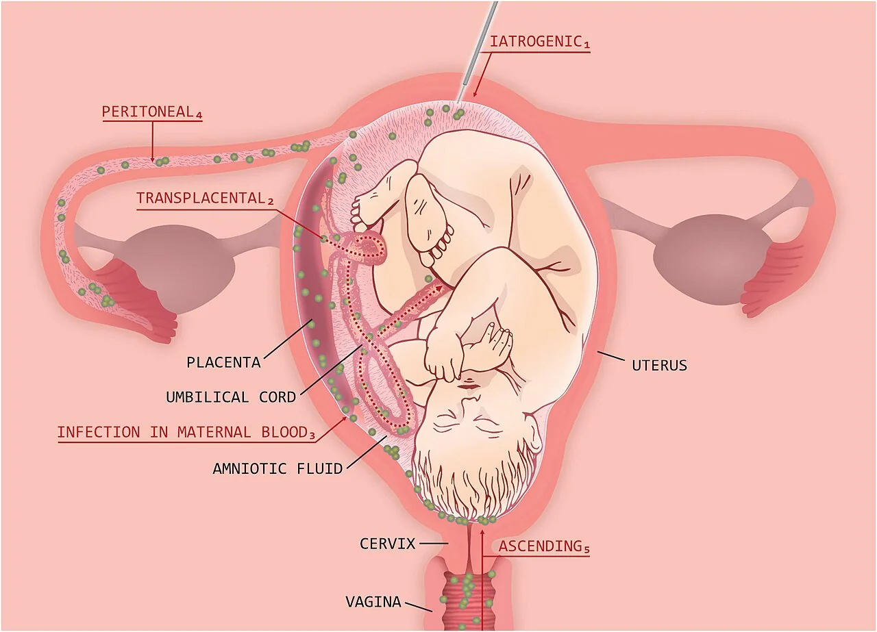
Infection is usually polymicrobial. The organisms most often isolated from amniotic fluid are the genital mycoplasmas, Ureaplasma urealyticum and Mycoplasma hominis, which are of low virulence but very frequently present. Group B streptococcus, Escherichia coli, Gardnerella vaginalis, Bacteroides species and Fusobacterium are also common. Listeria monocytogenes reaches the placenta haematogenously rather than by ascent, and is the classic cause after eating unpasteurised dairy products.
The inflammatory cascade is what causes most of the harm. Bacterial products stimulate decidual and membrane production of interleukin-1, interleukin-6, tumour necrosis factor and prostaglandins. Prostaglandins drive uterine contractions and cervical ripening, and matrix metalloproteinases weaken the membranes, which is the mechanism by which infection precipitates preterm labour and membrane rupture.1
Risk factors
- Prolonged rupture of membranes - the single most important factor, with risk rising with each hour
- Preterm prelabour rupture of membranes
- Prolonged labour, particularly a prolonged second stage
- Repeated digital vaginal examinations after membrane rupture - each one carries organisms upwards
- Internal fetal monitoring: fetal scalp electrodes and intrauterine pressure catheters
- Group B streptococcal colonisation and bacterial vaginosis
- Nulliparity, which is associated with longer labour
- Meconium-stained liquor, which impairs the bacteriostatic properties of amniotic fluid
- Invasive procedures: amniocentesis, chorionic villus sampling, cervical cerclage
- Immunosuppression, poorly controlled diabetes, smoking and alcohol use
Clinical features
The diagnosis is clinical. Maternal fever is the necessary feature, and at least one supporting feature is required.
| Feature | Threshold or description |
|---|---|
| Maternal fever | 38.0 °C or above on one occasion, or 38.0-38.9 °C on two occasions 30 minutes apart - required for the diagnosis |
| Maternal tachycardia | Above 100 beats per minute, in the absence of another explanation |
| Fetal tachycardia | Baseline above 160 beats per minute, often with reduced variability |
| Uterine tenderness | Tenderness between contractions rather than only during them |
| Amniotic fluid | Purulent or offensive-smelling liquor |
| Maternal leucocytosis | White cell count above 15 x10^9/L without recent corticosteroids |
Differential diagnosis
- Urinary tract infection and pyelonephritis - loin pain, dysuria and positive urinalysis; the commonest non-obstetric source of fever in pregnancy
- Epidural-related fever - low grade, no uterine tenderness, no fetal tachycardia beyond that explained by maternal temperature
- Respiratory infection, including influenza and COVID-19
- Appendicitis and other intra-abdominal sepsis
- Placental abruption - uterine tenderness and an abnormal CTG, but with bleeding and without fever
- Postpartum endometritis - the same disease process presenting after birth
- Non-infective causes of maternal tachycardia: pain, anxiety, dehydration, haemorrhage, thyrotoxicosis
Investigations
- Blood cultures before antibiotics, but never delaying them; bacteraemia is present in around 10%
- Full blood count, CRP, urea and electrolytes, liver function, lactate and coagulation screen as part of the sepsis pathway
- High vaginal swab and, if indicated, a placental swab after delivery
- Midstream urine for culture, to exclude a urinary source
- Continuous cardiotocography - fetal tachycardia with reduced variability is characteristic and may precede maternal signs
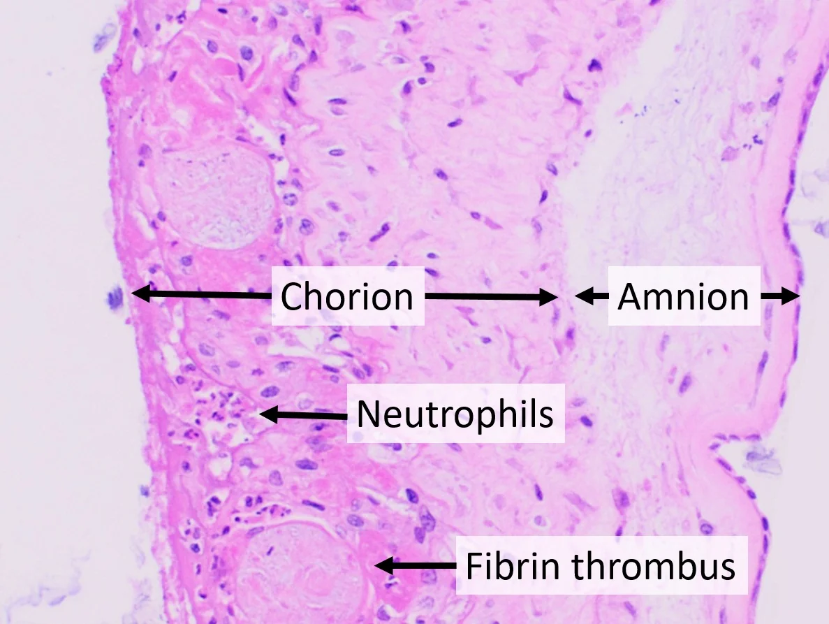
- Placental and membrane histology after birth, which is the diagnostic gold standard and demonstrates neutrophil infiltration; funisitis, inflammation of the umbilical cord, indicates a fetal inflammatory response
Amniocentesis for amniotic fluid glucose, Gram stain, culture and interleukin-6 gives the most definitive antenatal answer but is rarely performed in UK practice, since it is invasive and the clinical decision is usually clear enough without it. CRP has poor sensitivity and specificity for intra-amniotic infection and should not be used to rule it out.1
Management
Chorioamnionitis is managed as maternal sepsis until proven otherwise, and the definitive treatment is delivery. Both parts happen in parallel.
- Escalate immediately: senior obstetrician, anaesthetist, midwife coordinator and neonatal team; use the maternal early warning score and the sepsis pathway
- Broad-spectrum intravenous antibiotics within one hour of recognition, without waiting for culture results
- Oxygen, intravenous fluids and antipyretics; catheterise and monitor hourly urine output
- Take blood cultures, lactate and a full septic screen, but do not let this delay antibiotics
- Continuous fetal monitoring
- Expedite birth at any gestation - vaginal birth if it can be achieved reasonably quickly, caesarean section for obstetric indications or if the fetal condition demands it
- Do not give tocolysis; it is contraindicated because delaying delivery prolongs exposure of both mother and fetus to the infection
- Inform the neonatal team in advance so the baby can be assessed and treated for early-onset sepsis
A typical UK regimen is intravenous broad-spectrum cover of Gram-positive, Gram-negative and anaerobic organisms, for example co-amoxiclav, or a combination of a penicillin, gentamicin and metronidazole, with clindamycin substituted in penicillin allergy. Clindamycin is also added where a group A streptococcal or necrotising infection is suspected, because it suppresses exotoxin production. Follow local microbiology guidance, and continue antibiotics after delivery until the woman has been afebrile for 24-48 hours.2,3
Complications
| Group | Complications |
|---|---|
| Maternal acute | Sepsis and septic shock, bacteraemia in around 10%, acute kidney injury, ARDS, disseminated intravascular coagulation, maternal death |
| Maternal obstetric | Dysfunctional labour, higher caesarean rate, uterine atony and postpartum haemorrhage, wound infection, endometritis, pelvic abscess and septic pelvic thrombophlebitis |
| Fetal and neonatal | Preterm birth, early-onset neonatal sepsis, pneumonia, meningitis, stillbirth |
| Fetal inflammatory response syndrome | Periventricular leukomalacia, intraventricular haemorrhage, cerebral palsy, bronchopulmonary dysplasia and necrotising enterocolitis |
The fetal inflammatory response syndrome deserves emphasis because it explains outcomes that are otherwise puzzling. Fetal cytokines produced in response to intra-amniotic infection injure the developing white matter and lung directly, so an infant may sustain periventricular leukomalacia and later cerebral palsy without ever having a positive blood culture. This is one reason chorioamnionitis is treated so aggressively even when the mother appears only mildly unwell.1
Postpartum haemorrhage is common and often underestimated. An infected, inflamed myometrium contracts poorly, so uterine atony should be actively anticipated with an oxytocin infusion after delivery rather than treated once it occurs.
Postpartum endometritis
Endometritis is essentially the same disease occurring after birth, and chorioamnionitis is one of its principal risk factors. It complicates around 1-3% of vaginal births and considerably more caesarean sections, and typically presents on the second to fifth postnatal day.
- Fever, lower abdominal pain, a tender bulky uterus that is higher than expected for the postnatal day, and offensive lochia
- Risk factors are caesarean section, prolonged rupture of membranes, prolonged labour, retained products of conception, manual removal of the placenta and pre-existing chorioamnionitis
- Take blood cultures, vaginal and endocervical swabs and a midstream urine, then start broad-spectrum intravenous antibiotics
- Arrange pelvic ultrasound if retained products are suspected, and evacuate them once the woman is on antibiotics
- Consider septic pelvic thrombophlebitis where fever persists on adequate antibiotics with no collection identified
- Continue antibiotics until the woman has been afebrile and clinically well for 24-48 hours
The important practical point is that women are discharged early after birth, so endometritis often presents to primary care or to an emergency department rather than to the maternity unit. Any woman who has given birth within the last six weeks and presents with fever should be discussed with the obstetric team, because the diagnostic and physiological thresholds differ from those of the general population.2,4
Prevention
- Minimise vaginal examinations after rupture of membranes, and use strict aseptic technique for each one
- Erythromycin for 10 days in preterm prelabour rupture of membranes, which prolongs pregnancy and reduces neonatal infection; co-amoxiclav is avoided in this setting because the ORACLE trial found an association with neonatal necrotising enterocolitis
- Intrapartum antibiotic prophylaxis for group B streptococcus where indicated: previous baby with GBS disease, GBS bacteriuria or a positive swab in this pregnancy, preterm labour, or pyrexia in labour
- Benzylpenicillin 3 g intravenously then 1.5 g four-hourly until delivery is the standard UK GBS regimen
- Prompt induction of labour where membranes have ruptured at term and labour has not started spontaneously within 24 hours
- Treat bacterial vaginosis and urinary infection when detected in pregnancy
Red flags
Prognosis
With early recognition, prompt broad-spectrum antibiotics and timely delivery, maternal outcome is usually excellent, and most women recover fully without long-term sequelae. Delay is what converts a treatable infection into septic shock, and confidential enquiries into maternal deaths repeatedly identify failure to recognise sepsis and delay in giving antibiotics as the recurring themes.4
Neonatal outcome depends chiefly on gestation. At term, most babies exposed to chorioamnionitis are well and require only observation and, where indicated, antibiotics. At extreme prematurity, the combination of preterm birth and the fetal inflammatory response substantially increases the risk of chronic lung disease and adverse neurodevelopmental outcome, and this is worth explaining honestly to parents at the time.
Chorioamnionitis in one pregnancy is a risk factor for preterm birth in the next, largely because it identifies women prone to ascending infection or with a short cervix. A subsequent pregnancy warrants early booking, treatment of bacterial vaginosis, and consideration of cervical length surveillance.3
References
- Kim CJ, Romero R, Chaemsaithong P et al. Acute chorioamnionitis and funisitis: definition, pathologic features, and clinical significance. Am J Obstet Gynecol. 2015. Available here
- RCOG Green-top Guideline No. 64a. Bacterial sepsis in pregnancy. Available here
- NICE NG25. Preterm labour and birth. 2015 (updated 2022). Available here
- MBRRACE-UK. Saving Lives, Improving Mothers' Care. Available here
This article is written for revision and education. It is not clinical guidance and must not be used to make decisions about the care of a patient. Always check current NICE guidance and local protocols.