
Placenta Praevia
Key points
- Definition: the placenta lies directly over the internal cervical os; a placental edge within 20 mm of the os but not covering it is a low-lying placenta.
- Frequency: around 1 in 200 pregnancies at term, but roughly 5% of 20-week scans show a low-lying placenta and 90% of these resolve.
- Mechanism: the placenta does not move; the lower uterine segment forms and elongates in the third trimester, carrying the placental edge away from the os.
- Presentation: painless, unprovoked, bright red bleeding after 28 weeks, with a soft non-tender uterus and often a high or malpresenting presenting part.
- Rule: never perform a digital vaginal examination until placenta praevia has been excluded.
- Diagnosis: transvaginal ultrasound, which is safe and more accurate than the transabdominal approach; rescan at 32 and 36 weeks.
- Accreta risk: praevia over a previous caesarean scar carries a high risk of placenta accreta spectrum, rising with each previous caesarean.
- Birth: elective caesarean section at 36+0 to 37+0 weeks for uncomplicated praevia, earlier if there has been bleeding.
Introduction
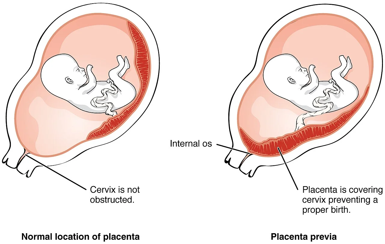
Placenta praevia is implantation of the placenta in the lower uterine segment such that it lies directly over the internal cervical os. It affects approximately 1 in 200 pregnancies at term and is a leading cause of antepartum and postpartum haemorrhage, of emergency hysterectomy, and of iatrogenic preterm birth.1
Its incidence is rising, and the reason is the rising caesarean section rate. Each caesarean leaves a scar in the lower segment which is a preferential site for subsequent implantation and which is also incapable of restraining trophoblastic invasion. The result is more praevia and, more importantly, more praevia complicated by placenta accreta spectrum, which is the condition that actually kills women.
Terminology and classification
RCOG abandoned the old four-grade classification in 2018 in favour of a two-term system based on the measured distance from the placental edge to the internal os. The grades are still encountered in older texts and in exam questions, so both are given here.
| Current term | Definition | Old grading |
|---|---|---|
| Normal | Placental edge more than 20 mm from the internal os | - |
| Low-lying placenta | Placental edge within 20 mm of the internal os but not covering it | Grade I (minor) and grade II (marginal) |
| Placenta praevia | Placenta lies directly over the internal os | Grade III (partial) and grade IV (complete or major) |
Risk factors
- Previous caesarean section - the risk rises with the number of previous sections
- Previous placenta praevia - recurrence risk of the order of 4-8%
- Previous uterine surgery or instrumentation - myomectomy, dilatation and curettage, surgical management of miscarriage, endometrial ablation
- Multiparity and advanced maternal age
- Multiple pregnancy - a larger placental surface area is more likely to reach the lower segment
- Assisted reproductive technology
- Smoking and cocaine use - placental hypertrophy in response to relative hypoxaemia
- Previous endometritis or Asherman's syndrome - damaged endometrium in the fundus favours low implantation
The common thread is damaged or suboptimal endometrium in the upper uterine cavity, which encourages the blastocyst to implant lower, or a large placenta which extends further from its implantation site.
Clinical features
Most cases are now diagnosed on ultrasound before any bleeding occurs, because the 20-week anomaly scan routinely reports placental site. Where praevia does present with bleeding, the pattern is characteristic.
| Feature | Placenta praevia | Placental abruption |
|---|---|---|
| Pain | Painless | Constant severe abdominal or back pain |
| Bleeding | Bright red, visible, often unprovoked or post-coital | Dark, may be minimal or entirely concealed |
| Uterus | Soft, non-tender, relaxed | Tense, tender, woody hard, irritable |
| Presenting part | High, unengaged, or malpresentation | Usually engaged and normal |
| Fetal condition | Usually normal until maternal compromise | Distress or death common, disproportionate to visible loss |
| Coagulopathy | Uncommon unless massive loss | Common - abruption is the leading obstetric cause of DIC |
| Ultrasound | Diagnostic | Frequently normal; abruption is a clinical diagnosis |
Bleeding in praevia occurs because the lower segment stretches and the inelastic placenta shears off the underlying decidua, opening maternal sinuses. The blood is therefore maternal, bright red, and painless, since there is no retroplacental collection to distend the myometrium. Bleeding characteristically begins in the third trimester, often around 28-32 weeks when lower segment formation accelerates.
Diagnosis
Placental site is assessed at the 18+0 to 20+6 anomaly scan. If the placenta is found to be low-lying or praevia, a transvaginal scan is arranged at around 32 weeks. If it remains low at 32 weeks, a further scan is performed at 36 weeks to plan the mode of birth.
Transvaginal ultrasound is the modality of choice. It is safe in praevia, contrary to intuition, because the probe is placed in the anterior fornix and the angle between the probe and the cervical canal means it does not enter the os. It is significantly more accurate than the transabdominal approach, which overdiagnoses praevia because of bladder filling, myometrial contractions and posterior placental shadowing.1
Screening for placenta accreta spectrum
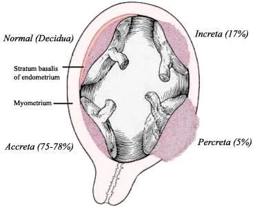
Placenta accreta spectrum describes abnormally adherent placentation, in which villi invade beyond the decidua because the decidua basalis is deficient, most often over a caesarean scar. It is graded by the depth of invasion.
| Previous caesarean sections | Approximate risk of accreta spectrum |
|---|---|
| None | Around 3% |
| One | Around 11% |
| Two | Around 40% |
| Three | Around 60% |
| Four or more | Around 65-70% |
Any woman with an anterior placenta praevia over a previous caesarean scar should have targeted greyscale and colour Doppler ultrasound looking for loss of the retroplacental clear zone, abnormal placental lacunae, bladder wall interruption, myometrial thinning and increased subplacental vascularity. MRI may be used where ultrasound is inconclusive or to assess parametrial and bladder involvement, but it does not replace ultrasound. Suspected accreta spectrum mandates delivery in a specialist centre by a multidisciplinary team.2
Differential diagnosis
In a woman presenting with painless third-trimester bleeding and no scan report to hand, the differential is the differential of antepartum haemorrhage generally, but the priorities differ.
- Placental abruption - painful, with a tense tender uterus; may coexist with praevia, so pain does not exclude it
- Vasa praevia - bleeding at rupture of membranes with immediate fetal compromise; the blood is fetal and the volume needed to exsanguinate the fetus is small
- Cervical causes - ectropion, polyp, cervicitis or carcinoma; typically small volume and often post-coital, but diagnosed on speculum examination only after praevia is excluded
- Vaginal trauma or infection
- Uterine rupture - severe pain, cardiovascular collapse and fetal bradycardia in a scarred uterus
- Bloody show - blood-stained mucus with the onset of labour, a diagnosis of exclusion
The important practical point is that these are distinguished by history, abdominal palpation and ultrasound, never by digital examination. If the placental site is unknown and the woman is bleeding, arrange an urgent scan and keep your fingers out of the vagina until you have the result.1
Antenatal management
- Counsel about the risk of bleeding, and advise immediate presentation if any bleeding, contractions or pain occur
- Advise avoiding penetrative intercourse, and discuss avoiding activities likely to provoke bleeding
- Screen for and treat anaemia - optimising haemoglobin antenatally is one of the few things that reliably improves the outcome of a major haemorrhage
- Ensure a valid group and save is available, and check for atypical antibodies which would delay crossmatching
- Antenatal corticosteroids between 34+0 and 35+6 weeks for women with praevia, and earlier if preterm birth is likely
- Outpatient management is reasonable for asymptomatic women who live close to the hospital, have immediate transport and constant support at home; inpatient care from around 34 weeks is offered where these conditions cannot be met
- Any woman who bleeds is admitted, and generally remains an inpatient
Tocolysis may be considered to allow corticosteroid administration in a woman with a small bleed, a stable maternal condition and a reassuring fetal condition, but it is contraindicated where bleeding is significant or the fetus is compromised.
Birth
Placenta praevia is an absolute indication for caesarean section: the placenta obstructs the presenting part and vaginal birth would mean delivering through it. A low-lying placenta with the edge more than 20 mm from the os can be delivered vaginally; between 0 and 20 mm the decision is individualised, taking into account the exact distance, whether the edge is thick or thin, and the woman's preference.
| Situation | Planned gestation |
|---|---|
| Uncomplicated placenta praevia or low-lying placenta | 36+0 to 37+0 weeks |
| History of antepartum bleeding or other risk factors for preterm birth | 34+0 to 36+6 weeks |
| Suspected placenta accreta spectrum | 35+0 to 36+6 weeks, in a specialist centre |
Planning the operation
- A consultant obstetrician and consultant anaesthetist present, or immediately available, for elective caesarean with praevia
- Regional anaesthesia is safe and is associated with less blood loss than general anaesthesia; the woman should be counselled that conversion to general anaesthesia may be needed
- Crossmatched blood available in theatre, with cell salvage considered and a major haemorrhage protocol in place
- Preoperative discussion and documented consent covering blood transfusion, uterine artery balloon occlusion or embolisation where available, and hysterectomy
- Consider a vertical uterine incision if the placenta lies anteriorly across the lower segment, to avoid incising through it
- Anticipate postpartum haemorrhage and have uterotonics, balloon tamponade and haemostatic sutures ready
Complications
| Group | Complications |
|---|---|
| Maternal antenatal | Recurrent antepartum haemorrhage, anaemia, prolonged hospital admission, venous thromboembolism from immobility |
| Intrapartum and postpartum | Massive haemorrhage, postpartum haemorrhage, blood transfusion and its complications, hysterectomy, bladder or ureteric injury, disseminated intravascular coagulation, maternal death |
| Placental | Placenta accreta, increta and percreta; retained placenta |
| Fetal and neonatal | Iatrogenic preterm birth and its consequences, malpresentation, fetal growth restriction, fetal anaemia if bleeding is severe |
Red flags
Prognosis
With modern imaging, planned caesarean birth and blood transfusion services, maternal mortality from placenta praevia in the UK is now very low, and most of the perinatal morbidity relates to preterm birth rather than to the bleeding itself. The great majority of women with a low-lying placenta at 20 weeks have an entirely normal pregnancy and birth.
The exception is placenta accreta spectrum, which remains a genuinely dangerous condition with a high rate of massive transfusion and hysterectomy, and which is the main driver of the recommendation that these women deliver in specialist centres with multidisciplinary teams. Recurrence risk of praevia in a subsequent pregnancy is around 4-8%, and any woman who has had a caesarean for praevia should be counselled that a further caesarean increases her risk of accreta spectrum next time.1,2
References
- RCOG Green-top Guideline No. 27a. Placenta praevia and placenta accreta: diagnosis and management. 2018. Available here
- Silver RM, Landon MB, Rouse DJ et al. Maternal morbidity associated with multiple repeat cesarean deliveries. Obstet Gynecol. 2006. Available here
- RCOG Green-top Guideline No. 52. Prevention and management of postpartum haemorrhage. Available here
This article is written for revision and education. It is not clinical guidance and must not be used to make decisions about the care of a patient. Always check current NICE guidance and local protocols.