
The Acute Abdomen: Assessment and Initial Management
Key points
- Acute abdomen: abdominal pain of recent onset severe enough to raise the question of urgent surgery. It is a presentation, not a diagnosis.
- Three pain types: visceral pain is dull, midline and poorly localised; parietal pain is sharp, localised and worse on movement; referred pain is felt at a site remote from the diseased organ.
- Localisation rule: foregut structures refer to the epigastrium, midgut to the periumbilical region and hindgut to the suprapubic region, because of the embryological level of their autonomic afferents.
- Peritonism: guarding, percussion tenderness, pain on coughing and a rigid abdomen indicate parietal peritoneal irritation and demand urgent senior review.
- Must not miss: ruptured abdominal aortic aneurysm, ectopic pregnancy, perforated viscus, mesenteric ischaemia, myocardial infarction and diabetic ketoacidosis.
- Essential tests: beta-hCG in every woman of childbearing age, plus FBC, U&Es, LFTs, amylase or lipase, CRP, glucose, group and save, and a venous gas with lactate.
- Imaging: contrast-enhanced CT of the abdomen and pelvis is the default in adults; ultrasound comes first for suspected biliary, gynaecological or paediatric pathology.
- Immediate care: A to E assessment, opioid analgesia, fluid resuscitation, nil by mouth, antibiotics if septic, and early surgical review. Analgesia does not mask the signs.
Introduction
The acute abdomen describes abdominal pain of recent onset that is severe enough to raise the possibility of urgent surgical intervention. It accounts for around 10% of emergency department attendances and is one of the commonest reasons for emergency admission to a surgical ward in the UK.1
It is a presentation rather than a diagnosis, and that distinction matters clinically. The task in front of you is rarely to arrive at a precise pathological label within the first hour. It is to answer three narrower questions: is this patient physiologically stable, does this abdomen need an operation, and if not, what does it need instead?
Roughly a third of patients admitted with acute abdominal pain are ultimately discharged with no specific diagnosis, a group historically labelled non-specific abdominal pain.2 That is not a failure of assessment. The purpose of the workup is to exclude the conditions that kill, not to name every cause of self-limiting pain.
The diagnostic reasoning rests on three pillars: the anatomy of the pain, the physiology of the patient, and the time course. A student who can say where the pain started, where it moved to, and how quickly it came on has already narrowed the differential more than any blood test will.
How abdominal pain is generated
Understanding the three types of abdominal pain is the single most useful piece of physiology in this topic, because it explains the classic migration patterns that examiners test.
Visceral pain
The abdominal viscera are innervated by unmyelinated C fibres travelling with sympathetic nerves. These are stimulated by stretch, distension, ischaemia and inflammation, but not by cutting or burning, which is why a surgeon can divide bowel in an awake patient under epidural anaesthesia without causing pain.
Because the afferents from each side converge bilaterally on the spinal cord, visceral pain is felt in the midline and is poorly localised. It is typically dull, cramping or colicky, and is often accompanied by autonomic features: nausea, vomiting, sweating and pallor. Patients with visceral pain characteristically cannot keep still and roll about, in contrast to those with peritonitis.
Where in the midline it is felt depends on the embryological origin of the organ, and this gives a rule worth committing to memory.
| Gut segment | Structures | Site of visceral pain | Arterial supply |
|---|---|---|---|
| Foregut | Distal oesophagus, stomach, duodenum as far as the ampulla, liver, gallbladder, spleen and pancreas | Epigastrium | Coeliac trunk |
| Midgut | Duodenum distal to the ampulla, jejunum, ileum, appendix, caecum and colon to the distal transverse colon | Periumbilical | Superior mesenteric artery |
| Hindgut | Distal transverse colon, descending and sigmoid colon, rectum to the dentate line | Suprapubic | Inferior mesenteric artery |
Parietal pain
The parietal peritoneum is innervated by somatic nerves from the overlying segmental spinal nerves and behaves like skin: the pain is sharp, severe and precisely localised over the inflamed area. It is aggravated by anything that moves peritoneum against peritoneum, which is why these patients lie still, dislike coughing, and report that the ambulance ride over the speed bumps was the worst part of the day.
The transition from visceral to parietal pain produces the migration classically described in appendicitis: dull periumbilical pain from the inflamed midgut appendix, which then shifts to sharp right iliac fossa pain once the inflammation reaches the adjacent parietal peritoneum.
Referred pain
- Shoulder tip pain (Kehr sign) - blood or another irritant under the diaphragm stimulates the phrenic nerve, whose C3, C4 and C5 roots also supply the shoulder. Think ruptured spleen, ruptured ectopic pregnancy or perforated viscus.
- Loin to groin pain - ureteric colic, referred along the T11 to L1 dermatomes to the scrotum or labium
- Right scapular pain - biliary colic and acute cholecystitis
- Back pain - retroperitoneal structures: pancreas, aorta and a posterior duodenal ulcer
- Epigastric pain arising in the chest - inferior myocardial infarction, lower lobe pneumonia and pericarditis all present to the surgical take
Causes by region
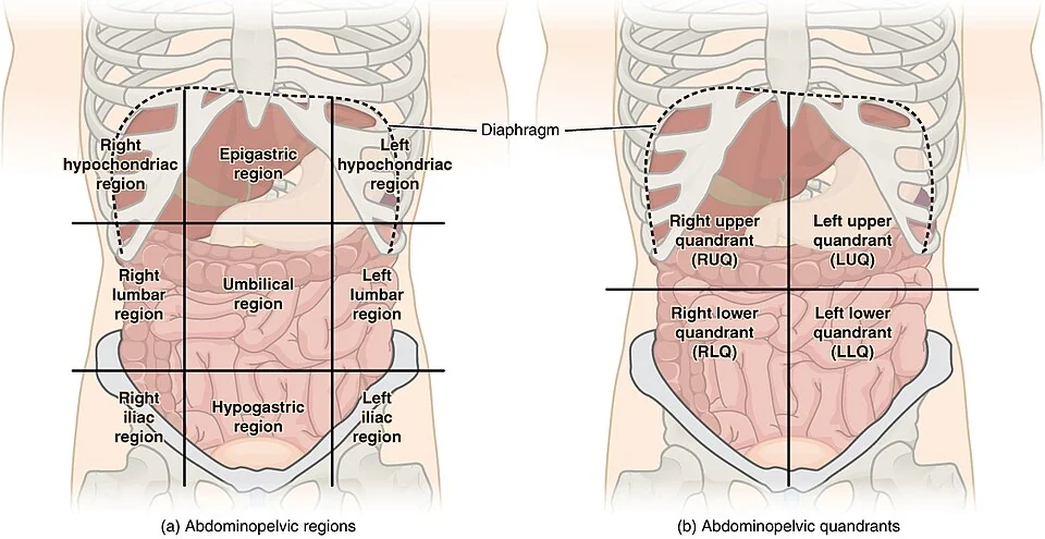
Once pain has localised, the region narrows the differential considerably. Learn this as a grid rather than as a list, because a grid is how you will use it at the bedside.
| Region | Causes |
|---|---|
| Right upper quadrant | Biliary colic, acute cholecystitis, ascending cholangitis, hepatitis, liver abscess, right basal pneumonia, Fitz-Hugh-Curtis syndrome |
| Epigastrium | Peptic ulcer disease, perforated ulcer, acute pancreatitis, gastritis, myocardial infarction, ruptured abdominal aortic aneurysm |
| Left upper quadrant | Splenic infarct or rupture, gastric ulcer, acute pancreatitis, left basal pneumonia |
| Central or periumbilical | Early appendicitis, small bowel obstruction, mesenteric ischaemia, ruptured abdominal aortic aneurysm, gastroenteritis |
| Right iliac fossa | Appendicitis, mesenteric adenitis, Crohn disease, caecal tumour, ovarian cyst accident, ectopic pregnancy, ureteric colic |
| Left iliac fossa | Diverticulitis, constipation, colorectal tumour, ovarian cyst accident, ectopic pregnancy, ureteric colic |
| Suprapubic | Urinary retention, cystitis, pelvic inflammatory disease, endometriosis, miscarriage |
| Diffuse | Perforation with generalised peritonitis, mesenteric ischaemia, obstruction, diabetic ketoacidosis, Addisonian crisis, sickle cell crisis |
Medical causes that present as a surgical abdomen
These are disproportionately represented in exam questions because missing them leads to an unnecessary laparotomy in a patient who is already unwell.
- Diabetic ketoacidosis - abdominal pain, vomiting and a raised white cell count, all of which resolve with insulin and fluid. Check capillary glucose and ketones in every acute abdomen.
- Inferior myocardial infarction - epigastric pain with nausea and sweating. An ECG is mandatory in anyone over 50 or with cardiovascular risk factors.
- Lower lobe pneumonia - diaphragmatic pleural irritation producing upper abdominal pain and guarding, especially in children
- Addisonian crisis - abdominal pain, vomiting, hypotension, hyponatraemia and hyperkalaemia
- Hypercalcaemia and acute intermittent porphyria - uncommon metabolic mimics that are nonetheless worth a moment of thought in unexplained recurrent pain
- Sickle cell vaso-occlusive crisis - the girdle syndrome may be indistinguishable from obstruction
- Herpes zoster - dermatomal pain that precedes the rash by several days
History
The single highest yield question is what the patient was doing at the moment the pain started, because the speed of onset maps closely onto the underlying mechanism.
| Onset | Mechanism | Typical causes |
|---|---|---|
| Instantaneous, maximal within seconds | Rupture, perforation or arterial occlusion | Perforated viscus, ruptured aortic aneurysm, mesenteric embolus, ruptured ectopic pregnancy |
| Rapid, over minutes to an hour | Obstruction of a hollow viscus, or torsion | Ureteric colic, biliary colic, volvulus, ovarian or testicular torsion |
| Gradual, over hours to days | Progressive inflammation or infection | Appendicitis, diverticulitis, cholecystitis, pancreatitis, pelvic inflammatory disease |
Points to cover
- Site, onset, character, radiation, timing, exacerbating and relieving factors and severity - the standard structure, but ask specifically where the pain started as well as where it is now
- Vomiting and its relationship to the pain - pain before vomiting favours a surgical cause, vomiting before pain favours gastroenteritis. Faeculent vomiting suggests distal obstruction; bilious vomiting excludes obstruction proximal to the ampulla.
- Bowel habit - absolute constipation to both faeces and flatus is the cardinal symptom of complete obstruction. Bloody diarrhoea suggests colitis, dysentery or ischaemia.
- Urinary symptoms - dysuria, frequency, haematuria and the possibility of retention
- A gynaecological history in every woman - last menstrual period, contraception, possibility of pregnancy, vaginal discharge or bleeding, previous ectopic pregnancy or pelvic inflammatory disease
- Previous abdominal surgery - adhesions are the commonest cause of small bowel obstruction in the UK, so a laparotomy scar changes the differential immediately
- Drugs - NSAIDs and corticosteroids (ulceration and masked signs), anticoagulants (bleeding), opioids (constipation) and immunosuppressants
- Cardiovascular history - atrial fibrillation moves mesenteric embolus sharply up the list, and known aneurysm or peripheral arterial disease raises rupture
- Red flag symptoms - weight loss, anorexia, change in bowel habit over weeks and rectal bleeding
Examination
Begin at the end of the bed. A patient lying rigidly still with shallow breathing has peritonitis until proven otherwise; a patient writhing and unable to settle has colic. This single observation is often more informative than palpation.
General and cardiorespiratory
- Full observations including respiratory rate and temperature, with a NEWS2 score calculated and acted on
- Signs of shock: tachycardia, hypotension, cool peripheries, prolonged capillary refill, reduced urine output and confusion
- Jaundice, anaemia, dehydration, cachexia and lymphadenopathy including Virchow node
- Chest examination and an ECG, since chest pathology commonly refers to the abdomen
Abdominal examination
- Inspection - distension, scars, visible peristalsis, hernial orifices, Grey Turner and Cullen signs and caput medusae
- Palpation - start away from the pain, with the patient looking at your face rather than at their abdomen. Assess for tenderness, guarding, rigidity and masses.
- Percussion - percussion tenderness is a gentler and more reliable sign of peritonism than rebound. Resonance over a distended abdomen suggests obstruction; shifting dullness suggests ascites or blood.
- Auscultation - absent bowel sounds suggest ileus or established peritonitis, and high-pitched tinkling sounds suggest mechanical obstruction. The sign is unreliable in isolation.
- Hernial orifices and external genitalia - an incarcerated femoral hernia and a testicular torsion are both missed by clinicians who do not look
- Digital rectal examination - for melaena, fresh blood, a rectal mass, faecal loading, or an empty ballooned rectum in obstruction
- Pelvic examination - where a gynaecological cause is likely, performed by an appropriately trained clinician with a chaperone
Named signs
| Sign | Elicited by | Suggests |
|---|---|---|
| Murphy sign | Pain and arrest of inspiration on palpation below the right costal margin, absent on the left | Acute cholecystitis |
| Rovsing sign | Palpation of the left iliac fossa causes pain in the right iliac fossa | Appendicitis |
| Psoas sign | Pain on passive extension of the right hip | Retrocaecal appendicitis or psoas abscess |
| Obturator sign | Pain on internal rotation of the flexed right hip | Pelvic appendicitis |
| Grey Turner sign | Flank bruising | Retroperitoneal haemorrhage, classically severe pancreatitis |
| Cullen sign | Periumbilical bruising | Retroperitoneal or intraperitoneal haemorrhage |
| Kehr sign | Left shoulder tip pain | Subdiaphragmatic irritation, classically splenic rupture |
| Boas sign | Hyperaesthesia below the right scapula | Acute cholecystitis |
Investigations
Bedside
- Urinary or serum beta-hCG in every woman of childbearing age. A negative test is what allows you to stop thinking about ectopic pregnancy. Omitting it is a common and serious error.
- Capillary glucose and ketones - to identify diabetic ketoacidosis
- Urine dipstick, interpreted with caution. Sterile pyuria and microscopic haematuria occur in appendicitis because the inflamed appendix lies against the ureter or bladder, so a positive dipstick does not exclude a surgical abdomen.
- 12-lead ECG - for myocardial infarction and atrial fibrillation
- Venous blood gas - a rapid lactate, potassium, haemoglobin and pH. A rising lactate with a metabolic acidosis in a patient whose abdomen looks unimpressive is the classic picture of mesenteric ischaemia.
- Bedside ultrasound - a focused assessment for free fluid, chiefly in trauma
Laboratory
- FBC - leucocytosis supports inflammation, but a normal white cell count never excludes it and is often normal in early appendicitis and in older or immunosuppressed patients
- U&Es - acute kidney injury from hypovolaemia, and electrolyte derangement from vomiting
- LFTs - an obstructive picture points to biliary pathology, and a raised bilirubin with fever and pain is Charcot triad
- Amylase or lipase - a level more than three times the upper limit of normal supports acute pancreatitis. Amylase is also modestly raised in perforation, mesenteric ischaemia and ectopic pregnancy, and may be normal in late presentation, so lipase is preferred where available.
- CRP - useful for tracking a trajectory rather than for making a diagnosis
- Clotting screen, group and save, and crossmatch if bleeding or theatre is a possibility
- Blood cultures if febrile, taken before antibiotics
Imaging
Contrast-enhanced CT of the abdomen and pelvis is the default investigation in an adult with an undifferentiated acute abdomen. It is sensitive for perforation, obstruction, collections, ischaemia and aneurysm, and it changes management in a substantial proportion of patients. Concern about radiation dose should not delay it in an acutely unwell adult, though it carries more weight in young patients and in pregnancy.3
- Erect chest radiograph - can show free subdiaphragmatic gas, but its sensitivity for perforation is only around 70%, so a normal film does not exclude one. It is quick, and it also identifies pneumonia.
- Abdominal radiograph - now used selectively, chiefly for suspected obstruction or a suspected foreign body. It has no role in the routine assessment of undifferentiated abdominal pain.
- Ultrasound - first line for suspected biliary disease, gynaecological pathology, pregnancy and children, because it avoids ionising radiation and images the gallbladder better than CT does
- CT angiography - for suspected mesenteric ischaemia or aortic pathology, with an arterial phase
- MRCP - for suspected common bile duct stones when ultrasound is inconclusive
- Diagnostic laparoscopy - both diagnostic and therapeutic, and particularly useful in young women in whom appendicitis and gynaecological pathology cannot be separated clinically
Initial management
Resuscitation runs in parallel with assessment, not after it. The sequence below should be underway before the diagnosis is known.
- A to E assessment with oxygen if hypoxic or shocked, and continuous monitoring
- Two large-bore cannulae, with bloods taken at the time of insertion, including group and save
- Fluid resuscitation with a balanced crystalloid such as Hartmann solution, in boluses of 250 to 500 mL with reassessment after each. Patients with obstruction or peritonitis may be litres depleted through third-space losses.4
- Analgesia - titrated intravenous morphine with an antiemetic, given early
- Nil by mouth, with a nasogastric tube on free drainage if there is obstruction or persistent vomiting
- Catheterise and monitor hourly urine output in anyone shocked or going to theatre
- Antibiotics if there is sepsis or an identified source, following local policy and within one hour where the sepsis criteria are met5
- Venous thromboembolism prophylaxis, unless bleeding or imminent surgery contraindicates it
- Early senior surgical review, with critical care involved early in anyone with organ dysfunction
Deciding who needs an operation
The decision to operate is a clinical one, and the following broadly indicate that an abdomen will not settle without surgery:
- Generalised peritonitis with a rigid abdomen
- Free intraperitoneal gas from a perforated viscus
- Haemodynamic instability from suspected intra-abdominal haemorrhage, including a ruptured aortic aneurysm or ruptured ectopic pregnancy
- Bowel obstruction with signs of strangulation or ischaemia
- An irreducible, tender hernia
- Failure to improve, or deterioration, on conservative management
Many conditions once treated operatively are now managed conservatively in the first instance: uncomplicated diverticulitis, adhesional small bowel obstruction without ischaemia, selected appendicitis, and localised collections that can be drained radiologically. The default is no longer the knife, but nor is prolonged temporising in a patient who is not improving.
Groups where the presentation is atypical
Older adults
Older patients present later, with less pain, less fever and a lower white cell count, and are more likely to have a serious underlying diagnosis. Perforation may produce only mild tenderness. Mortality from emergency laparotomy rises steeply with age and frailty, so the threshold for CT should be low and the threshold for senior review lower still.
Pregnancy
The gravid uterus displaces the appendix upwards and laterally, so appendicitis may present with right upper quadrant pain in the third trimester. Leucocytosis is physiological in pregnancy and therefore unhelpful. Ultrasound and MRI are the imaging modalities of choice, and the obstetric team must be involved from the outset. Ectopic pregnancy should be assumed in any woman with abdominal pain and a positive beta-hCG until an intrauterine pregnancy is demonstrated.
Immunosuppression and corticosteroids
Corticosteroids, chemotherapy and biologic therapy blunt both the inflammatory response and the physical signs. A patient on long-term prednisolone may have a perforated viscus with an almost normal abdomen and only a modest tachycardia. Neutropenic patients may develop neutropenic enterocolitis, which is managed medically, and both the white cell count and the CRP may be misleadingly low.
Children
Mesenteric adenitis, intussusception, testicular torsion and constipation dominate the paediatric differential. Younger children localise poorly, and a lower lobe pneumonia is a genuinely common cause of abdominal pain in this group. Always examine the testes and the hernial orifices.
Complications
The complications of an acute abdomen arise by three routes: the underlying disease, the physiological insult, and the treatment itself.
- Sepsis and septic shock from peritoneal contamination or an untreated collection
- Hypovolaemic shock from haemorrhage or from third-space losses into obstructed bowel
- Acute kidney injury, driven by hypovolaemia and compounded by NSAIDs and contrast
- Bowel infarction and short bowel syndrome where ischaemia is diagnosed late
- Abdominal compartment syndrome in massive distension or after damage-control surgery
- Post-operative complications - ileus, anastomotic leak, wound dehiscence, incisional hernia and adhesions, which in turn generate future obstruction
- Delirium, deconditioning and loss of independence, particularly in frail patients after a prolonged admission
Red flags
Prognosis
Outcome depends far more on the physiological state of the patient and on the delay to definitive treatment than on the specific diagnosis. Most patients admitted with acute abdominal pain recover without an operation, and non-specific abdominal pain is self-limiting.
The high-risk group is those undergoing emergency laparotomy. When the National Emergency Laparotomy Audit began, 30-day mortality was around 11%, and it has since fallen through a set of unglamorous process changes: consultant surgeon and anaesthetist present in theatre, documented risk assessment before the operation, arrival in theatre within a time frame matched to urgency, and post-operative critical care for high-risk patients.7
That fall matters for how you think about the acute abdomen as a student. The determinants of survival are largely organisational and lie in the first few hours: recognising the sick patient, resuscitating them, obtaining imaging promptly, calculating a risk score, and escalating early. Almost all of that happens before anyone reaches an operating theatre, and much of it can be done by the most junior member of the team.
References
- Royal College of Surgeons of England. Emergency surgery: standards for unscheduled surgical care. Available here
- Gans SL, Pols MA, Stoker J, Boermeester MA. Guideline for the diagnostic pathway in patients with acute abdominal pain. Digestive Surgery. 2015. Available here
- Royal College of Radiologists. iRefer: making the best use of clinical radiology. Available here
- NICE CG174. Intravenous fluid therapy in adults in hospital. 2013, updated 2017. Available here
- NICE NG51. Sepsis: recognition, diagnosis and early management. 2016, updated 2024. Available here
- Manterola C, Vial M, Moraga J, Astudillo P. Analgesia in patients with acute abdominal pain. Cochrane Database of Systematic Reviews. 2011. Available here
- National Emergency Laparotomy Audit. Annual reports. Available here
This article is written for revision and education. It is not clinical guidance and must not be used to make decisions about the care of a patient. Always check current NICE guidance and local protocols.