
Abdominal Aortic Aneurysm
Key points
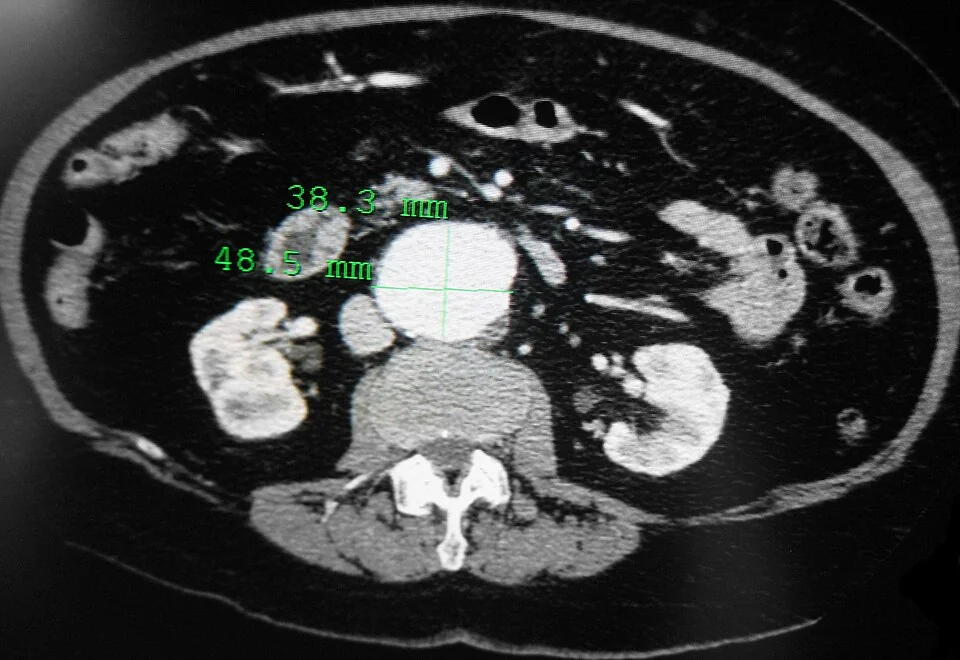
- Definition: permanent focal dilatation of the aorta to at least 1.5 times its normal diameter. In practice an infrarenal aortic diameter of 3 cm or more.
- Epidemiology: present in around 1 to 2% of UK men aged 65 to 74, and four to six times commoner in men than women.
- Strongest risk factor: smoking. Age, male sex, hypertension and a first-degree relative with an aneurysm follow. Diabetes is consistently associated with a lower risk.
- Why size matters: by the law of Laplace, wall tension rises with radius, so each increment in diameter accelerates further expansion and rupture risk.
- Screening: the NHS programme offers a single abdominal ultrasound to men in the year they turn 65. It has roughly halved AAA-related mortality in screened men.
- Surveillance: 3.0 to 4.4 cm is rescanned yearly, 4.5 to 5.4 cm every three months, and 5.5 cm or more is referred to vascular surgery within two weeks.
- Repair threshold: diameter of 5.5 cm or more, growth of more than 1 cm in a year, or a symptomatic aneurysm at any size.
- Rupture: abdominal or back pain with hypotension and a pulsatile mass. Do not delay for imaging in an unstable patient - call the vascular team.
Introduction and definitions
An aneurysm is a permanent, localised dilatation of an artery to at least 1.5 times its expected diameter. Since the normal infrarenal aorta measures roughly 2 cm in men and slightly less in women, the working definition of an abdominal aortic aneurysm is an infrarenal aortic diameter of 3 cm or more. Around 90% are infrarenal, which is fortunate, because that position makes them accessible to both open and endovascular repair.
Two distinctions are worth having clear:
- True aneurysm - all three layers of the arterial wall (intima, media and adventitia) are involved. This describes almost all abdominal aortic aneurysms.
- False aneurysm (pseudoaneurysm) - a breach in the wall contained only by adventitia or surrounding tissue, typically after trauma, arterial puncture or anastomotic breakdown
- Fusiform - symmetrical dilatation involving the whole circumference, which is the usual morphology
- Saccular - an eccentric outpouching from one side of the wall, more often associated with infection or trauma
The clinical problem is that an abdominal aortic aneurysm is silent until it kills. Around 80% of ruptures are fatal, and the majority of those deaths occur before the patient reaches an operating theatre. Everything about the management of the condition - screening, surveillance, thresholds for elective repair - is an attempt to intervene during the silent phase.
Pathophysiology
Aneurysm formation is a degenerative process in the tunica media rather than a simple consequence of atherosclerosis, though the two share risk factors and frequently coexist.
- Loss of elastin and collagen from the media, the proteins that give the aorta its strength and recoil. Elastin content is already lowest in the infrarenal aorta, which is one reason aneurysms form there.
- Increased activity of matrix metalloproteinases, particularly MMP-2 and MMP-9, which degrade the extracellular matrix, with a relative deficiency of their tissue inhibitors
- Chronic transmural inflammation with lymphocyte and macrophage infiltration of the media and adventitia
- Loss of vascular smooth muscle cells by apoptosis, so the matrix is not repaired
- Progressive dilatation, at which point the mechanics take over
The law of Laplace explains why aneurysms behave the way they do. For a cylinder, wall tension is proportional to the pressure multiplied by the radius. As the aorta dilates, the tension in its already weakened wall rises, which causes further dilatation, which raises the tension again. Growth therefore accelerates with size - a 4 cm aneurysm grows at roughly 2 to 3 mm a year, while a 5.5 cm aneurysm may grow at 5 mm or more - and this positive feedback is the whole reason surveillance intervals shorten as the aneurysm enlarges.
Risk factors
- Smoking - by far the strongest modifiable risk factor, increasing risk several-fold and accelerating expansion. Stopping smoking is the only intervention proven to slow growth.
- Increasing age - prevalence rises steeply after 60
- Male sex - four to six times commoner in men, though women who develop one rupture at a smaller diameter and have worse outcomes
- Family history - a first-degree relative with an aneurysm roughly doubles the risk, and screening of siblings is considered
- Hypertension and hyperlipidaemia
- Chronic obstructive pulmonary disease, independent of smoking
- Connective tissue disorders - Marfan syndrome and vascular (type IV) Ehlers-Danlos syndrome, which typically produce aneurysms at a younger age and in the thoracic aorta
- Existing arterial disease elsewhere - peripheral arterial disease, popliteal aneurysm and previous aneurysm repair
- Infection - a mycotic aneurysm, classically salmonella or staphylococcal, and historically syphilitic aortitis
Clinical features
Unruptured aneurysm
Most are asymptomatic and found incidentally on screening or on imaging performed for another reason. When symptoms do occur they are:
- A pulsatile, expansile abdominal mass in the epigastrium or periumbilical region. The distinction between expansile and merely transmitted pulsation is important: place both hands on either side of the mass and see whether they are pushed apart (expansile, aneurysmal) or upwards together (transmitted from a normal aorta beneath an overlying mass).
- Vague abdominal, flank or back pain, from stretching of surrounding tissue. New or worsening pain in a known aneurysm suggests rapid expansion or impending rupture and is a surgical emergency.
- Distal embolisation - showers of thrombus or atheromatous debris from the aneurysm sac causing blue toes with palpable pedal pulses, sometimes called trash foot
- Compressive symptoms - early satiety, ureteric obstruction, or lower limb venous congestion
- Rarely, an aortocaval fistula causing high-output cardiac failure with an abdominal bruit and a machinery murmur, or an aortoenteric fistula presenting with gastrointestinal bleeding, usually after previous aortic graft surgery
Physical examination is unreliable. Abdominal palpation detects perhaps 30 to 50% of aneurysms in obese patients and rather more in thin ones, so a normal examination is not reassuring and is not a substitute for ultrasound.
Ruptured aneurysm
The distinction between retroperitoneal and intraperitoneal rupture matters. Around 80% rupture posteriorly into the retroperitoneum, where surrounding tissue can tamponade the bleeding temporarily and give a window of hours in which to operate. Anterior rupture into the peritoneal cavity is uncontained and usually rapidly fatal.
Screening and surveillance
The NHS Abdominal Aortic Aneurysm Screening Programme invites men for a single abdominal ultrasound in the year they turn 65. Men over 65 who have not been screened can self-refer. Women are not screened, because prevalence is much lower and screening has not been shown to be cost-effective, though women who do develop aneurysms rupture at smaller diameters.
The programme has been shown in randomised trials, most notably the Multicentre Aneurysm Screening Study, to roughly halve AAA-related mortality in screened men, and it is one of the more clearly beneficial screening programmes in the NHS.1
| Diameter | Classification | Action | Approximate annual rupture risk |
|---|---|---|---|
| Under 3.0 cm | Normal | Discharged from the screening programme, no further scans | Negligible |
| 3.0 to 4.4 cm | Small aneurysm | Ultrasound surveillance every 12 months | Under 1% |
| 4.5 to 5.4 cm | Medium aneurysm | Ultrasound surveillance every 3 months | About 1 to 3% |
| 5.5 cm or more | Large aneurysm | Refer to vascular surgery to be seen within 2 weeks | Rising steeply - around 10% or more above 6 cm, and over 30% above 8 cm |
Growth of more than 1 cm in a year at any diameter, and any symptomatic aneurysm, also warrant urgent vascular referral irrespective of absolute size.2
Investigations
- Abdominal ultrasound - the screening and surveillance investigation. It is cheap, quick, requires no radiation or contrast, and measures the anteroposterior diameter accurately. It cannot reliably assess the suprarenal aorta or diagnose rupture.
- CT angiography of the aorta - the definitive investigation. It defines the relationship to the renal and visceral arteries, the neck length and angulation, iliac anatomy and access vessels, and is required for planning any repair. It is also the investigation of choice in suspected rupture in a haemodynamically stable patient.
- MR angiography - an alternative where iodinated contrast must be avoided, though less practical in the acute setting
- Bloods - FBC, U&Es, clotting, group and crossmatch at least six units in suspected rupture, plus a venous gas for haemoglobin and lactate
- ECG, echocardiography and lung function tests as part of preoperative cardiopulmonary assessment, since the population has a high burden of coexisting ischaemic heart disease
- Cardiopulmonary exercise testing in many units, to stratify risk before elective repair
Management
Conservative management and risk factor modification
For aneurysms below the repair threshold, treatment is surveillance plus aggressive cardiovascular risk reduction. This is not a passive strategy: these patients have a high burden of cardiovascular disease, and more of them die of myocardial infarction than of aneurysm rupture.
- Smoking cessation - the only intervention shown to slow aneurysm growth, as well as reducing rupture risk and perioperative complications
- Blood pressure control to target, following NICE hypertension guidance
- A high-intensity statin and an antiplatelet agent, for cardiovascular risk reduction rather than for any direct effect on the aneurysm
- Diabetes and weight management, and encouragement of regular exercise
- Adherence to the surveillance programme, and clear safety-netting advice about what symptoms should prompt immediate attendance
- DVLA notification - car and motorcycle drivers must notify the DVLA if the aneurysm is 6 cm or more and are disqualified at 6.5 cm or more; the thresholds for lorry and bus drivers are lower. Check the current guidance, as these rules are revised.4
Note that neither beta-blockers, doxycycline, ACE inhibitors nor statins have been shown in trials to slow aneurysm growth, despite plausible mechanisms. Do not present any of them as aneurysm-directed therapy.
Elective repair
| Open repair | EVAR | |
|---|---|---|
| Procedure | Laparotomy or retroperitoneal approach, aortic cross-clamping, and inlay of a tube or bifurcated graft | A stent graft is deployed within the aneurysm sac via the femoral arteries, excluding it from the circulation |
| Anaesthesia and recovery | General anaesthesia, intensive care, 7 to 10 days in hospital, weeks to months of recovery | Can be done under regional anaesthesia, 1 to 3 days in hospital, rapid recovery |
| Perioperative mortality | Higher, around 3 to 5% | Lower, around 1 to 2% |
| Long-term outcome | Durable. Late graft-related complications are uncommon. | The early survival advantage is lost by around 2 to 4 years, and late aneurysm-related mortality is higher |
| Follow-up | Minimal | Lifelong imaging surveillance is mandatory to detect endoleak, migration and sac expansion |
| Anatomical requirements | Few | Requires an adequate infrarenal neck length and angulation, and suitable iliac access vessels |
NICE guidance NG156 took a position that surprised many at the time: for an unruptured infrarenal aneurysm, it recommends considering open surgical repair as the first choice, and offering EVAR only where open repair is unsuitable because of anaesthetic or medical risk or abdominal comorbidity. The reasoning is the long-term data from the EVAR-1 trial, in which the early advantage of EVAR reversed over time because of late rupture and reintervention.2 This remains debated in the vascular community, and practice varies, so present it as a NICE recommendation with an acknowledged controversy rather than as settled consensus.
Ruptured aneurysm
- Immediate vascular surgical referral - this is a phone call, not a referral letter, and should happen before imaging is arranged
- Two large-bore cannulae, urgent crossmatch of at least six units, and activation of the major haemorrhage protocol
- Permissive hypotension. Do not resuscitate to a normal blood pressure - raising it dislodges the clot tamponading the rupture. Aim to maintain cerebration and a radial pulse, with a systolic pressure around 70 to 90 mmHg, until the aorta is controlled.
- Analgesia, oxygen and correction of coagulopathy, with tranexamic acid where local protocol supports it
- CT angiography only if the patient is stable enough, to determine suitability for endovascular repair
- Repair by EVAR or open surgery. The IMPROVE trial found no significant difference in 30-day mortality between an endovascular-first strategy and open repair, but better three-year survival and quality of life with an endovascular strategy, and it is increasingly the default where anatomy and logistics permit.5
- Honest early discussion about ceilings of care, since many patients are frail and operative mortality is high. A decision not to operate is a legitimate and sometimes kind one, made by a senior clinician with the family.
Complications
Of the aneurysm
- Rupture, the dominant risk and the reason for the whole surveillance apparatus
- Distal embolisation causing acute limb or digital ischaemia
- Thrombosis of the sac with acute aortic occlusion, which is rare but catastrophic
- Aortocaval fistula, causing high-output cardiac failure, leg oedema and an abdominal bruit
- Aortoenteric fistula, usually secondary to a previous graft, presenting with a herald gastrointestinal bleed followed by exsanguinating haemorrhage
- Compression of adjacent structures - ureters, duodenum, inferior vena cava
Of repair
- Cardiac events - myocardial infarction is the commonest cause of perioperative death after open repair, driven by the physiological stress of aortic cross-clamping
- Acute kidney injury, from clamping above the renals, hypotension and contrast
- Colonic ischaemia, from interruption of the inferior mesenteric artery. Bloody diarrhoea after aortic surgery is ischaemic colitis until proven otherwise.
- Spinal cord ischaemia and paraplegia, rare in infrarenal repair but a recognised risk
- Graft infection and aortoenteric fistula, late but devastating
- Endoleak after EVAR - persistent blood flow into the sac. Type I (at the proximal or distal seal) and type III (graft defect or component separation) are the high-pressure leaks that need prompt intervention; type II, from retrograde flow via lumbar or the inferior mesenteric artery, is the commonest and is usually observed.
- Stent migration, limb occlusion and the need for reintervention, which is the price of the lower early mortality of EVAR
- Incisional hernia, sexual dysfunction and prolonged ileus after open repair
Red flags
Prognosis
Untreated, the natural history is determined by size. Small aneurysms rupture rarely and most patients die of something else, usually cardiac. Above 5.5 cm the annual rupture risk climbs steadily, and above 8 cm it exceeds 30% a year.
Rupture carries an overall mortality of around 80%. Roughly half of patients die before reaching hospital, and of those who reach an operating theatre, 30 to 50% do not survive to discharge. This is the figure that justifies both screening and elective repair, because elective repair carries an operative mortality of only 1 to 5%.
After successful elective repair, long-term survival is good and approaches that of the age-matched population, though it remains limited by the coexisting cardiovascular disease that these patients almost universally have. The practical consequence is that managing an aneurysm well means managing cardiovascular risk as seriously as managing the aorta - and, for EVAR patients, that lifelong surveillance is not optional, because an unattended endoleak reintroduces exactly the risk the operation was meant to remove.
References
- Public Health England / NHS. Abdominal aortic aneurysm screening programme. Available here
- NICE NG156. Abdominal aortic aneurysm: diagnosis and management. 2020. Available here
- UK Small Aneurysm Trial Participants. Mortality results for randomised controlled trial of early elective surgery or ultrasonographic surveillance for small abdominal aortic aneurysms. The Lancet. 1998. Available here
- DVLA. Assessing fitness to drive: a guide for medical professionals. Available here
- IMPROVE Trial Investigators. Comparative clinical effectiveness and cost effectiveness of endovascular strategy v open repair for ruptured abdominal aortic aneurysm: three year results of the IMPROVE randomised trial. BMJ. 2017. Available here
This article is written for revision and education. It is not clinical guidance and must not be used to make decisions about the care of a patient. Always check current NICE guidance and local protocols.