
Acute Pancreatitis: Diagnosis, Severity Scoring and Management
Key points
- Acute pancreatitis: acute inflammation of the pancreas caused by premature intracellular activation of digestive enzymes, leading to autodigestion of the gland.
- Two dominant causes: gallstones (around 50%) and alcohol (around 25%) account for three quarters of UK cases; the mnemonic GET SMASHED covers the rest.
- Diagnosis: requires two of three: characteristic epigastric pain radiating to the back; amylase or lipase more than 3 times the upper limit of normal; or characteristic imaging.
- Amylase caveat: the level does not correlate with severity, and can be normal in late presentation, alcohol-related disease and hypertriglyceridaemia. Lipase is more sensitive and specific.
- Severity scoring: Glasgow-Imrie (PANCREAS), Ranson and APACHE II; a CRP above 150 mg/L at 48 hours is a simple and useful marker of severe disease.
- Management: supportive - aggressive IV fluid resuscitation, analgesia, and early enteral nutrition; antibiotics are not given prophylactically.
- Gallstone pancreatitis: urgent ERCP if there is coexisting cholangitis or persistent biliary obstruction, and cholecystectomy during the same admission once recovered.
- Complications: early - systemic inflammatory response, ARDS, acute kidney injury; late - pancreatic necrosis, infected necrosis and pseudocyst.
Introduction
Acute pancreatitis is an acute inflammatory process of the pancreas, resulting from premature activation of digestive enzymes within acinar cells, which causes the gland to autodigest itself.1 The resulting local inflammation can trigger a systemic inflammatory response with multi-organ failure.
It is a common surgical emergency, with a UK incidence of roughly 30-40 per 100,000 per year and rising. The clinical course is strikingly bimodal: around 80% of cases are mild and self-limiting, settling within a week with supportive care, while the remaining 20% develop severe disease with necrosis, organ failure and a mortality of 15-30%. Identifying which patients are heading in that direction is the central clinical task.
Causes
Gallstones and alcohol together account for around 75% of cases in the UK. The classic mnemonic for the remainder is GET SMASHED:
| Letter | Cause | Notes |
|---|---|---|
| G | Gallstones | The commonest cause (around 50%). A stone impacts at the ampulla, obstructing pancreatic outflow. More common in women |
| E | Ethanol (alcohol) | Around 25%. More common in men; causes direct acinar toxicity and ductal protein plugs |
| T | Trauma | Blunt abdominal trauma, classically handlebar or seatbelt injury compressing the pancreas against the vertebral column |
| S | Steroids | Corticosteroids |
| M | Mumps and other infections | Also coxsackievirus, EBV, CMV, HIV, and Mycoplasma |
| A | Autoimmune | IgG4-related autoimmune pancreatitis, which responds to corticosteroids |
| S | Scorpion sting | Famous in examinations, vanishingly rare in the UK (Tityus trinitatis, Trinidad) |
| H | Hypercalcaemia, Hypertriglyceridaemia, Hypothermia | Triglycerides above 11 mmol/L; hypercalcaemia from any cause including hyperparathyroidism |
| E | ERCP | Post-ERCP pancreatitis complicates around 3-5% of procedures |
| D | Drugs | Azathioprine, thiazides, furosemide, sodium valproate, sulfonamides, tetracyclines, oestrogens, GLP-1 agonists, mesalazine, didanosine |
Other causes include pancreatic or ampullary malignancy (always consider in an older patient with no obvious cause), pancreas divisum, sphincter of Oddi dysfunction, vasculitis, and hereditary pancreatitis from PRSS1, SPINK1 or CFTR mutations. Around 10-15% remain idiopathic after investigation, though many of these prove to be microlithiasis or biliary sludge.
Pathophysiology
Whatever the trigger, the final common pathway is premature conversion of trypsinogen to trypsin inside acinar cells, overwhelming protective mechanisms such as SPINK1. Activated trypsin then activates the entire cascade of pancreatic proenzymes - elastase, phospholipase A2 and others - causing proteolysis, fat necrosis and haemorrhage. Released inflammatory mediators drive a systemic inflammatory response syndrome (SIRS) with capillary leak, third-space fluid loss, hypovolaemia and, in severe cases, ARDS, acute kidney injury and multi-organ failure. Saponification of fat by lipases binds calcium, contributing to the hypocalcaemia seen in severe disease.
Clinical features
- Severe epigastric pain radiating through to the back, of relatively rapid onset, constant and often severe. Classically relieved by sitting forward and worsened by lying flat or by eating
- Nausea and profuse vomiting, which typically does not relieve the pain
- Epigastric tenderness with guarding; bowel sounds may be reduced or absent from a paralytic ileus
- Fever, tachycardia and hypotension from SIRS and third-space fluid loss
- Jaundice, suggesting a gallstone cause with biliary obstruction
- Tachypnoea and hypoxia, which may indicate a pleural effusion or evolving ARDS
- Dehydration, oliguria and, in severe cases, shock and confusion
Signs of severe haemorrhagic pancreatitis
Two eponymous signs indicate retroperitoneal haemorrhage tracking to the skin. Both are rare, late and denote severe disease, and their absence means nothing:
- Grey Turner sign - bruising of the flanks
- Cullen sign - bruising around the umbilicus (periumbilical)
Investigations
Making the diagnosis
The revised Atlanta criteria require two of the following three:2
- Characteristic abdominal pain - epigastric, radiating to the back
- Serum amylase or lipase more than 3 times the upper limit of normal
- Characteristic findings on imaging (CT, MRI or ultrasound)
Establishing the cause and assessing severity
- Liver function tests: an ALT above 150 U/L strongly suggests a gallstone cause. A raised bilirubin and ALP suggest ongoing biliary obstruction
- Abdominal ultrasound within 24 hours - to identify gallstones and biliary dilatation. This is essential in every patient, as it determines whether cholecystectomy is needed
- Full blood count (leucocytosis, and haematocrit as a marker of haemoconcentration), urea and electrolytes, CRP, calcium, glucose, LDH, albumin, triglycerides and arterial blood gas
- CRP above 150 mg/L at 48 hours is a simple and widely used marker of severe disease
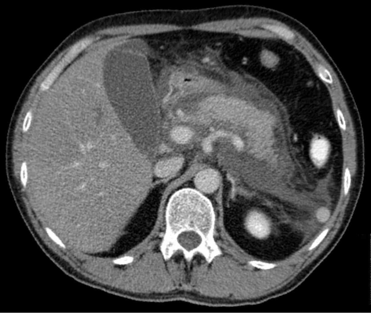
- Contrast-enhanced CT abdomen - not required for diagnosis and best deferred to 72 hours or later, as necrosis takes time to become radiologically apparent. Indicated where the diagnosis is uncertain, or to assess for necrosis and complications in a patient failing to improve
- MRCP or endoscopic ultrasound if biliary obstruction is suspected but ultrasound is inconclusive, or to investigate idiopathic pancreatitis for microlithiasis or tumour
Severity scoring
Several scores predict severity. The Glasgow-Imrie score is widely used in the UK, applied at 48 hours, with a score of 3 or more indicating severe pancreatitis warranting critical care input. The mnemonic is PANCREAS:
| Letter | Criterion |
|---|---|
| P | PaO₂ below 8 kPa |
| A | Age over 55 years |
| N | Neutrophils - white cell count above 15 × 10⁹/L |
| C | Calcium below 2 mmol/L |
| R | Renal function - urea above 16 mmol/L |
| E | Enzymes - LDH above 600 U/L or AST above 200 U/L |
| A | Albumin below 32 g/L |
| S | Sugar - glucose above 10 mmol/L |
Other systems include the Ranson criteria (assessed at admission and 48 hours), APACHE II (usable at any time), the BISAP score, and the Balthazar CT severity index. Note that amylase does not appear in any severity score.
Differential diagnosis
- Perforated peptic ulcer - sudden onset, board-like rigidity, free air on erect chest X-ray; amylase may also be raised
- Acute cholecystitis and biliary colic - right upper quadrant pain, Murphy sign positive
- Acute mesenteric ischaemia - pain out of proportion to signs, in an arteriopath or patient with atrial fibrillation; amylase can be raised
- Ruptured or leaking abdominal aortic aneurysm - always consider in an older patient with abdominal or back pain and shock
- Myocardial infarction, particularly inferior MI presenting with epigastric pain - do an ECG
- Bowel obstruction and strangulated hernia
- Diabetic ketoacidosis, which can itself cause abdominal pain and a raised amylase
- Ectopic pregnancy - do a pregnancy test in every woman of childbearing age
- Basal pneumonia and, less commonly, renal colic
Management
There is no specific treatment for acute pancreatitis - management is supportive, aimed at maintaining organ perfusion, controlling pain, providing nutrition and treating the cause and any complications.3
Initial supportive management
- ABCDE assessment with high-flow oxygen if hypoxic, and early consideration of critical care for severe disease
- Aggressive IV fluid resuscitation - the single most important intervention. Third-space losses are large. Use crystalloid (Hartmann's solution is preferred over 0.9% saline), guided by urine output (target 0.5 mL/kg/hour), haemodynamics and lactate. Both under- and over-resuscitation are harmful
- Analgesia - often requiring opioids; do not withhold them for fear of sphincter of Oddi spasm, as this concern is not evidence-based
- Antiemetics and a nasogastric tube if there is vomiting or ileus
- Catheterise and monitor fluid balance closely in severe disease
- Correct electrolytes, particularly calcium, magnesium and potassium, and manage hyperglycaemia
- VTE prophylaxis
Nutrition
The historical practice of prolonged 'pancreatic rest' with nil by mouth has been abandoned. Early enteral nutrition is now standard, ideally oral feeding within 24-72 hours as tolerated, or nasogastric/nasojejunal feeding if oral intake is not possible.1 Enteral feeding maintains gut mucosal integrity and reduces bacterial translocation, infected necrosis and mortality. Parenteral nutrition is reserved for those who cannot tolerate enteral feeding at all.
Antibiotics
Prophylactic antibiotics are not recommended, even in severe or necrotising pancreatitis, as they do not improve outcomes and promote resistance and fungal infection. Antibiotics are given only for proven or strongly suspected infection, such as infected necrosis, cholangitis or an extrapancreatic infection.
Managing the cause
- Gallstone pancreatitis: urgent ERCP with sphincterotomy (within 72 hours) is indicated if there is coexisting acute cholangitis or persistent biliary obstruction. It is not required for uncomplicated gallstone pancreatitis where the stone has passed. Cholecystectomy should be performed during the same admission, or within 2 weeks, once the patient has recovered - delaying it risks recurrent pancreatitis
- Alcohol: manage withdrawal with a benzodiazepine and thiamine (Pabrinex), and provide alcohol cessation support
- Hypertriglyceridaemia: insulin infusion, fibrates and occasionally plasmapheresis
- Hypercalcaemia: fluids and treatment of the underlying cause, usually hyperparathyroidism
- Drugs: stop the causative agent
- Autoimmune pancreatitis: corticosteroids, with dramatic response
Managing local complications
Sterile necrosis is managed conservatively. Infected necrosis - suspected when a patient deteriorates or develops sepsis after the first week, and confirmed by gas within the collection on CT or by fine-needle aspiration - requires antibiotics and intervention. The modern approach is the "step-up" strategy: percutaneous or endoscopic drainage first, escalating to minimally invasive necrosectomy only if needed, and delaying intervention beyond 4 weeks where possible to allow the collection to wall off. This has substantially lower mortality than early open necrosectomy.
Complications
Early (systemic)
- Systemic inflammatory response syndrome and multi-organ failure - the leading cause of early death
- Acute respiratory distress syndrome (ARDS), pleural effusions and atelectasis
- Acute kidney injury from hypovolaemia
- Hypocalcaemia from fat saponification, and hypomagnesaemia
- Hyperglycaemia from impaired insulin secretion
- Disseminated intravascular coagulation and shock
- Ileus and abdominal compartment syndrome
Late (local)
| Complication | Features |
|---|---|
| Acute peripancreatic fluid collection | Within 4 weeks, no defined wall; most resolve spontaneously |
| Pancreatic pseudocyst | A collection of pancreatic fluid with a non-epithelialised fibrous wall, forming after around 4 weeks. Often asymptomatic; may cause pain, early satiety or obstruction. Most resolve; drain (endoscopically) if symptomatic, infected or enlarging |
| Pancreatic necrosis | Devitalised pancreatic or peripancreatic tissue, seen as non-enhancing areas on contrast CT |
| Infected necrosis | Suspect with deterioration or sepsis after the first week; gas in the collection on CT. Requires antibiotics and step-up drainage or necrosectomy. Carries the highest mortality |
| Walled-off necrosis | Mature, encapsulated necrosis after around 4 weeks |
| Pancreatic abscess | A circumscribed collection of pus |
| Vascular | Splenic vein thrombosis (which can cause gastric varices and left-sided portal hypertension), portal vein thrombosis, and pseudoaneurysm with risk of catastrophic haemorrhage |
| Chronic sequelae | Progression to chronic pancreatitis, exocrine insufficiency with steatorrhoea, and endocrine insufficiency with diabetes |
Red flags
Prognosis
Around 80% of episodes are mild, resolving within a week with supportive care and carrying a mortality under 1%. The remaining 20% develop moderate or severe disease, where mortality rises to 15-30%, and reaches 30-40% with infected necrosis and multi-organ failure.2
Mortality follows a bimodal distribution: early deaths (within the first week) result from the systemic inflammatory response and multi-organ failure, while late deaths result from infected necrosis and sepsis. Persistent organ failure beyond 48 hours is the strongest single predictor of death - transient organ failure that resolves carries a much better outlook.
Longer-term, around a third of patients develop exocrine insufficiency and a quarter develop diabetes after severe pancreatitis, and recurrent attacks may progress to chronic pancreatitis. Recurrence is largely preventable by treating the cause: performing cholecystectomy during the index admission for gallstone pancreatitis, and achieving alcohol abstinence, are the two interventions with the greatest impact on long-term outcome.
References
- NICE NG104. Pancreatitis. 2018 (updated 2020). Available here
- Banks PA et al. Classification of acute pancreatitis - 2012: revision of the Atlanta classification. Gut. 2013. Available here
- Working Group IAP/APA. Evidence-based guidelines for the management of acute pancreatitis. Pancreatology. 2013. Available here
- Hellerhoff, CC BY-SA 3.0, via Wikimedia Commons. Available here
- NICE Clinical Knowledge Summaries (CKS). Pancreatitis - acute. 2023. Available here
- Blamey SL et al. Prognostic factors in acute pancreatitis (Glasgow-Imrie criteria). Gut. 1984. Available here
- van Santvoort HC et al. A step-up approach or open necrosectomy for necrotizing pancreatitis. N Engl J Med. 2010. Available here
This article is written for revision and education. It is not clinical guidance and must not be used to make decisions about the care of a patient. Always check current NICE guidance and local protocols.