
Acute Kidney Injury: Staging, Causes and Immediate Management
Key points
- Acute kidney injury (AKI): an abrupt decline in glomerular filtration over hours to days, detected by a rising creatinine or falling urine output. It is a syndrome with many causes, not a diagnosis in itself.
- KDIGO criteria: a creatinine rise of 26 micromol/L or more within 48 hours, a rise to 1.5 times baseline or more within 7 days, or urine output under 0.5 mL/kg/hour for more than 6 hours. Any one is sufficient.
- Three anatomical categories: pre-renal (reduced perfusion - the commonest, around 40-70%), intrinsic renal (damage to the parenchyma), and post-renal (obstruction). This framework drives the entire work-up.
- Commonest picture in hospital: an older patient with pre-existing CKD who becomes hypovolaemic or septic while taking an ACE inhibitor, a diuretic and an NSAID - the so-called triple whammy.
- Essential early investigations: U&Es, urine dipstick (blood and protein point to intrinsic disease), fluid status assessment, and renal ultrasound within 24 hours if obstruction is suspected or no cause is apparent.
- Management: treat the cause, restore perfusion, stop nephrotoxic drugs, relieve obstruction, and identify and treat life-threatening complications - above all hyperkalaemia.
- Indications for urgent dialysis: remembered as AEIOU - refractory Acidosis, Electrolyte disturbance (hyperkalaemia), Intoxication, refractory fluid Overload, and Uraemic complications (encephalopathy or pericarditis).
- Prognosis: AKI is associated with substantially increased inpatient mortality and, even when creatinine recovers, an increased long-term risk of CKD - so every episode needs follow-up.
Introduction
Acute kidney injury is an abrupt deterioration in renal function over hours to days, resulting in the retention of nitrogenous waste, dysregulation of fluid and electrolyte balance, and impaired acid-base homeostasis.1 The older term 'acute renal failure' has been abandoned deliberately, because it implied a binary event, whereas even modest rises in creatinine carry prognostic significance.
AKI is remarkably common and is detected in around 15 to 20% of hospital admissions, rising far higher in critical care.2 It matters for three reasons: it is associated with a substantial increase in inpatient mortality; a significant proportion of cases are avoidable, arising from dehydration, sepsis and nephrotoxic prescribing; and even apparently full biochemical recovery leaves an increased long-term risk of chronic kidney disease.
Definition and staging
The KDIGO criteria are the internationally accepted definition and are used by the automated e-alert systems now embedded in UK laboratory reporting.1 AKI is present if any one of the following is met:
- A rise in serum creatinine of 26 micromol/L or more within 48 hours
- A rise in serum creatinine to 1.5 times the baseline or more, known or presumed to have occurred within the preceding 7 days
- A urine output of less than 0.5 mL/kg/hour for more than 6 consecutive hours
| Stage | Serum creatinine | Urine output |
|---|---|---|
| Stage 1 | 1.5 to 1.9 times baseline, or a rise of 26 micromol/L or more | Under 0.5 mL/kg/hour for 6 to 12 hours |
| Stage 2 | 2.0 to 2.9 times baseline | Under 0.5 mL/kg/hour for 12 hours or more |
| Stage 3 | 3.0 times baseline or more, or a rise to 354 micromol/L or above, or the initiation of renal replacement therapy (or, in under-18s, an eGFR fall to under 35) | Under 0.3 mL/kg/hour for 24 hours or more, or anuria for 12 hours or more |
Causes
Classifying by anatomical site is the most useful bedside approach, because each category has a distinct investigation and management pathway. In practice the categories frequently overlap - prolonged pre-renal hypoperfusion progresses to intrinsic acute tubular necrosis if uncorrected.
Pre-renal (reduced renal perfusion)
The commonest category, accounting for the majority of hospital-acquired AKI. The renal parenchyma is initially structurally normal and function recovers rapidly if perfusion is restored - which is what makes early recognition so valuable.
| Mechanism | Examples |
|---|---|
| True hypovolaemia | Haemorrhage, vomiting and diarrhoea, burns, excessive diuresis, poor oral intake, pancreatitis, diabetic ketoacidosis |
| Reduced cardiac output | Cardiogenic shock, decompensated heart failure, massive pulmonary embolism, cardiac tamponade - together termed cardiorenal syndrome |
| Systemic vasodilation | Sepsis, anaphylaxis, liver cirrhosis with splanchnic vasodilation, and drugs causing hypotension |
| Renal vascular disease | Bilateral renal artery stenosis (or unilateral with a single functioning kidney), renal artery dissection or thromboembolism, aortic dissection |
| Drugs impairing autoregulation | NSAIDs block the prostaglandin-mediated vasodilation of the afferent arteriole; ACE inhibitors and ARBs block angiotensin II-mediated constriction of the efferent arteriole. Both reduce glomerular filtration pressure. Calcineurin inhibitors (ciclosporin, tacrolimus) cause afferent vasoconstriction. |
| Hepatorenal syndrome | Functional renal failure in advanced liver disease, from extreme splanchnic vasodilation with intense intrarenal vasoconstriction |
Intrinsic renal
| Site | Cause | Clues |
|---|---|---|
| Tubules | Acute tubular necrosis (ATN) - the commonest intrinsic cause. Ischaemic (prolonged pre-renal hypoperfusion, sepsis, surgery) or nephrotoxic (aminoglycosides, iodinated contrast, cisplatin, amphotericin, myoglobin in rhabdomyolysis, haemoglobin, light chains, ethylene glycol) | Muddy brown granular casts on urine microscopy; does not respond to fluid challenge; urine sodium high and urine osmolality low |
| Interstitium | Acute interstitial nephritis (AIN) - an allergic reaction, most often to drugs: penicillins and cephalosporins, NSAIDs, proton pump inhibitors, rifampicin, allopurinol, sulfonamides, mesalazine. Also infection and sarcoidosis. | Fever, rash and eosinophilia in a minority (the classic triad is often incomplete), sterile pyuria and white cell casts, and a raised eosinophil count. Treatment is drug withdrawal, sometimes with corticosteroids. |
| Glomeruli | Glomerulonephritis in all its forms, including rapidly progressive (crescentic) GN from ANCA-associated vasculitis, anti-GBM disease and lupus nephritis | Haematuria and proteinuria on dipstick, red cell casts and dysmorphic red cells on microscopy, hypertension, systemic features. Requires urgent nephrology input and often biopsy. |
| Vessels | Vasculitis, thrombotic microangiopathy (haemolytic uraemic syndrome, thrombotic thrombocytopenic purpura), cholesterol embolism after arterial instrumentation, malignant hypertension, scleroderma renal crisis | Thrombocytopenia with a haemolytic blood film and schistocytes in thrombotic microangiopathy; livedo and peripheral emboli in cholesterol embolism |
| Intratubular obstruction | Myeloma (light chain cast nephropathy), tumour lysis syndrome (urate and phosphate), aciclovir, methotrexate, sulfadiazine | Very high calcium, anaemia and bone pain in myeloma; recent chemotherapy in tumour lysis |
Post-renal (obstruction)
Obstruction is the category most important not to miss, because it is often rapidly and completely reversible with a catheter or a nephrostomy - and because prolonged obstruction causes permanent damage. Bear in mind that obstruction must be bilateral (or unilateral in a single functioning kidney) to raise the creatinine.
- Bladder outlet obstruction - benign prostatic hyperplasia (much the commonest cause overall), prostate cancer, urethral stricture, and the frequently forgotten blocked urinary catheter
- Ureteric obstruction - bilateral calculi, pelvic or retroperitoneal malignancy, retroperitoneal fibrosis, ureteric stricture, iatrogenic ligation during pelvic surgery
- Intraluminal - clot, sloughed papillae, or a stone at the pelviureteric junction
- Neurogenic bladder - spinal cord pathology, diabetic autonomic neuropathy, and anticholinergic drugs causing acute retention
Risk factors
NICE recommends actively identifying patients at risk, because prevention is far more effective than treatment.2
- Age 65 or over
- Pre-existing chronic kidney disease - the single strongest risk factor; an eGFR under 60 markedly increases susceptibility
- Heart failure, liver disease and diabetes mellitus
- Sepsis and hypovolaemia of any cause
- Nephrotoxic drugs - NSAIDs, ACE inhibitors and ARBs, diuretics, aminoglycosides, calcineurin inhibitors
- Iodinated contrast within the past week
- Recent major surgery, particularly cardiac and vascular surgery, and emergency laparotomy
- Symptoms or history of urological obstruction
- Previous AKI
- Cognitive impairment or dependence for fluid intake, which predisposes to unrecognised dehydration
Clinical features
Most AKI is detected biochemically before it becomes symptomatic, often through an automated e-alert.4 When features are present they arise either from the underlying cause, from fluid and electrolyte disturbance, or from uraemia.
- Oliguria or anuria - though a substantial proportion of AKI is non-oliguric, so normal urine output is reassuring only in part
- Features of the cause - thirst, postural dizziness and reduced skin turgor in hypovolaemia; fever and rigors in sepsis; loin pain and haematuria in stones; a palpable bladder and poor stream in obstruction; rash and arthralgia in vasculitis
- Fluid overload - peripheral and pulmonary oedema, raised JVP, orthopnoea and hypoxia, particularly once oliguria is established or fluids have been given liberally
- Uraemic features - anorexia, nausea and vomiting, fatigue, pruritus, hiccups, restless legs, confusion and, at the severe end, asterixis, seizures, uraemic encephalopathy and a pericardial rub from uraemic pericarditis
- Hyperkalaemic features - usually silent, but may cause muscle weakness, paraesthesiae or palpitations, and can present as cardiac arrest
- Kussmaul (deep, sighing) respiration in severe metabolic acidosis
Investigations
Bedside
- Urine dipstick, in every patient, before catheterisation where possible - this is the single most discriminating bedside test. Blood and protein together suggest intrinsic glomerular disease and should prompt urgent nephrology discussion; a bland dipstick favours a pre-renal or post-renal cause. Leucocytes and nitrites suggest infection.
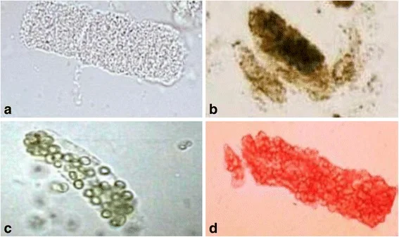
- Urine microscopy - muddy brown granular casts in ATN, red cell casts and dysmorphic red cells in glomerulonephritis, white cell casts in interstitial nephritis or pyelonephritis, crystals in tumour lysis or ethylene glycol
- ECG - to look for the changes of hyperkalaemia (tented T waves, small or absent P waves, broad QRS, and ultimately a sine wave pattern)
- Fluid balance chart, daily weights and observations
- Bladder scan - for residual volume, to identify retention
Laboratory
- U&Es - creatinine, urea and potassium; compare against every available previous result to establish a baseline and the rate of change
- Venous blood gas or bicarbonate - for metabolic acidosis, and a rapid potassium in the unstable patient
- Full blood count and CRP - anaemia may suggest CKD or myeloma; thrombocytopenia raises thrombotic microangiopathy; eosinophilia suggests interstitial nephritis
- Bone profile - hypercalcaemia suggests myeloma or malignancy; hyperphosphataemia and hyperkalaemia with a high urate suggest tumour lysis
- Creatine kinase - if there is any suggestion of a fall, immobility, seizure, compartment syndrome or statin use, to detect rhabdomyolysis
- Blood cultures and a septic screen where infection is possible
- Liver function and clotting
- Immunology and myeloma screen, where intrinsic renal disease is suspected - ANA, ANCA, anti-GBM antibodies, complement C3 and C4, immunoglobulins, serum free light chains and serum/urine electrophoresis, plus ASOT and hepatitis and HIV serology as indicated
| Index | Pre-renal | ATN |
|---|---|---|
| Urine sodium | Low (under 20 mmol/L) - avid tubular sodium reabsorption is intact | High (over 40 mmol/L) - damaged tubules cannot reabsorb sodium |
| Urine osmolality | High (over 500 mosm/kg) - concentrating ability preserved | Low (under 350 mosm/kg) - concentrating ability lost |
| Fractional excretion of sodium | Under 1% | Over 2% |
| Urine:plasma urea or creatinine ratio | High | Low |
| Response to fluid challenge | Creatinine and urine output improve | Little or no improvement |
Imaging
- Renal tract ultrasound within 24 hours is recommended if obstruction is suspected, if there is no identified cause, or if the patient is not improving as expected. It looks for hydronephrosis, kidney size and echogenicity (small echogenic kidneys indicate CKD), and stones or masses.2
- Ultrasound within 6 hours if pyonephrosis (an obstructed, infected kidney) is suspected - fever and loin pain with obstruction is a urological emergency needing immediate decompression
- Ultrasound is not always needed where the cause is clearly pre-renal and the patient responds promptly to treatment
- CT for suspected retroperitoneal pathology, malignancy or stone disease where ultrasound is inconclusive - avoid contrast where possible
- Renal biopsy - reserved for suspected intrinsic renal disease of uncertain cause, particularly rapidly progressive glomerulonephritis, where the result will change treatment
Management
There is no specific drug that reverses AKI. Management is supportive and directed at the cause: restore perfusion, remove the insult, relieve obstruction, and treat complications while the kidney recovers.
Treat the cause and restore perfusion
- Hypovolaemia - give intravenous crystalloid (a balanced solution such as Hartmann's, or 0.9% sodium chloride), titrated in boluses with reassessment of fluid status after each. Aim to restore euvolaemia, not to force a diuresis. Beware of over-filling the oliguric patient.
- Sepsis - treat per the sepsis pathway with prompt antibiotics and source control; renal recovery follows haemodynamic recovery
- Cardiogenic causes - fluids will worsen matters; the priority is improving cardiac output, and diuresis may be needed despite the AKI
- Obstruction - catheterise for bladder outlet obstruction (and flush or replace an existing catheter that may be blocked), and involve urology for nephrostomy or ureteric stenting in upper tract obstruction
- Intrinsic disease - discuss urgently with nephrology; immunosuppression (corticosteroids, cyclophosphamide, rituximab, plasma exchange) may be needed for vasculitis or anti-GBM disease, and delay costs nephrons
Treat the complications
| Complication | Management |
|---|---|
| Hyperkalaemia | See the dedicated callout below - this is the most immediately life-threatening complication |
| Fluid overload and pulmonary oedema | Sit up, give oxygen, restrict fluid and sodium, and give intravenous loop diuretic (e.g. furosemide) - noting that diuretics treat the overload but do not treat the AKI itself and do not improve renal outcomes. If refractory, this is an indication for renal replacement therapy. |
| Metabolic acidosis | Treat the underlying cause and restore perfusion. Sodium bicarbonate may be considered in severe acidosis under specialist advice; refractory severe acidosis is an indication for dialysis. |
| Uraemia | Nausea and anorexia are managed symptomatically; encephalopathy or pericarditis are indications for urgent dialysis |
| Hyperphosphataemia | Dietary restriction and phosphate binders if prolonged |
| Bleeding risk | Uraemia causes platelet dysfunction; consider this before procedures |
| Nutrition | Involve a dietitian - AKI is a catabolic state, and protein and calorie needs are often underestimated |
Renal replacement therapy
Escalation and follow-up
- Discuss with nephrology where there is stage 3 AKI, suspected intrinsic renal disease (especially a positive dipstick for blood and protein), an unclear cause, failure to improve, or a possible indication for RRT2
- Involve urology urgently for obstruction requiring decompression
- Monitor U&Es at least daily while AKI is evolving, along with fluid balance and weight
- On recovery, review and restart held medications deliberately - ACE inhibitors and other drugs are frequently omitted permanently by accident, or restarted too early; document a clear plan
- Arrange follow-up renal function after discharge, since AKI confers an increased long-term risk of CKD, and check for persistent proteinuria
- Document the episode clearly in the discharge summary, including the cause, the drugs stopped and whether they should be restarted
Complications
- Hyperkalaemia with arrhythmia and cardiac arrest
- Pulmonary oedema from fluid overload, often iatrogenic
- Metabolic acidosis
- Uraemic complications - encephalopathy, seizures, pericarditis and pericardial effusion, platelet dysfunction with bleeding
- Hyperphosphataemia and hypocalcaemia
- Increased susceptibility to infection, and to the toxicity of drugs that are renally cleared
- Progression to chronic kidney disease - a well-established association even after apparent recovery, and the reason follow-up matters
- End-stage renal disease requiring long-term dialysis in a minority
- Substantially increased short- and long-term mortality, and increased subsequent cardiovascular risk
Red flags
Prognosis
Outcome depends principally on the cause, the stage and the comorbidity of the patient rather than on the creatinine itself. Pre-renal AKI recognised and corrected early usually recovers completely within days. Established ATN typically recovers over 1 to 3 weeks, often passing through a polyuric recovery phase as tubular function returns before concentrating ability does - a phase during which fluid and electrolyte losses can be substantial and need replacing.
AKI nonetheless carries a substantial excess inpatient mortality, reaching well above 30 to 40% in patients with stage 3 AKI in critical care. Much of this reflects the severity of the underlying illness - sepsis, cardiac failure, major surgery - rather than the renal failure alone, but AKI is an independent contributor.
The most under-appreciated point is what happens afterwards. An episode of AKI, even one that appears to resolve fully, is associated with a significantly increased long-term risk of chronic kidney disease, end-stage renal disease, cardiovascular events and death.3 A proportion of patients never return to their true baseline. This is why every patient needs their renal function and proteinuria rechecked after discharge, their medication restarted deliberately rather than by default, and the episode documented so that future prescribers know they are dealing with a susceptible kidney.
References
- KDIGO Clinical Practice Guideline for Acute Kidney Injury. Kidney International Supplements. 2012. Available here
- NICE NG148. Acute kidney injury: prevention, detection and management. 2019. Available here
- Chawla LS, Eggers PW, Star RA, Kimmel PL. Acute kidney injury and chronic kidney disease as interconnected syndromes. New England Journal of Medicine. 2014. Available here
- NICE Clinical Knowledge Summaries. Acute kidney injury. Available here
- BNF. Prescribing in renal impairment. Available here
- UK Kidney Association. Clinical practice guidelines - treatment of acute hyperkalaemia in adults. Available here
- Mohsenin V., CC BY 4.0, via Wikimedia Commons. Available here
This article is written for revision and education. It is not clinical guidance and must not be used to make decisions about the care of a patient. Always check current NICE guidance and local protocols.