
Pericarditis: Diagnosis and Management
Key points
- Acute pericarditis: inflammation of the pericardium, most often viral or idiopathic in the UK.
- The pain: sharp and pleuritic, worse lying flat, relieved by sitting forward, and it may radiate to the trapezius ridge.
- The sign: a pericardial friction rub - scratchy, best heard at the left sternal edge with the patient leaning forward in expiration.
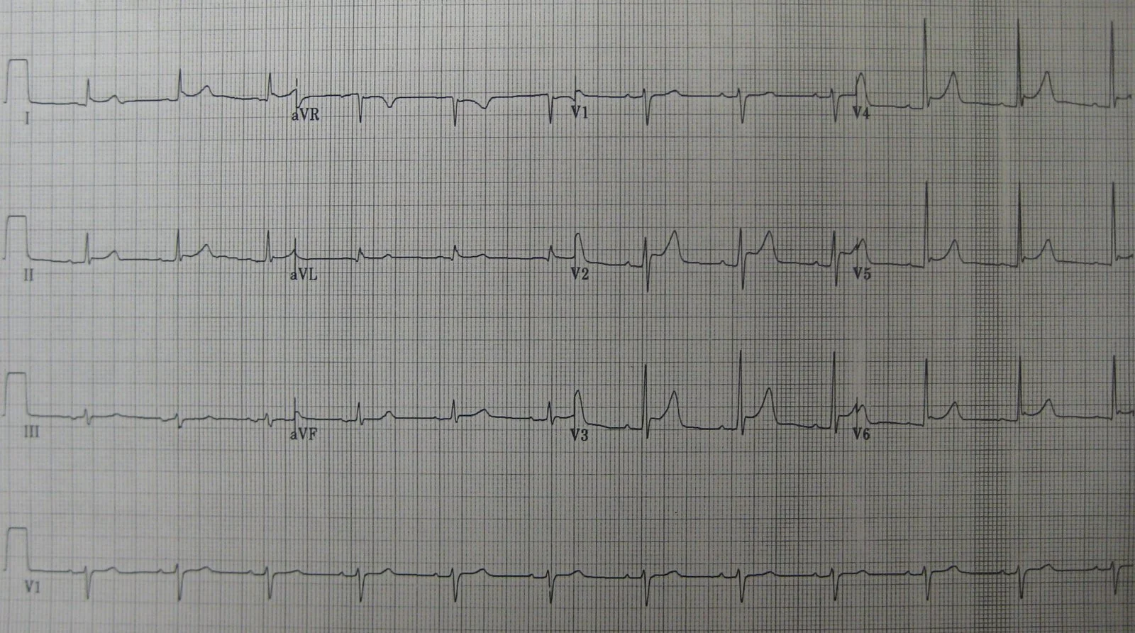
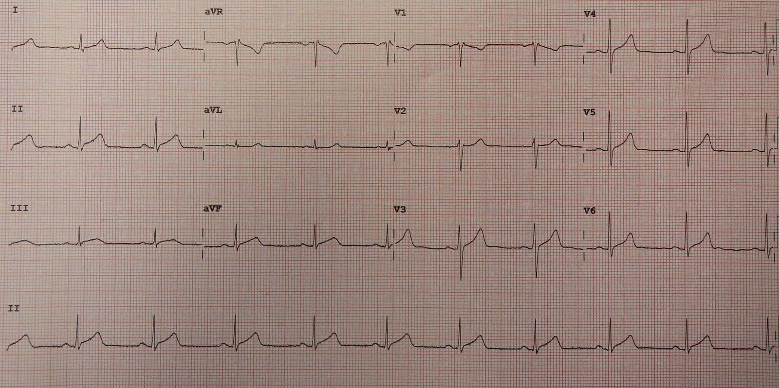
- The ECG: widespread concave saddle-shaped ST elevation with PR depression, and reciprocal ST depression with PR elevation in aVR.
- Diagnosis: requires two of four criteria: characteristic pain, a rub, characteristic ECG changes, or a new pericardial effusion.
- Management: high-dose NSAID or aspirin, plus colchicine for 3 months - colchicine roughly halves the recurrence rate.
- Avoid steroids: corticosteroids increase the risk of recurrence and are reserved for specific indications.
- Complications: pericardial effusion, cardiac tamponade, recurrence in up to 30%, and constrictive pericarditis.
Introduction
The pericardium is a two-layered sac around the heart: a visceral layer adherent to the epicardium and a fibrous parietal layer, separated by 15-50 mL of serous fluid. Acute pericarditis is inflammation of this sac, with or without an accompanying effusion.1
It accounts for around 5% of emergency department attendances with non-ischaemic chest pain, and its importance in exams is that it is the great mimic of myocardial infarction. Both present with chest pain and ST elevation, and both are common - but one goes to the catheter laboratory and one does not.
Where the inflammation extends into the underlying myocardium, the term myopericarditis is used. This is signalled by a raised troponin, and it changes the prognosis and the advice about exercise.
Aetiology
In high-income countries, the great majority of cases are viral or idiopathic - and in practice these are the same category, since a viral cause is rarely confirmed. Globally, tuberculosis is the commonest cause.1
| Category | Examples |
|---|---|
| Idiopathic | Around 80-90% of UK cases; presumed viral |
| Viral | Coxsackie B, echovirus, adenovirus, influenza, EBV, HIV, SARS-CoV-2 |
| Bacterial | Tuberculosis (the leading cause worldwide), Staphylococcus, Streptococcus, pneumococcus |
| Post-myocardial infarction | Early peri-infarction pericarditis (1-3 days), or Dressler's syndrome (2-6 weeks) |
| Metabolic | Uraemia in advanced renal failure; hypothyroidism |
| Autoimmune | SLE, rheumatoid arthritis, systemic sclerosis, sarcoidosis, familial Mediterranean fever |
| Neoplastic | Metastatic lung and breast cancer, lymphoma, mesothelioma |
| Iatrogenic and traumatic | Post-cardiac surgery, radiotherapy, catheter ablation, chest trauma |
| Drugs | Hydralazine, isoniazid, procainamide, phenytoin, some immune checkpoint inhibitors |
Clinical features
The history is the most useful part of the assessment, and the character of the pain is what separates pericarditis from ACS.
| Feature | Pericarditis | Acute coronary syndrome |
|---|---|---|
| Character | Sharp, stabbing, pleuritic | Heavy, crushing, constricting |
| Posture | Worse lying flat, relieved sitting forward | Not affected by position |
| Breathing | Worse on inspiration and coughing | Not usually affected |
| Onset | Often over hours, may follow a viral illness | Abrupt |
| Radiation | Trapezius ridge is near-specific | Jaw, neck, arms |
| Duration | Days, persistent | Usually continuous but hours |
Radiation to the trapezius ridge is worth remembering because it is close to specific for pericardial pain. It occurs because the phrenic nerve, which supplies the parietal pericardium, arises from C3-C5 - the same roots supplying the trapezius.
Associated features include a low-grade fever, and a preceding coryzal or gastrointestinal illness in the days to weeks before. Ask specifically about breathlessness, which may indicate a developing effusion, and about the causes in the table above: renal failure, malignancy, recent cardiac surgery, TB risk factors, and new medications.
Clinical examination
The classic sign is a pericardial friction rub: a superficial, scratchy or creaking sound, likened to walking on fresh snow. It is generated by the inflamed layers moving against each other.
The rest of the examination is directed at detecting an effusion and tamponade: heart rate and blood pressure, JVP, heart sounds, and signs of the underlying cause such as a rheumatological rash, lymphadenopathy or uraemic features. Muffled heart sounds and a raised JVP in a hypotensive patient are tamponade until proven otherwise.
Diagnosis
The European Society of Cardiology defines acute pericarditis by the presence of at least two of four criteria:1
- Pericarditic chest pain - sharp, pleuritic, worse lying flat
- Pericardial friction rub
- New widespread ST elevation or PR depression on the ECG
- Pericardial effusion, new or worsening
Supporting features are raised inflammatory markers and evidence of pericardial inflammation on CT or cardiac MRI.
Differential diagnosis
- Acute coronary syndrome - the critical distinction; see the ECG features below
- Pulmonary embolism - pleuritic pain and breathlessness, but with hypoxia and VTE risk factors
- Pneumonia with pleurisy - fever, focal signs, consolidation on chest X-ray
- Aortic dissection - tearing pain to the back with a pulse deficit
- Myocarditis - overlaps considerably; raised troponin with impaired ventricular function
- Musculoskeletal chest pain - reproducible on palpation
- Gastro-oesophageal reflux - burning, food and posture related
- Benign early repolarisation - a normal variant that mimics the ECG changes
Investigations
ECG
The ECG is abnormal in around 60% of cases and classically evolves through four stages, though patients rarely present neatly at one of them.2
| Stage | Timing | Findings |
|---|---|---|
| I | First hours to days | Widespread concave (saddle-shaped) ST elevation with PR depression; ST depression and PR elevation in aVR |
| II | First week | ST segments normalise; T waves flatten |
| III | Weeks | Widespread T wave inversion |
| IV | Weeks to months | Normalisation, though T inversion may persist |
Bloods
- CRP and ESR - raised in most cases; CRP is also used to guide the duration of treatment
- Troponin - a raised level indicates myopericarditis. This is important, since it changes the exercise advice and the follow-up.
- FBC - leucocytosis; lymphopenia may suggest a viral cause
- U&Es - to identify uraemic pericarditis
- TFTs - hypothyroidism is an uncommon but reversible cause
- Blood cultures if febrile, and autoimmune screen or TB testing where the history suggests it
Imaging
- Chest X-ray - usually normal. A globular, 'water bottle' cardiac silhouette appears only with a large effusion, generally over 300 mL.
- Transthoracic echocardiogram - recommended in all cases, to detect an effusion, assess for tamponade, and evaluate ventricular function
- Cardiac MRI - shows pericardial inflammation and late gadolinium enhancement; useful where the diagnosis is uncertain or myocarditis is suspected
- CT chest - for pericardial thickening, calcification, and to look for malignancy or TB
Management
Most patients with idiopathic or viral pericarditis are managed as outpatients and recover fully. Treatment aims to relieve pain, resolve inflammation, and - critically - reduce the risk of recurrence.
First-line therapy
The combination of an anti-inflammatory and colchicine is standard, and both components matter:1
- NSAID or aspirin at anti-inflammatory dose - for example ibuprofen 600 mg three times daily, or aspirin 750-1000 mg three times daily, continued for 1-2 weeks and then tapered. Aspirin is preferred after myocardial infarction.
- Colchicine 0.5 mg once or twice daily for 3 months - added to the NSAID, not instead of it. Colchicine approximately halves the rate of recurrence and shortens symptom duration.3
- Gastroprotection with a proton pump inhibitor alongside the NSAID
- Exercise restriction until symptoms have resolved and inflammatory markers have normalised - and for at least 3 months in athletes, or where there is myopericarditis
CRP is used to guide duration: continue the NSAID until symptoms resolve and CRP normalises, then taper rather than stopping abruptly.
Treating the cause
Purulent bacterial pericarditis requires urgent drainage and intravenous antibiotics. Tuberculous pericarditis needs standard antituberculous therapy, with adjunctive corticosteroids considered. Uraemic pericarditis is an indication for intensified dialysis. Malignant effusions may require drainage and a pericardial window.
Who needs admission
Most patients do not. Admit those with any poor prognostic feature, since these predict a specific underlying cause or a complicated course:
- Fever above 38°C
- Subacute onset over weeks rather than hours
- Large pericardial effusion (more than 20 mm echo-free space) or cardiac tamponade
- Failure to respond to NSAIDs and colchicine after 7 days
- Myopericarditis - raised troponin
- Immunosuppression
- Anticoagulant therapy - because of haemorrhagic effusion risk
- Recent trauma
Complications
Pericardial effusion and tamponade
Fluid accumulating in the pericardial space raises intrapericardial pressure. Because the fibrous pericardium is not acutely distensible, the rate of accumulation matters more than the volume: 150 mL appearing over minutes can cause tamponade, while 1000 mL accumulating over months may not.
Rising pressure impairs diastolic filling of the right heart, reducing preload, stroke volume and cardiac output. This is cardiac tamponade, and it is a medical emergency.
Recurrent pericarditis
Recurrence affects 15-30% of patients treated without colchicine, and roughly half that with it. Management is a repeat course of NSAID plus a longer course of colchicine, typically 6 months. Refractory cases may need azathioprine, intravenous immunoglobulin or anakinra.
Constrictive pericarditis
Chronic inflammation can leave the pericardium thickened, fibrosed and sometimes calcified, restricting diastolic filling. It is rare after idiopathic pericarditis (under 1%) but far more common after tuberculous, purulent or post-surgical disease. Features are those of right heart failure with a raised JVP, Kussmaul's sign, a pericardial knock, ascites and peripheral oedema. Definitive treatment is surgical pericardiectomy.
Red flags
Prognosis
The outlook for idiopathic and viral pericarditis is good. Most patients improve within days of starting treatment and recover completely, and serious complications are uncommon.1
Recurrence is the main problem, affecting up to 30% of those treated with an NSAID alone. This is the single best argument for adding colchicine, and the reason it should not be omitted just because the patient feels better - the ICAP and CORP trials both showed a roughly halved recurrence rate.3
Prognosis is determined by the underlying cause rather than the pericarditis itself. Tuberculous, purulent and malignant pericarditis all carry substantially worse outcomes and a much higher risk of progressing to constriction.
References
- Adler Y, Charron P, Imazio M et al. ESC Guidelines for the diagnosis and management of pericardial diseases. European Heart Journal. 2015. Available here
- Chiabrando JG, Bonaventura A, Vecchié A et al. Management of acute and recurrent pericarditis. Journal of the American College of Cardiology. 2020. Available here
- Imazio M, Brucato A, Cemin R et al. A randomized trial of colchicine for acute pericarditis (ICAP). New England Journal of Medicine. 2013. Available here
- NICE Clinical Knowledge Summaries. Pericarditis. Available here
- BNF. Colchicine - indications and dosing. Available here
- NICE NG185. Acute coronary syndromes. 2020, updated 2025. Available here
- Troughton RW, Asher CR, Klein AL. Pericarditis. The Lancet. 2004. Available here
This article is written for revision and education. It is not clinical guidance and must not be used to make decisions about the care of a patient. Always check current NICE guidance and local protocols.