
Hyperkalaemia
Key points
- Hyperkalaemia: serum potassium above 5.5 mmol/L. Graded mild 5.5-5.9, moderate 6.0-6.4, severe 6.5 or above - and severity is defined by the ECG, not the number alone.1
- Why it kills: a raised extracellular potassium reduces the resting membrane potential gradient, impairing cardiac depolarisation and conduction, and causing VF, asystole or PEA arrest.
- Commonest causes: acute kidney injury and CKD, drugs (ACE inhibitors, ARBs, spironolactone, NSAIDs, trimethoprim), acidosis, tissue breakdown (rhabdomyolysis, tumour lysis) and Addison's disease.
- Always exclude pseudohyperkalaemia: haemolysed sample, prolonged tourniquet, fist clenching, delayed processing, or a very high platelet or white cell count. Repeat the sample - but never delay treatment in an unwell patient.
- ECG progression: tall tented T waves → flattened/absent P waves and prolonged PR → widened QRS → sine wave → VF/asystole. Any ECG change means severe hyperkalaemia regardless of level.
- Step 1 - protect the heart: IV calcium gluconate 10% 30 mL (or calcium chloride) if there are ECG changes. It stabilises the myocardium within minutes but does NOT lower potassium, and may need repeating.
- Step 2 - shift potassium into cells: 10 units soluble insulin in 25 g glucose IV (the mainstay), plus nebulised salbutamol 10-20 mg. Monitor for rebound hypoglycaemia for at least 6 hours.
- Step 3 - remove potassium: haemodialysis is definitive. Potassium binders (patiromer, sodium zirconium cyclosilicate) are used for non-acute and chronic control.
Introduction
Hyperkalaemia is the most common life-threatening electrolyte abnormality encountered in hospital practice, and one of the few that can kill within minutes. It is defined as a serum potassium above 5.5 mmol/L, and it is a recurring cause of preventable cardiac arrest.1
Around 98% of total body potassium is intracellular, so the serum level represents a very small fraction of the total and is exquisitely sensitive to shifts across cell membranes. Two systems maintain it: rapid transcellular shift (driven by insulin, beta-2 adrenergic stimulation and pH) and slower renal excretion (driven principally by aldosterone acting on the distal nephron). Almost every cause of hyperkalaemia is a failure of one of these two mechanisms - or simply too much potassium entering the circulation at once.
Causes
Reduced renal excretion
- Acute kidney injury - the commonest single cause in acute practice
- Chronic kidney disease, particularly stages 4-5
- Drugs affecting the renin-angiotensin-aldosterone axis - ACE inhibitors, ARBs, aldosterone antagonists (spironolactone, eplerenone), direct renin inhibitors
- Potassium-sparing diuretics - amiloride
- NSAIDs - reduce renal perfusion and renin release
- Trimethoprim (and high-dose co-trimoxazole) - blocks the epithelial sodium channel, acting rather like amiloride; a very common and easily forgotten cause
- Calcineurin inhibitors - ciclosporin, tacrolimus
- Heparin - suppresses aldosterone synthesis
- Adrenal insufficiency (Addison's disease) - aldosterone deficiency; look for the accompanying hyponatraemia
- Type 4 renal tubular acidosis - hyporeninaemic hypoaldosteronism, classically in diabetic nephropathy
Shift out of cells
- Metabolic acidosis - particularly diabetic ketoacidosis, where hydrogen ions move into cells in exchange for potassium
- Insulin deficiency - DKA patients have a high serum potassium but a depleted total body potassium, which is why potassium falls dramatically once insulin is started
- Beta-blockers - impair beta-2-mediated cellular uptake
- Digoxin toxicity - inhibits the Na⁺/K⁺-ATPase pump
- Suxamethonium - depolarising muscle relaxant; dangerous in burns, crush injury and neuromuscular disease
- Hyperosmolality and severe hyperglycaemia
- Rare: hyperkalaemic periodic paralysis
Increased potassium load or release
- Rhabdomyolysis - massive release from damaged muscle; potassium may rise before renal function deteriorates
- Tumour lysis syndrome - after chemotherapy for bulky, chemosensitive tumours
- Massive haemolysis and large-volume blood transfusion (stored red cells leak potassium)
- Extensive burns, crush injury and tissue necrosis
- Excessive potassium supplementation - oral or, more dangerously, intravenous
- Dietary excess - only significant in the presence of impaired excretion; note 'low-sodium' salt substitutes are potassium chloride and are a genuine trap in CKD patients
Clinical features
Hyperkalaemia is frequently asymptomatic until the point of cardiac arrest, which is precisely what makes it dangerous. It is usually detected on blood tests rather than suspected clinically, so a low threshold for checking potassium in at-risk patients is essential.
- Often none - and the absence of symptoms is no reassurance
- Generalised muscle weakness and fatigue, which may progress to flaccid paralysis
- Paraesthesiae and perioral numbness
- Palpitations, or awareness of an irregular pulse
- Nausea and vomiting
- Reduced deep tendon reflexes
- Bradycardia or arrhythmia
- Cardiac arrest - VF, pulseless electrical activity or asystole; hyperkalaemia is one of the reversible 'four Hs' of cardiac arrest
- Features of the underlying cause - oliguria in AKI, pigmenturia in rhabdomyolysis, pigmentation and hypotension in Addison's disease
Investigations
- 12-lead ECG - immediately. This is the single most important investigation and determines urgency of treatment. It should be done while awaiting laboratory confirmation, not after it
- Venous or arterial blood gas - gives a potassium result within a minute, invaluable when the laboratory result is awaited
- Repeat laboratory U&Es - to confirm and to exclude pseudohyperkalaemia
- Creatinine and eGFR - to identify AKI or CKD as the cause
- Bicarbonate and blood gas - for metabolic acidosis
- Glucose - hyperglycaemia and DKA as a cause, and a baseline before giving insulin
- Calcium, phosphate and magnesium - hypocalcaemia worsens the cardiac effects of hyperkalaemia
- Creatine kinase - if rhabdomyolysis is suspected
- FBC, LDH, urate and phosphate - for tumour lysis syndrome and haemolysis
- Cortisol and short Synacthen test - if Addison's disease is suspected, particularly with coexisting hyponatraemia
- Digoxin level - if the patient takes digoxin
- A careful drug review - this is an investigation in its own right, and frequently identifies the cause
ECG changes
The changes are progressive, and reflect the effect of potassium on myocardial repolarisation and then conduction:
- Tall, peaked ('tented') T waves - narrow-based and symmetrical; the earliest change
- Flattening then loss of P waves, with PR interval prolongation
- Widening of the QRS complex - a genuinely ominous sign indicating impaired conduction
- Merging of the widened QRS with the T wave to form a sine wave - pre-terminal
- Ventricular fibrillation, pulseless electrical activity or asystole
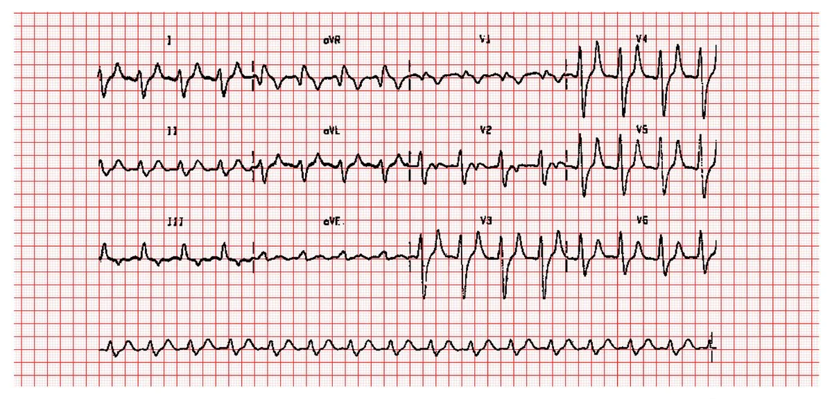
Two caveats are worth holding onto. A normal ECG does not exclude significant hyperkalaemia - some patients arrest with few preceding changes. And ECG changes correlate imperfectly with the serum level, particularly in chronic kidney disease where patients tolerate higher levels.
Management
Treatment follows a logical three-step sequence: protect the heart, shift potassium into cells, then remove it from the body. Each step does something the others do not, and the order matters.2
| Step | Treatment | Onset / duration | Notes |
|---|---|---|---|
| 1. Protect the myocardium | Calcium gluconate 10%, 30 mL IV over 5-10 min (or calcium chloride 10%, 10 mL via central access) | Onset 1-3 min, lasts 30-60 min | Given if there are ECG changes. Stabilises the cardiac membrane but does NOT lower potassium. Repeat if changes persist. Use with great caution in digoxin toxicity - give slowly, diluted |
| 2. Shift into cells | 10 units soluble insulin (Actrapid) in 25 g glucose (e.g. 50 mL of 50% or 125 mL of 20%) IV over 15-30 min | Onset 15-30 min, lasts 4-6 h | The mainstay. Lowers K⁺ by ~0.6-1.0 mmol/L. Monitor blood glucose hourly for at least 6 hours - delayed hypoglycaemia is common and is the main hazard |
| 2. Shift into cells | Salbutamol 10-20 mg nebulised | Onset 15-30 min | Adjunct, not a substitute for insulin-glucose. Causes tachycardia and tremor; less effective in patients on beta-blockers |
| 2. (Selected) | Sodium bicarbonate | Variable | Only if there is significant metabolic acidosis; not routine, and evidence of benefit is limited |
| 3. Remove from the body | Haemodialysis | Immediate and definitive | The definitive treatment, especially in AKI/CKD, refractory or severe hyperkalaemia. Involve renal team early |
| 3. Remove from the body | Potassium binders - sodium zirconium cyclosilicate or patiromer | Hours | For non-life-threatening and chronic hyperkalaemia; NICE-approved. Allow continuation of prognostically important RAAS inhibitors in heart failure and CKD |
| 3. Remove from the body | Loop diuretic (furosemide) | Hours | Only useful if the patient is volume-replete with preserved urine output |
Treating the underlying cause
- Stop the offending drugs - ACE inhibitors, ARBs, spironolactone, amiloride, NSAIDs, trimethoprim, potassium supplements and salt substitutes. Review this explicitly; it is the commonest reversible factor
- Treat acute kidney injury - restore volume, relieve obstruction, stop nephrotoxins
- Treat DKA with fluid and insulin, anticipating that potassium will fall rapidly and will need replacing
- Aggressive fluid resuscitation in rhabdomyolysis and tumour lysis
- Hydrocortisone and fludrocortisone in adrenal insufficiency
- Dietary potassium restriction and dietitian input in CKD
- Cardiac monitoring throughout for any patient with severe hyperkalaemia or ECG changes
- In cardiac arrest, hyperkalaemia is one of the reversible 'four Hs' - give IV calcium and insulin-glucose during resuscitation and consider urgent dialysis
Complications
- Fatal arrhythmia - ventricular fibrillation, pulseless electrical activity and asystole; the reason hyperkalaemia is treated as an emergency
- Bradyarrhythmia and complete heart block
- Cardiac arrest - hyperkalaemia is a reversible cause and should be actively considered in any arrest in a renal or dialysis patient
- Flaccid paralysis and respiratory muscle weakness in severe cases
- Rebound hypoglycaemia after insulin-glucose treatment - common, sometimes delayed by several hours, and a genuine cause of harm if glucose is not monitored
- Rebound hyperkalaemia as the effect of shifting agents wears off, if the underlying cause has not been addressed
- Complications of treatment - tissue necrosis from extravasated calcium chloride, tachyarrhythmia from salbutamol, fluid overload from bicarbonate
- Undertreatment of prognostically important drugs - patients with heart failure or CKD may have ACE inhibitors or spironolactone withheld unnecessarily, losing survival benefit; potassium binders exist precisely to avoid this
Red flags
Prognosis
Acute hyperkalaemia treated promptly has an excellent outcome - the electrolyte abnormality itself is entirely reversible, and appropriate treatment normalises cardiac conduction within minutes to hours. The danger lies almost entirely in delayed recognition, which is why an ECG and a blood gas should be obtained the moment a high potassium is reported rather than waiting for a confirmatory laboratory sample.
Prognosis is largely determined by the underlying cause rather than the potassium level. Hyperkalaemia from a reversible drug effect in a patient with normal kidneys carries a very different outlook from hyperkalaemia complicating established AKI, sepsis or tumour lysis syndrome. In hospitalised patients, hyperkalaemia is a marker of illness severity and is independently associated with increased mortality - not because the potassium itself is usually fatal once treated, but because it signals significant renal, adrenal or metabolic derangement.
Chronic hyperkalaemia presents a different problem. In patients with CKD and heart failure, the drugs that cause it - ACE inhibitors, ARBs and mineralocorticoid receptor antagonists - are precisely the drugs that improve survival. Historically these were reduced or stopped, at real prognostic cost. The availability of modern potassium binders (patiromer and sodium zirconium cyclosilicate) has changed this, allowing many patients to remain on optimal RAAS blockade with controlled potassium. Recurrent hyperkalaemia should therefore prompt a considered strategy - dietary review, binder therapy, correction of acidosis and diuretic adjustment - rather than reflexive withdrawal of prognostically valuable treatment.
References
- UK Kidney Association. Clinical Practice Guideline: Treatment of Acute Hyperkalaemia in Adults. 2023. Available here
- Resuscitation Council UK. Advanced Life Support - special circumstances: electrolyte abnormalities. Available here
- NICE Clinical Knowledge Summaries. Hyperkalaemia. Available here
- NICE TA599 / TA623. Sodium zirconium cyclosilicate and patiromer for treating hyperkalaemia. Available here
- NICE NG148. Acute kidney injury: prevention, detection and management. 2019. Available here
- BNF. Calcium gluconate, insulin and salbutamol - use in hyperkalaemia. Available here
- Michael Rosengarten BEng MD, McGill (CardioNetworks ECGpedia), CC BY-SA 3.0, via Wikimedia Commons. Available here
This article is written for revision and education. It is not clinical guidance and must not be used to make decisions about the care of a patient. Always check current NICE guidance and local protocols.