
Intussusception
Key points
- Definition: telescoping of one segment of bowel into the lumen of the adjacent distal segment, causing obstruction and progressive ischaemia of the invaginated bowel.
- Who gets it: the commonest cause of intestinal obstruction between 6 months and 2 years, peaking around 5-10 months, with a male to female ratio of about 2:1.
- Site: ileocolic in around 90% - the terminal ileum telescoping through the ileocaecal valve into the colon.
- Cause: idiopathic in most infants, following hypertrophy of Peyer's patches after a viral illness. A pathological lead point is more likely under 3 months or over 2 years.
- Classic triad: colicky abdominal pain, a sausage-shaped mass and redcurrant jelly stool - but all three are present in fewer than a quarter of cases, and the stool is a late sign.
- The presentation students miss: pallor and profound lethargy between episodes, which can dominate the picture and be mistaken for sepsis or encephalopathy.
- Diagnosis: abdominal ultrasound - the target sign in transverse section and the pseudokidney sign in longitudinal section.
- Treatment: resuscitate, then air enema reduction under fluoroscopy, successful in 75-90%. Surgery for failed reduction, perforation or peritonitis.
Introduction
Intussusception occurs when a proximal segment of bowel - the intussusceptum - invaginates into the lumen of the segment immediately distal to it, the intussuscipiens. Peristalsis then drags it further in, taking the mesentery with it.
It is the commonest cause of intestinal obstruction in infancy, and it is entirely curable if treated in time. Untreated it progresses through venous congestion to arterial compromise, infarction, perforation and death within a few days.
The clinical difficulty is that the textbook picture - the classic triad of colic, a sausage-shaped mass and redcurrant jelly stool - occurs in fewer than a quarter of children, and the redcurrant jelly is a late feature signifying mucosal necrosis. Waiting for the triad is waiting for ischaemia. What should prompt the diagnosis instead is a young child with intermittent inconsolable screaming, pallor, and unexplained lethargy in between.
Aetiology
Idiopathic
In 75-90% of children under 2 no anatomical lead point is found. The accepted explanation is hypertrophy of Peyer's patches in the terminal ileum following a viral infection - adenovirus and rotavirus being the usual culprits - creating a bulge that peristalsis then propels forwards. This fits the epidemiology neatly: the peak age matches the peak of lymphoid tissue prominence, and cases cluster seasonally with viral illness.
Pathological lead points
A structural lead point is found in perhaps 5-10% overall, and should be actively suspected at the extremes of age - under 3 months or over 2 years - and in recurrent cases.
- Meckel's diverticulum - the commonest pathological lead point
- Intestinal polyp, including in Peutz-Jeghers syndrome
- Henoch-Schönlein purpura - a submucosal haematoma acting as the lead point, characteristically causing an ileoileal rather than ileocolic intussusception, which is harder to see and cannot be reduced by enema
- Lymphoma - particularly in an older child, where intussusception may be the presenting feature of a Burkitt lymphoma
- Cystic fibrosis - inspissated intestinal contents
- Intestinal duplication cyst or haemangioma
- Postoperative, after abdominal surgery, usually small-bowel and often presenting as prolonged ileus
Pathophysiology
Understanding the sequence explains both the symptoms and their timing.
- The intussusceptum is drawn into the distal segment, dragging its mesentery with it. Peristaltic waves advance it further, and each wave produces a paroxysm of severe colic - which is why the pain is intermittent and periodic rather than constant.
- The compressed mesenteric veins obstruct first, since they are thin-walled. The invaginated bowel becomes congested and oedematous.
- Oedema increases the bulk of the intussusceptum, worsening the compression - a self-perpetuating cycle, and the reason the condition never resolves once established.
- Arterial inflow is eventually compromised, producing mucosal ischaemia. Sloughed mucosa mixed with blood and mucus is passed as the classic redcurrant jelly stool - a sign that ischaemia has already occurred.
- Transmural necrosis follows, with perforation, peritonitis and septic shock.
- Meanwhile mechanical obstruction causes vomiting, third-space fluid loss and dehydration, which is often severe by the time of presentation.
Clinical features
- Paroxysmal severe colic - episodes every 15-20 minutes in which the child screams, draws the knees up to the chest, and goes pale and sweaty, each lasting a few minutes
- Pallor and lethargy between episodes - initially the child seems normal between paroxysms, but as the illness progresses they become increasingly quiet, floppy and withdrawn
- Vomiting - non-bilious at first, becoming bilious as the obstruction progresses
- Redcurrant jelly stool - blood and mucus, present in around half of cases and characteristically late. Perform a rectal examination: blood on the glove may be the only evidence.
- A sausage-shaped mass, typically palpable in the right upper quadrant or along the line of the transverse colon, and best felt between paroxysms
- Dance's sign - an unusually empty right iliac fossa, since the caecum has been drawn out of it
- Later features: abdominal distension, dehydration, shock, peritonism and fever
Examination
Examine the child between paroxysms, when the abdomen is soft enough to palpate properly, and repeat the examination if the first is unhelpful - the findings change substantially over a few hours.
- Observations and hydration: heart rate, capillary refill, mucous membranes, and conscious level. Tachycardia with a soft abdomen is a warning that third-space losses are already significant.
- Inspection: distension, and visible peristalsis in a thin infant
- Palpation: feel systematically for the sausage-shaped mass, which lies transversely in the right upper quadrant or across the epigastrium and is often described as having a concave upper border
- The right iliac fossa, which may feel abnormally empty as the caecum is drawn out of it
- Signs of peritonism - guarding, rigidity, rebound - which change the management from enema to theatre
- Rectal examination, which may reveal blood or mucus on the glove long before any is passed spontaneously, and very occasionally a palpable intussusceptum
- The groin and scrotum in every case, since an incarcerated inguinal hernia produces a near-identical picture and is found only by looking
- The whole skin, for the purpuric rash of Henoch-Schönlein purpura on the buttocks and extensor surfaces
Differential diagnosis
| Diagnosis | Distinguishing features |
|---|---|
| Gastroenteritis | Diarrhoea predominates, pain is less severe and non-paroxysmal, contact history. Caution: early intussusception is frequently mislabelled as gastroenteritis.3 |
| Malrotation with volvulus | Younger, bilious vomiting from the outset, rapid deterioration, upper gastrointestinal contrast study diagnostic |
| Incarcerated inguinal hernia | Tender irreducible groin swelling - always examine the groin and scrotum |
| Appendicitis | Older child, constant right iliac fossa pain with localised tenderness and guarding, low-grade fever7 |
| Constipation | Chronic history, palpable faecal loading, no pallor or lethargy |
| Meckel's diverticulitis or bleed | Painless rectal bleeding, or right-sided pain; may itself be the lead point |
| Henoch-Schönlein purpura | Purpuric rash on the buttocks and extensor surfaces, arthralgia, haematuria - and may coexist with intussusception |
| Sepsis or meningitis | Fever, non-blanching rash, meningism; the lethargic presentation overlaps |
| Testicular or ovarian torsion | Localising signs on examining the genitalia or pelvis |
Investigations
Ultrasound
Abdominal ultrasound is the first-line investigation and is highly sensitive and specific in experienced hands. It should be arranged urgently rather than being deferred to a daytime list.
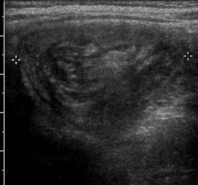
- Target or doughnut sign in transverse section - concentric rings of bowel wall
- Pseudokidney sign in longitudinal section
- Free intraperitoneal fluid, trapped fluid within the intussusceptum, and absent Doppler flow in the invaginated wall all suggest ischaemia and predict failed enema reduction
- Identification of a pathological lead point, which changes management towards surgery
- Ultrasound also excludes several of the differentials in the same examination
Other investigations
- Abdominal radiograph - not needed for diagnosis, but useful where perforation is suspected. It may show small bowel obstruction, a soft tissue mass, paucity of gas in the right lower quadrant, or free air.
- FBC, U&Es, CRP, blood gas with lactate, glucose and group and save - to assess dehydration, electrolyte disturbance and evidence of ischaemia
- Blood cultures if febrile or septic
- Contrast or air enema, which is diagnostic and, in most cases, also therapeutic
Management
Resuscitation first
- Nil by mouth with a nasogastric tube on free drainage
- Intravenous access and a 20 mL/kg bolus of 0.9% sodium chloride if shocked, repeated as needed, followed by maintenance and deficit replacement5
- Analgesia - these children are in severe pain and are often undertreated. Use a weight-based opioid where colic is severe.1
- Broad-spectrum intravenous antibiotics before any attempt at reduction, since reduction can translocate bacteria
- Involve the paediatric surgical team from the outset, and transfer to a centre with paediatric surgery and interventional radiology if not already there
Non-operative reduction
Air (pneumatic) enema reduction under fluoroscopic guidance is the first-line treatment, with success rates of 75-90%. Air is insufflated into the rectum under controlled pressure, pushing the intussusceptum back to its normal position; successful reduction is confirmed by free flow of air into the terminal ileum. Hydrostatic reduction with contrast or with ultrasound-guided saline is used in some centres with comparable results.
Surgery
- Indicated for failed enema reduction, perforation, peritonitis, shock, or a pathological lead point
- Manual reduction at laparotomy or laparoscopy, by squeezing the intussusceptum backwards rather than pulling it out, which risks tearing ischaemic bowel
- Resection with primary anastomosis if the bowel is non-viable, cannot be reduced, or contains a lead point that must be removed
- Appendicectomy is often performed at the same time if the caecum is mobilised
Afterwards
- Observe in hospital for 24 hours after successful enema reduction, since most recurrences occur early
- Feeding is reintroduced once the child is comfortable and the abdomen is soft
- Recurrence occurs in around 10% after non-operative reduction, usually within the first 72 hours, and can be treated with a repeat enema. Recurrence is less common after surgical reduction.
- Repeated recurrence should prompt investigation for a pathological lead point
- Safety net the family: return immediately if the colic, pallor, vomiting or lethargy recur
Complications
- Bowel ischaemia and infarction, requiring resection
- Perforation and peritonitis, either from the disease or during attempted enema reduction
- Septic shock and multi-organ failure
- Dehydration and electrolyte disturbance from vomiting and third-space loss
- Recurrence - around 10% after enema reduction
- Short bowel syndrome after extensive resection, though this is uncommon
- Adhesive small bowel obstruction as a late consequence of laparotomy
Prognosis
With prompt diagnosis and treatment the outlook is excellent, and mortality in the UK is well under 1%. Children reduced by enema typically go home the following day and have no long-term consequences at all.4
Outcome is essentially a function of time to diagnosis. Delays of more than 24 hours substantially increase the rate of failed enema reduction, the need for bowel resection, and the risk of perforation - and delay usually happens because early intussusception was labelled as colic, gastroenteritis or a viral illness.
A small number of children present atypically and account for most of the diagnostic delays: infants under 3 months, in whom a pathological lead point is likely and the classical colic may be absent; children over 2 years, in whom lymphoma must be considered; postoperative small-bowel intussusception, which masquerades as prolonged ileus; and the child with Henoch-Schonlein purpura, whose abdominal pain is easily attributed to the vasculitis itself. In each of these, ultrasound is the way out of the uncertainty.
The practical lesson is to lower the threshold rather than wait for confirmation. In an infant with paroxysmal screaming, pallor and vomiting, an urgent ultrasound costs very little, and the alternative - waiting for redcurrant jelly stool to declare the diagnosis - means waiting for the bowel to become ischaemic.
References
- Advanced Life Support Group. Advanced Paediatric Life Support: A Practical Approach to Emergencies. Available here
- UKHSA. Rotavirus: the Green Book, chapter 27b. Available here
- NICE CG84. Diarrhoea and vomiting caused by gastroenteritis in under 5s: diagnosis and management. 2009. Available here
- British Association of Paediatric Surgeons. Congenital Anomalies Surveillance System (BAPS-CASS). Available here
- NICE NG29. Intravenous fluid therapy in children and young people in hospital. 2015, updated 2020. Available here
- NICE NG143. Fever in under 5s: assessment and initial management. 2019, updated 2021. Available here
- NICE Clinical Knowledge Summaries. Appendicitis. Available here
This article is written for revision and education. It is not clinical guidance and must not be used to make decisions about the care of a patient. Always check current NICE guidance and local protocols.