
Rhabdomyolysis
Key points
- Rhabdomyolysis: breakdown of skeletal muscle with release of intracellular contents - myoglobin, creatine kinase (CK), potassium, phosphate and urate - into the circulation.
- Causes: trauma and crush injury, prolonged immobilisation ('found down'), extreme exertion, seizures, drugs and toxins (notably statins and illicit stimulants), and extreme temperature. Often multifactorial.
- The classic triad: myalgia, weakness and dark ('tea-coloured') urine - but present together in a minority of cases; many patients are asymptomatic and picked up only on blood tests.
- Diagnosis: a CK more than 5 times the upper limit of normal (often quoted as CK > 1000 U/L, against a normal of roughly 150-200 U/L) in the appropriate clinical context.
- The dipstick trick: urine dipstick is positive for blood, because it cannot distinguish haemoglobin from myoglobin, but microscopy shows no red blood cells - this mismatch is the exam giveaway for myoglobinuria.
- Key danger: AKI (myoglobin is directly nephrotoxic to renal tubules) and life-threatening hyperkalaemia, which can occur before renal function has even started to fall.
- Management: aggressive intravenous crystalloid fluid resuscitation is the cornerstone, titrated to urine output, alongside treatment of hyperkalaemia and identification of the underlying cause.
- Compartment syndrome: a cause and a consequence of rhabdomyolysis - measure compartment pressures and involve orthopaedics early if suspected.
Introduction
Rhabdomyolysis is the breakdown of striated skeletal muscle with leakage of intracellular contents into the systemic circulation. The damaged sarcolemma releases creatine kinase, myoglobin, potassium, phosphate and urate, producing a characteristic biochemical picture and, in a substantial minority, acute kidney injury.1
It is common in acute medicine and surgery, ranging from an asymptomatic incidental finding on blood tests (for example after a fall with a long lie, or in a patient started on a statin) to a life-threatening emergency with severe hyperkalaemia and anuric AKI after a crush injury. The severity spans this entire spectrum, and the priority is always the same: identify it early and start fluids before the creatinine has even had time to rise.
Aetiology
Muscle injury occurs through three overlapping mechanisms: direct trauma, excessive energy demand or ischaemia, and direct toxic or metabolic injury to the myocyte. Many patients have more than one contributing cause - for example an elderly patient found down on the floor after a fall, who is also on a statin and has been unable to reach fluids for hours.2
Traumatic and compressive
- Crush injury - road traffic collisions, building collapse, industrial accidents
- Prolonged immobilisation - a fall with a 'long lie' (especially in the elderly), unconsciousness from overdose or intoxication, surgery in a fixed position
- Compartment syndrome - both a cause (compression) and a consequence (swelling within the compartment) of rhabdomyolysis
- Extensive burns and electrical injury, including lightning strikes
Exertional
- Strenuous exercise, particularly in the unconditioned, in hot conditions, or with inadequate hydration - marathon runners, military recruits, new gym attendees doing unaccustomed eccentric exercise
- Status epilepticus and prolonged seizure activity
- Severe agitation or dystonia - e.g. in delirium tremens or psychiatric illness
Drugs and toxins
- Statins, especially at high dose, in renal impairment, in hypothyroidism, or combined with a fibrate or macrolide (interactions that raise statin levels)
- Illicit drugs - cocaine, amphetamines and MDMA (through a combination of vasoconstriction, hyperthermia and seizures), and heroin
- Alcohol, particularly binge intoxication with a long lie
- Malignant hyperthermia - triggered by volatile anaesthetics and suxamethonium in genetically susceptible patients
- Neuroleptic malignant syndrome and serotonin syndrome - antipsychotics and serotonergic drugs respectively
- Carbon monoxide poisoning and snake envenomation
Temperature-related
- Heat stroke and severe hyperthermia
- Severe hypothermia
Infective, inflammatory and metabolic
- Viral myositis - influenza and other common viruses, particularly in children
- Severe electrolyte disturbance - hypokalaemia, hypophosphataemia and hyponatraemia impair myocyte energy metabolism
- Inherited metabolic myopathies - McArdle disease (a glycogen storage disorder) and disorders of fatty acid oxidation, which should be suspected in recurrent rhabdomyolysis with minimal exertion, especially in younger patients
- Autoimmune myositis - polymyositis and dermatomyositis
Clinical features
The classic triad of myalgia, muscle weakness and dark urine is well known but is present together in only around 10% of patients. Presentation is often non-specific, and a high index of suspicion is needed in at-risk patients.
- Muscle pain, tenderness and swelling - typically affecting the proximal muscle groups (thighs, calves, shoulders, lower back), though it can be localised to the site of injury or absent altogether
- Muscle weakness
- Dark, tea- or cola-coloured urine - from myoglobinuria; often the presenting complaint and always worth asking about directly
- Malaise, nausea, vomiting and low-grade fever - non-specific systemic symptoms
- Reduced urine output if AKI has developed
- Symptoms of the underlying cause - confusion or agitation (drugs, delirium tremens), a history of a fall or long lie, a seizure witnessed by others, exertion in the heat
- Many patients, particularly those with drug-induced or mild exertional rhabdomyolysis, are entirely asymptomatic and are identified only because a CK was checked
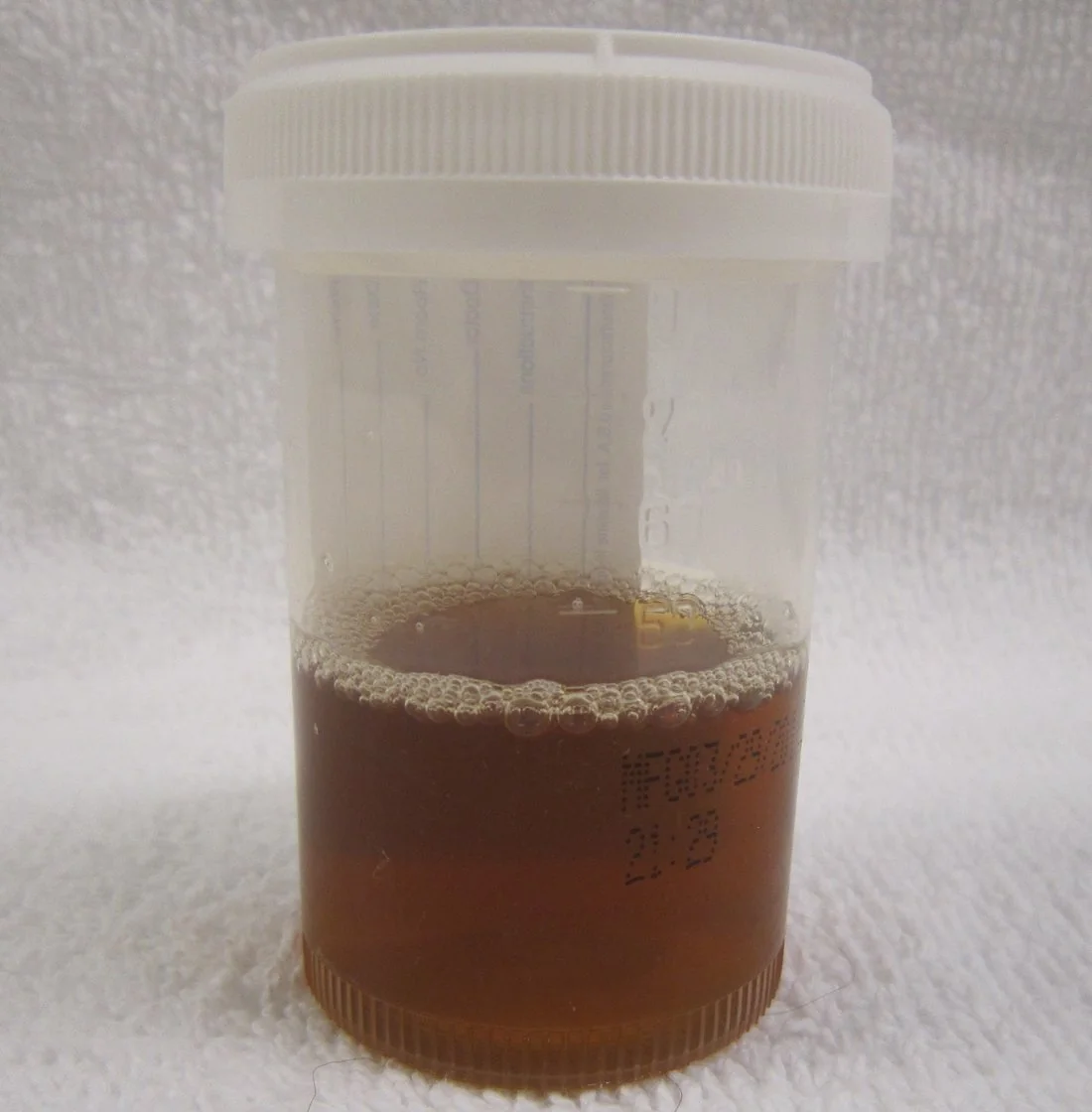
Examination should assess the affected muscle groups for swelling, tenderness and reduced power, and specifically look for signs of compartment syndrome - a tense, swollen compartment with pain disproportionate to examination and pain on passive stretch is an emergency (see Complications).
Differential diagnosis
Rhabdomyolysis itself is usually straightforward to confirm biochemically once suspected. The differential lies more in identifying the cause, and in distinguishing myoglobinuria from other causes of discoloured urine or a positive dipstick for blood.
| Cause | Distinguishing features |
|---|---|
| Myoglobinuria (rhabdomyolysis) | Markedly raised CK, history of muscle injury/exertion/drugs, myalgia |
| Haemoglobinuria (intravascular haemolysis) | Anaemia, raised bilirubin and LDH, low haptoglobin, schistocytes on film; CK normal |
| Porphyria | Abdominal pain, neuropsychiatric features, urine darkens on standing; raised urinary porphobilinogen |
True glomerular haematuria (e.g. from glomerulonephritis) is distinguished by red cells and casts on urine microscopy, alongside a normal CK.
Investigations
Essential investigations
- Creatine kinase (CK) - the key diagnostic test. Diagnostic threshold is generally taken as more than 5 times the upper limit of normal, often quoted as CK > 1000 U/L (normal roughly 150-200 U/L, higher in athletic or heavily muscled individuals). CK rises within 2-12 hours of injury, peaks at 24-72 hours, and falls by roughly 40-50% per day once the insult has stopped - a failure to fall as expected suggests ongoing muscle injury (e.g. undiagnosed compartment syndrome)
- U&Es and creatinine - to detect AKI and, critically, hyperkalaemia, which can develop rapidly and disproportionately to the degree of renal impairment because potassium is released directly from damaged muscle
- Urine dipstick and microscopy - dipstick positive for blood with no red cells seen on microscopy confirms myoglobinuria rather than haematuria
- Venous or arterial blood gas - for a rapid potassium result and to assess for metabolic acidosis
- ECG - to look for changes of hyperkalaemia (tented T waves, widened QRS, loss of P waves) - this should not be delayed while awaiting laboratory potassium
- Calcium, phosphate and urate - hypocalcaemia occurs early (calcium is deposited in damaged muscle and complexed by released phosphate), hyperphosphataemia and hyperuricaemia follow from cell lysis
- Clotting screen and FBC - to screen for disseminated intravascular coagulation, a recognised complication of severe rhabdomyolysis
Further and targeted investigations
- Creatine kinase-MB or troponin - CK-MB is unhelpful (it rises alongside total CK from skeletal muscle) but a troponin can help if cardiac injury is genuinely suspected
- Urinary myoglobin - rarely measured in practice; it is unstable and often undetectable despite ongoing myoglobinuria, so a negative result does not exclude the diagnosis
- Imaging or compartment pressure measurement - if compartment syndrome is suspected clinically
- Toxicology screen - if a drug cause is suspected
- CT or further imaging of the injured area after major trauma
- Creatine kinase and further metabolic/genetic work-up - if there is recurrent rhabdomyolysis with modest exertion, raising suspicion of an underlying metabolic myopathy
Management
Management rests on three pillars: aggressive fluid resuscitation, correction of life-threatening electrolyte disturbance, and treatment of the underlying cause. The single most important early intervention is starting intravenous fluids as soon as rhabdomyolysis is suspected, without waiting for laboratory confirmation.
Fluid resuscitation
- Intravenous 0.9% saline is the mainstay, started early and at high volume (typically several litres over the first 24 hours in significant cases) to restore intravascular volume, maintain renal perfusion, and flush myoglobin through the tubules before it precipitates
- Titrate to a target urine output - commonly around 200-300 mL/hour, adjusted for the clinical context and reassessed regularly to avoid fluid overload
- Fluids should be started before hospital arrival where possible in major crush injury (this is a standard part of pre-hospital and disaster medicine teaching), and continued through any surgery or extrication
- Reassess frequently - elderly patients, those with cardiac or renal comorbidity, and those with major crush injury need careful monitoring of fluid balance to avoid pulmonary oedema
Managing hyperkalaemia
Treated as an emergency using the standard protocol: IV calcium gluconate to stabilise the cardiac membrane if there are ECG changes, insulin-dextrose and nebulised salbutamol to shift potassium intracellularly, with dialysis reserved for severe or refractory cases. Serial ECGs and potassium levels should be checked frequently in the first 24-48 hours, as levels can rise rapidly.
Urinary alkalinisation
Alkalinising the urine with intravenous sodium bicarbonate (target urinary pH > 6.5) reduces the precipitation of myoglobin as tubular casts and reduces the toxicity of free iron released from the haem moiety, and may be used in severe rhabdomyolysis (CK typically > 5,000-10,000 U/L). Evidence for a mortality or renal benefit over saline alone is limited, and it is used less routinely than in the past; it should be avoided if there is significant hypocalcaemia, since alkalosis worsens ionised hypocalcaemia and risks tetany or arrhythmia.
Renal replacement therapy
Indicated for established AKI with the standard indications - refractory hyperkalaemia, severe acidosis, fluid overload unresponsive to diuretics, or uraemic complications. Renal function typically recovers over weeks once the acute insult has resolved, since the injury is predominantly acute tubular necrosis, which is reversible.
Treating the cause and other measures
- Stop the causative drug (e.g. statin) and treat withdrawal, overdose or the underlying medical condition
- Early surgical or orthopaedic involvement where there is a crush injury or suspected compartment syndrome
- Avoid further nephrotoxins - NSAIDs, iodinated contrast and other nephrotoxic drugs where possible
- Analgesia for muscle pain
- Monitor calcium - it typically corrects itself as the underlying process resolves and calcium is mobilised back out of damaged muscle; calcium replacement is usually avoided unless the patient is symptomatic (tetany, arrhythmia) because of the risk of subsequent rebound hypercalcaemia and metastatic calcification
Complications
- Acute kidney injury - the major complication, occurring in around 15-50% of cases depending on severity, from a combination of myoglobin cast formation obstructing the tubules, direct tubular toxicity from free iron released from myoglobin's haem group, and renal vasoconstriction
- Hyperkalaemia - can be severe and rapidly progressive, causing life-threatening cardiac arrhythmia; may occur even before renal function has significantly deteriorated because potassium is released directly from lysed muscle
- Hypocalcaemia - early, from calcium deposition into injured muscle; can cause tetany or contribute to arrhythmia in severe cases
- Rebound hypercalcaemia - during the recovery phase, as calcium is released from damaged muscle and there is upregulation of vitamin D activation in response to the earlier hypocalcaemia
- Metabolic acidosis - from the breakdown products released and from AKI
- Compartment syndrome - swelling within a closed fascial compartment can raise pressure enough to compromise perfusion, which both perpetuates and results from muscle injury; requires urgent fasciotomy if confirmed
- Disseminated intravascular coagulation - a recognised complication of severe rhabdomyolysis, from release of thromboplastin-like substances from damaged muscle
- Cardiac arrhythmia - from hyperkalaemia or hypocalcaemia
Red flags
Prognosis
The prognosis is highly variable and depends principally on the cause, severity, and how early treatment is started. Mild, isolated rhabdomyolysis (for example after unaccustomed exercise or an uncomplicated statin reaction) typically resolves fully within days to a couple of weeks with hydration and removal of the trigger, and renal function is usually unaffected.
Where AKI develops, it is usually due to acute tubular necrosis, which is typically reversible - most patients recover renal function over 1 to 3 weeks, though a minority, particularly those with severe crush injury, pre-existing renal impairment or delayed treatment, require temporary or occasionally permanent dialysis.
Mortality is low in isolated rhabdomyolysis but rises substantially in the context of major trauma, multi-organ crush syndrome, or severe electrolyte disturbance causing cardiac arrest - early, aggressive fluid resuscitation is the single intervention shown to improve outcomes, which is why it is started on clinical suspicion rather than after laboratory confirmation.
References
- Torres PA, Helmstetter JA, Kaye AM, Kaye AD. Rhabdomyolysis: pathogenesis, diagnosis, and treatment. Ochsner Journal. 2015. Available here
- Stanley M, Adigun R. Rhabdomyolysis. StatPearls. 2023. Available here
- Chavez LO, Leon M, Einav S, Varon J. Beyond muscle destruction: a systematic review of rhabdomyolysis for clinical practice. Critical Care. 2016. Available here
- NICE Clinical Knowledge Summaries. Rhabdomyolysis (referenced within acute kidney injury guidance). Available here
- Bosch X, Poch E, Grau JM. Rhabdomyolysis and acute kidney injury. New England Journal of Medicine. 2009. Available here
- Resuscitation Council UK. Treatment algorithm for hyperkalaemia. Available here
- James Heilman, MD, CC BY-SA 3.0, via Wikimedia Commons. Available here
This article is written for revision and education. It is not clinical guidance and must not be used to make decisions about the care of a patient. Always check current NICE guidance and local protocols.