
Prostate Cancer
Key points
- Prostate cancer: the commonest cancer in men in the UK. Over 95% are adenocarcinomas, and around 70-80% arise in the peripheral zone - which is why they are palpable on DRE but cause few urinary symptoms early.
- Risk factors: increasing age, Black ethnicity (roughly twice the risk, presenting younger), family history, and BRCA2 mutations.
- Presentation: often asymptomatic, detected via PSA testing. Advanced disease presents with LUTS, haematuria, or bone pain from sclerotic metastases to the axial skeleton.
- PSA: prostate-specific but not cancer-specific. Raised by BPH, infection, retention, catheterisation, DRE, ejaculation and cycling; halved by 5-alpha reductase inhibitors (double the value to interpret).
- Diagnostic pathway: multiparametric MRI first, scored PI-RADS/Likert 1-5, then targeted transperineal biopsy if suspicious. MRI-first spares many men an unnecessary biopsy.1
- Grading: Gleason score - the two commonest patterns added (e.g. 3+4=7), now reported as Grade Group 1-5. Combined with PSA and T stage to give a risk category.
- Management: ranges from active surveillance in low-risk disease to radical prostatectomy or radiotherapy in localised disease, and androgen deprivation therapy in advanced disease.
- Metastases: characteristically osteoblastic (sclerotic) bone metastases - the opposite of most solid tumours - causing a raised ALP with a normal calcium.
Introduction
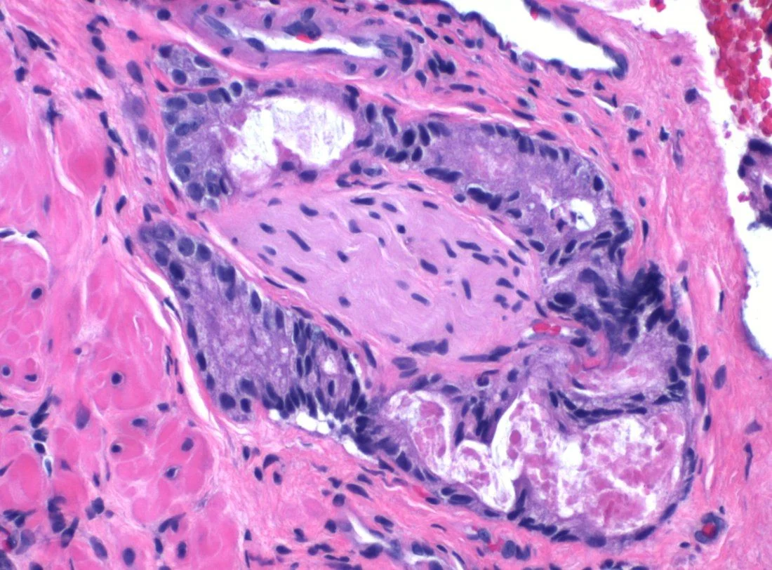
Prostate cancer is the most commonly diagnosed cancer in men in the UK, with over 50,000 new cases each year, and the second commonest cause of male cancer death. The great majority - over 95% - are acinar adenocarcinomas arising from the glandular epithelium.2
Its defining clinical characteristic is enormous variability in behaviour. Many prostate cancers are indolent and would never cause symptoms or shorten life if left entirely alone, while others metastasise early and kill. Autopsy studies find prostate cancer in a substantial proportion of older men who died of unrelated causes. This is why the central problem in prostate cancer is not detection but distinguishing the cancers that need treating from those that do not - and why overdiagnosis and overtreatment dominate discussions about screening.
Risk factors
- Increasing age - the dominant risk factor; rare below 50 and rising steeply thereafter
- Black ethnicity - approximately twice the incidence and mortality, with presentation at a younger age. UK guidance recognises this, and Black men should be counselled that their risk is higher and may warrant earlier discussion of PSA testing
- Family history - risk roughly doubles with an affected first-degree relative, and rises further with more affected relatives or early-onset disease
- Genetic predisposition - BRCA2 (substantially increased risk and more aggressive disease), BRCA1, and Lynch syndrome (HNPCC)
- Tall stature, obesity and metabolic syndrome - associated with more aggressive disease
- Diet - high intake of processed and red meat and dairy has been implicated, though evidence is weak and inconsistent
- Anabolic steroid use and, historically, exogenous testosterone
Note what is not a risk factor: benign prostatic hyperplasia does not cause prostate cancer, although the two commonly coexist because both are diseases of ageing. Vasectomy is not an established risk factor.
Clinical features
Early prostate cancer is usually asymptomatic. Most cases in the UK are now detected through PSA testing prompted either by symptoms of coexisting BPH or by patient request.
Local disease
- Often none
- Lower urinary tract symptoms - hesitancy, poor stream, frequency, nocturia; usually reflecting coexisting BPH rather than the cancer itself
- Haematuria or haematospermia
- Erectile dysfunction
- Perineal or suprapubic discomfort
Locally advanced and metastatic disease
- Bone pain - particularly the lumbar spine, pelvis, ribs and femur; the axial skeleton is affected preferentially. This is the classic presentation of previously undiagnosed metastatic disease
- Pathological fracture
- Spinal cord compression - back pain with leg weakness, sensory level, and bladder or bowel dysfunction; an oncological emergency
- Weight loss, anorexia and fatigue
- Anaemia from bone marrow infiltration
- Lower limb oedema from pelvic lymphadenopathy
- Renal failure from bilateral ureteric obstruction by local extension or nodes
- Rectal symptoms from direct invasion (uncommon)
Digital rectal examination
A normal DRE does not exclude prostate cancer, but abnormal findings are important. Suspicious features are:
- Hard, craggy or nodular texture (a benign gland is smooth and firm)
- Asymmetry between the lobes
- Loss of the median sulcus
- Fixed, immobile gland or extension beyond the capsule
- Any of these findings warrants urgent referral irrespective of the PSA result
Investigations
Prostate-specific antigen (PSA)
PSA is a glycoprotein produced by prostatic epithelium. It is organ-specific but not cancer-specific, and its limitations must be understood before it is requested.
Imaging and biopsy
- Multiparametric MRI (mpMRI) of the prostate is performed FIRST, before any biopsy. This is a major change in practice: MRI-first identifies clinically significant cancers more accurately and allows around a quarter of men to avoid biopsy altogether
- The MRI is scored 1 to 5 on a Likert or PI-RADS scale. A score of 1-2 makes significant cancer unlikely and biopsy may be safely omitted after discussion; 3-5 prompts biopsy
- Prostate biopsy - now usually transperineal rather than transrectal, because it carries a substantially lower risk of sepsis and samples the anterior prostate better. Biopsies are targeted to MRI-identified lesions, often with systematic cores
- Staging imaging for intermediate and high-risk disease: bone scintigraphy (isotope bone scan) for skeletal metastases, and CT or MRI of the abdomen and pelvis for nodal disease. PSMA PET-CT is increasingly used and is more sensitive for both nodal and metastatic disease
Other tests
- U&Es - to detect obstructive renal impairment
- FBC - anaemia from marrow infiltration
- Alkaline phosphatase (ALP) - raised with bone metastases, with a normal calcium because the lesions are osteoblastic (bone-forming) rather than lytic
- Testosterone - baseline before starting androgen deprivation therapy
- Bone profile and calcium - hypercalcaemia is uncommon in prostate cancer, unlike most bony metastatic disease
Grading and staging
Gleason grading and Grade Groups
The Gleason system grades the architectural pattern of the glands from 1 to 5 (higher being less differentiated). The score reports the most common pattern plus the second most common pattern, so a Gleason score of 3+4=7 differs prognostically from 4+3=7 - the first number matters more. Modern reporting converts this into Grade Groups 1 to 5.
| Grade Group | Gleason score | Interpretation |
|---|---|---|
| 1 | ≤ 6 (3+3) | Low grade; often suitable for active surveillance |
| 2 | 7 (3+4) | Favourable intermediate risk |
| 3 | 7 (4+3) | Unfavourable intermediate risk - worse than 3+4 |
| 4 | 8 (4+4, 3+5, 5+3) | High grade |
| 5 | 9-10 | Very high grade; most aggressive |
Risk stratification
Localised disease is stratified into low, intermediate and high risk by combining PSA, Grade Group and T stage, and this determines management:
- Low risk: PSA under 10, Grade Group 1, and stage T1-T2a
- Intermediate risk: PSA 10-20, or Grade Group 2-3, or stage T2b
- High risk: PSA over 20, or Grade Group 4-5, or stage T2c or above
- T staging: T1 impalpable and not visible; T2 confined within the prostate; T3 extends through the capsule (T3a) or invades seminal vesicles (T3b); T4 invades adjacent structures such as bladder or rectum
Management
Treatment depends on risk category, stage, life expectancy, comorbidity and patient preference. Because many prostate cancers are indolent, doing nothing actively is a legitimate and often correct choice, and shared decision-making is central.1
Localised disease
| Option | Who it is for | Key points and adverse effects |
|---|---|---|
| Active surveillance | Low-risk and selected favourable intermediate-risk disease, with the intention of treating radically if it progresses | Monitoring with PSA, repeat MRI and biopsy. Avoids or defers the harms of treatment; requires the patient to accept living with an untreated cancer |
| Watchful waiting | Older men or those with significant comorbidity and limited life expectancy, where the intention is symptom control only, not cure | Less intensive monitoring; treatment is hormonal and palliative if symptoms develop |
| Radical prostatectomy | Fit men with localised disease and a life expectancy over 10 years | Erectile dysfunction (very common) and urinary incontinence (usually stress incontinence, often improving over 12 months). PSA should become undetectable afterwards |
| Radical radiotherapy (external beam or brachytherapy) | Localised or locally advanced disease; an alternative to surgery | Often combined with neoadjuvant/adjuvant androgen deprivation in intermediate and high-risk disease. Causes proctitis, cystitis, erectile dysfunction and a small long-term risk of second malignancy (bladder, rectal) |
Advanced and metastatic disease
Prostate cancer growth is androgen-dependent, so the mainstay of advanced disease is androgen deprivation therapy (ADT), usually combined with additional agents.
- GnRH (LHRH) agonists - goserelin, leuprorelin. These initially stimulate the pituitary before downregulating it, causing a testosterone surge or 'tumour flare' in the first 1-2 weeks. Cover with an anti-androgen (e.g. bicalutamide) started before or alongside the first dose, particularly where there is spinal metastatic disease, because flare can precipitate cord compression
- GnRH antagonists - degarelix; suppress testosterone immediately without a flare, useful where there is impending cord compression
- Anti-androgens - bicalutamide, and the newer agents enzalutamide and apalutamide
- Abiraterone - blocks androgen synthesis (CYP17); given with prednisolone
- Chemotherapy - docetaxel - used with ADT in metastatic hormone-sensitive disease and in castration-resistant disease
- Bilateral orchidectomy - surgical castration; rapid and permanent, now rarely used but still an option
- Bone protection - bisphosphonates or denosumab to reduce skeletal events; radiotherapy for painful bone metastases; radium-223 for symptomatic bone-predominant disease
- Palliative care involvement for symptom control
Complications
- Metastatic spinal cord compression - the key oncological emergency; prostate cancer is one of the commonest causes
- Bone metastases - characteristically osteoblastic (sclerotic), causing pain, pathological fracture and marrow failure
- Bladder outlet obstruction and urinary retention
- Bilateral ureteric obstruction causing hydronephrosis and renal failure
- Anaemia and pancytopenia from marrow infiltration
- Lymphoedema of the lower limbs from pelvic nodal disease
- Erectile dysfunction and urinary incontinence - from the disease and, more often, from its treatment
- Osteoporosis and cardiovascular disease from long-term androgen deprivation
- Radiotherapy toxicity - radiation proctitis, cystitis, and second malignancies
- Castration-resistant progression - disease that progresses despite castrate testosterone levels
- Psychological impact - anxiety and depression, both from diagnosis and from treatment effects on sexual function and body image
Red flags
Prognosis
Overall survival in prostate cancer is good, and better than for most solid tumours - around 78% of men in England survive 10 years or more, and for localised disease survival approaches that of the general population. Many men with low-risk disease will die with prostate cancer rather than of it, which is precisely why active surveillance and watchful waiting are appropriate rather than negligent.
Prognosis is determined principally by Grade Group, PSA and stage at diagnosis. Localised low-risk disease (Grade Group 1) has an excellent outlook with 10-year cancer-specific survival in excess of 95%, and long-term follow-up of active surveillance cohorts shows very low prostate cancer mortality. High-grade (Grade Group 4-5) and metastatic disease behave very differently: median survival in metastatic disease has improved substantially with the addition of docetaxel, abiraterone and the newer androgen receptor agents to standard ADT, but it remains incurable, and castration-resistant disease eventually develops in most.
Ethnic disparities in outcome are important and examinable. Black men have roughly double the incidence and mortality of prostate cancer, present younger and often with more advanced disease. Alongside this, the dominant harm in prostate cancer care remains overtreatment of indolent disease - erectile dysfunction and incontinence inflicted on men whose cancer would never have troubled them. The MRI-first diagnostic pathway and structured risk stratification exist specifically to reduce that harm, and understanding this tension is central to understanding the condition.
References
- NICE NG131. Prostate cancer: diagnosis and management. 2019, updated 2021. Available here
- Cancer Research UK. Prostate cancer statistics. Available here
- NICE NG12. Suspected cancer: recognition and referral. 2015, updated 2023. Available here
- NHS Prostate Cancer Risk Management Programme (PCRMP). PSA testing and informed choice. Available here
- Ahmed HU, El-Shater Bosaily A, Brown LC et al. PROMIS: diagnostic accuracy of multiparametric MRI in prostate cancer. Lancet. 2017. Available here
- Hamdy FC, Donovan JL, Lane JA et al. ProtecT trial: 10-year outcomes after monitoring, surgery or radiotherapy. NEJM. 2016. Available here
- NICE NG12 / Metastatic spinal cord compression (CG75). Recognition and management. Available here
- Mikael Häggström, MD, CC0, via Wikimedia Commons. Available here
This article is written for revision and education. It is not clinical guidance and must not be used to make decisions about the care of a patient. Always check current NICE guidance and local protocols.