
Arrhythmias: A Systematic Approach
Key points
- Arrhythmia: any heart rhythm that is not normal sinus rhythm, arising from abnormal impulse formation, abnormal conduction, or re-entry.
- The four questions: is the patient stable? Is the rate fast or slow? Is the QRS narrow or broad? Is the rhythm regular or irregular?
- Narrow complex: QRS under 120 ms means the ventricles are activated through the normal His-Purkinje system, so the rhythm must originate at or above the AV node.
- Broad complex: QRS 120 ms or more. Assume ventricular tachycardia until proven otherwise, particularly if there is structural heart disease.
- Adverse features: shock, syncope, myocardial ischaemia and heart failure. Any one of these mandates synchronised DC cardioversion in tachycardia, or atropine and pacing in bradycardia.
- Reversible causes: electrolyte disturbance, ischaemia, hypoxia, drugs, thyroid disease and sepsis. Treat these before or alongside the rhythm itself.
- Investigations: a 12-lead ECG during symptoms is the single most valuable test. If intermittent, escalate through Holter, patch, event recorder and implantable loop recorder.
- Key principle: treat the patient, not the monitor. An asymptomatic run of a rhythm and a peri-arrest patient with the same rhythm need very different responses.
Introduction
An arrhythmia is any cardiac rhythm other than normal sinus rhythm. The term covers everything from a benign ectopic beat felt as a single skipped heartbeat to ventricular fibrillation, and the range is wide enough that students often try to memorise each rhythm as a separate entity.
That approach fails under exam pressure and at the bedside. A far more reliable method is to answer four questions in order, because the answers narrow the possibilities to a handful and, crucially, the first answer determines whether there is time to think at all.1
- Is the patient stable? Adverse features change the management immediately, whatever the rhythm.
- Is the rate fast or slow? Above 100 beats per minute is a tachyarrhythmia; below 60 is a bradyarrhythmia.
- Is the QRS narrow or broad? This localises the origin of the rhythm above or below the AV node.
- Is the rhythm regular or irregular? This is what separates atrial fibrillation from the other narrow complex tachycardias.
This article is the framework. The individual rhythms - atrial fibrillation, atrial flutter, supraventricular tachycardia, ventricular tachycardia, long QT syndrome and the bradyarrhythmias - each have their own article, and the detail of diagnosis and definitive management sits there.
Normal conduction
Understanding where an arrhythmia comes from requires knowing the normal path an impulse takes, because every abnormal rhythm is a deviation from it.
- The sinoatrial (SA) node, in the upper right atrium, depolarises spontaneously at 60-100 per minute and sets the rate. It is richly supplied by both sympathetic and parasympathetic fibres, which is why the sinus rate varies with respiration, exercise and fear.
- The impulse spreads across both atria, producing the P wave.
- The atrioventricular (AV) node is the only electrical connection between atria and ventricles in a normal heart. It deliberately delays conduction, producing the PR interval and allowing atrial contraction to fill the ventricles before they contract.
- The bundle of His divides into right and left bundle branches, and then into the Purkinje fibres, which distribute the impulse rapidly throughout the ventricular myocardium. This rapid, organised spread is what produces a narrow QRS.
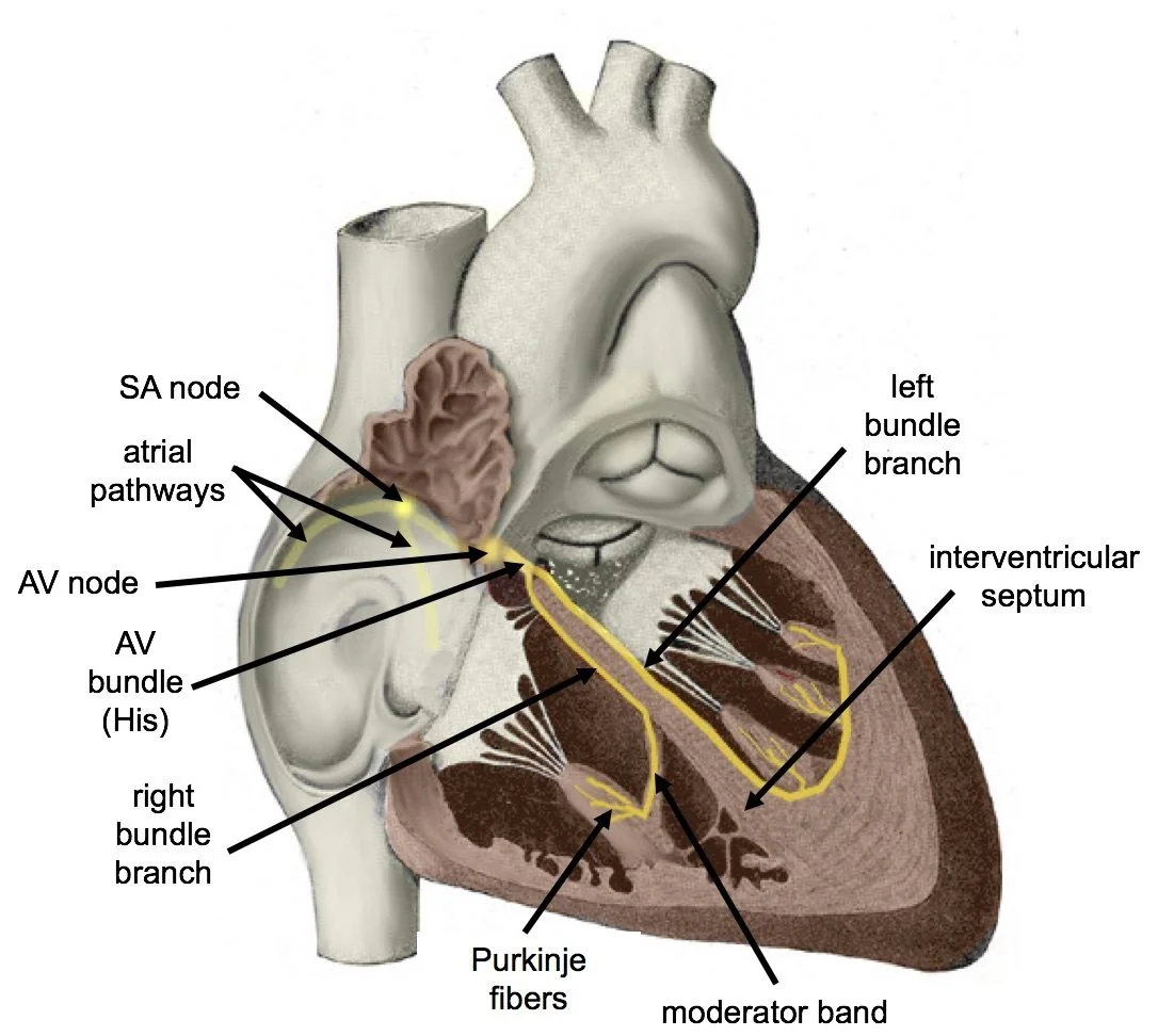
Every part of the conducting system has automaticity, but at progressively slower intrinsic rates. This hierarchy is a safety mechanism: if a higher pacemaker fails, a lower one takes over, though at a slower rate.
| Pacemaker | Intrinsic rate | QRS width | Reliability |
|---|---|---|---|
| Sinoatrial node | 60-100/min | Narrow | Normal |
| Atrial tissue | 60-80/min | Narrow | Reasonable |
| AV node and junction | 40-60/min | Narrow | Reasonably reliable |
| Ventricular (His-Purkinje and below) | 20-40/min | Broad | Unreliable - may fail without warning |
Mechanisms
Arrhythmias arise by three mechanisms. Knowing which applies explains why particular treatments work and why others are useless.
Re-entry
By far the commonest mechanism. It requires two pathways with different conduction speeds and refractory periods, joined into a circuit, and a trigger such as an ectopic beat that blocks in one pathway and travels down the other. The impulse then arrives back at the blocked pathway from the other end, finds it recovered, and conducts retrogradely, setting up a self-sustaining loop.
Re-entry explains atrioventricular nodal re-entrant tachycardia (dual pathways within the AV node), atrioventricular re-entrant tachycardia (an accessory pathway such as in Wolff-Parkinson-White syndrome), atrial flutter (a macro-re-entrant circuit around the tricuspid annulus), and most ventricular tachycardia (a circuit around a region of myocardial scar).
Because the circuit is anatomically fixed, re-entrant rhythms are typically regular, start and stop abruptly, and can be terminated by anything that interrupts the circuit, which is the principle behind vagal manoeuvres, adenosine, cardioversion and catheter ablation.
Abnormal automaticity
Myocardial tissue outside the SA node depolarises spontaneously and takes over. This is favoured by ischaemia, catecholamines, hypokalaemia, hypomagnesaemia, digoxin toxicity and mechanical stretch. Examples include atrial tachycardia, accelerated idioventricular rhythm after reperfusion, and multifocal atrial tachycardia in severe respiratory disease.
Automatic rhythms typically warm up and cool down rather than starting and stopping abruptly, and they do not respond to cardioversion in the way re-entrant rhythms do, because there is no circuit to break.
Triggered activity
Afterdepolarisations occur during or just after repolarisation and, if large enough, trigger a new action potential. Early afterdepolarisations occur when repolarisation is prolonged - the mechanism of torsades de pointes in long QT syndrome. Delayed afterdepolarisations occur in calcium-overloaded cells, and underlie digoxin toxicity arrhythmias and catecholaminergic polymorphic ventricular tachycardia.
Classification
The practical classification is the one that maps onto the ECG in front of you.
| Rate | QRS | Rhythm | Likely diagnoses |
|---|---|---|---|
| Fast | Narrow | Regular | Sinus tachycardia, AVNRT, AVRT, atrial flutter with fixed block, atrial tachycardia |
| Fast | Narrow | Irregular | Atrial fibrillation, atrial flutter with variable block, multifocal atrial tachycardia |
| Fast | Broad | Regular | Ventricular tachycardia, SVT with bundle branch block or aberrancy, antidromic AVRT |
| Fast | Broad | Irregular | Atrial fibrillation with bundle branch block, polymorphic VT or torsades, pre-excited AF |
| Slow | Narrow | Regular | Sinus bradycardia, junctional escape rhythm, second degree block with fixed conduction ratio |
| Slow | Narrow or broad | Irregular | Second degree AV block (Mobitz I or II), sinus arrest with escape beats, slow AF |
| Slow | Broad | Regular | Complete heart block with ventricular escape, idioventricular rhythm |
Aetiology and reversible causes
Arrhythmias rarely occur in a vacuum. Identifying the precipitant is part of the diagnosis, and in many cases correcting it is the treatment.
Cardiac causes
- Ischaemic heart disease - acute ischaemia, and scar from previous infarction providing the substrate for re-entrant VT
- Structural heart disease - cardiomyopathy, valvular disease, and any cause of atrial or ventricular dilatation
- Heart failure - through stretch, neurohormonal activation and electrolyte shifts from diuretics
- Inflammatory disease - myocarditis and pericarditis
- Congenital and inherited disease - accessory pathways, long QT syndrome, Brugada syndrome, arrhythmogenic right ventricular cardiomyopathy
- Post-cardiac surgery - atrial fibrillation is very common in the first few days
Non-cardiac causes
- Electrolyte disturbance - hypokalaemia, hyperkalaemia, hypomagnesaemia and hypocalcaemia. Check and correct these in every arrhythmia.
- Endocrine - thyrotoxicosis (classically causing atrial fibrillation), phaeochromocytoma
- Hypoxia and respiratory disease - COPD, pulmonary embolism, obstructive sleep apnoea
- Sepsis and any critical illness
- Drugs - beta-agonists, digoxin, antiarrhythmics themselves (proarrhythmia), and any QT-prolonging drug
- Stimulants and toxins - caffeine, alcohol (including holiday heart syndrome), cocaine, amphetamines
- Anaemia, hypovolaemia and pain - common causes of sinus tachycardia that must be treated rather than rate-controlled
Clinical features
Symptoms depend on the rate, the duration, the presence of atrioventricular synchrony, and the state of the underlying heart. The same rhythm can be an incidental finding in one patient and cause collapse in another.
Symptoms
- Palpitations - the commonest presenting complaint, though many arrhythmias are asymptomatic
- Presyncope and syncope - syncope without warning in an arrhythmia is a red flag for a life-threatening rhythm
- Breathlessness - loss of atrial contribution to filling, or rate-related heart failure
- Chest pain - a fast rate increases myocardial oxygen demand and shortens diastolic filling of the coronaries
- Fatigue and reduced exercise tolerance - common in bradyarrhythmias and in persistent atrial fibrillation
- Nothing at all - found incidentally on an ECG, a blood pressure machine, or a wearable device
History
The history is often more diagnostic than any test, because most patients are in sinus rhythm by the time they are seen.
- Onset and offset - abrupt on/off suggests a re-entrant tachycardia; gradual warm-up and cool-down suggests sinus tachycardia or an automatic focus
- Regularity - ask the patient to tap out the rhythm on the desk. It is a surprisingly reliable way to separate atrial fibrillation from a regular tachycardia.
- Rate - fast and pounding, or slow and heavy
- Triggers - exertion, emotion, alcohol, caffeine, posture, or the startle response and swimming (which point to long QT syndrome)
- Terminating manoeuvres - if Valsalva or cold water stops it, a re-entrant tachycardia involving the AV node is likely
- Associated features - chest pain, breathlessness, syncope, polyuria after an episode (a feature of AVNRT, from atrial natriuretic peptide release)
- Drug history - including over the counter medicines, herbal preparations and recreational drugs
- Family history - sudden cardiac death under 40, unexplained drowning, or unexplained single-vehicle road traffic accidents
Clinical examination
Examination establishes stability first, and only then looks for the cause and the diagnosis.
- Pulse - rate, rhythm, volume and character. Take it for a full minute in an irregular rhythm, and compare the apical rate with the radial rate: a pulse deficit occurs in atrial fibrillation because some beats are too weak to reach the wrist.
- Blood pressure - hypotension is an adverse feature demanding immediate action
- JVP - cannon a waves occur when the atrium contracts against a closed tricuspid valve, seen in complete heart block and in ventricular tachycardia with AV dissociation. Regular cannon waves occur in junctional rhythm.
- Heart sounds - variable intensity of the first heart sound is another sign of AV dissociation. Listen for murmurs suggesting valve disease or cardiomyopathy.
- Signs of heart failure - raised JVP, basal crackles, peripheral oedema, third heart sound
- Perfusion - conscious level, capillary refill, skin temperature and urine output
- Thyroid status, anaemia and evidence of sepsis - looking for the precipitant
Investigations
The 12-lead ECG
A 12-lead ECG during symptoms is the definitive investigation and is worth considerable effort to obtain. A rhythm strip alone frequently cannot distinguish VT from SVT with aberrancy, and a single-lead wearable trace is not sufficient for a diagnosis that will commit a patient to lifelong anticoagulation or a device.
Even when the patient is back in sinus rhythm, the resting ECG carries diagnostic information:
- Delta wave and short PR interval - Wolff-Parkinson-White syndrome
- Prolonged QTc - congenital or acquired long QT syndrome
- Coved ST elevation in V1-V2 - Brugada syndrome
- Pathological Q waves - previous infarction, providing the substrate for scar-related VT
- Left ventricular hypertrophy - hypertensive heart disease or hypertrophic cardiomyopathy
- Epsilon waves and T inversion in V1-V3 - arrhythmogenic right ventricular cardiomyopathy
- Bundle branch block or bifascicular block - conducting system disease
Capturing an intermittent arrhythmia
Match the monitoring duration to the symptom frequency. There is no value in a 24-hour tape for symptoms occurring monthly.2
| Symptom frequency | Investigation | Notes |
|---|---|---|
| Daily | 24-hour Holter monitor | Also quantifies ectopic burden and assesses rate control in AF |
| Every few days | 48-hour to 7-day Holter or adhesive patch monitor | Patch monitors are better tolerated for longer wear |
| Weekly to monthly | External event recorder or 14-30 day patch | Patient-activated or auto-triggered |
| Less than monthly, or syncope | Implantable loop recorder | Records for up to 3 years; the investigation of choice in unexplained syncope with suspected arrhythmia |
| Exercise-related | Exercise tolerance test | For catecholaminergic VT, exercise-induced VT, and rate response assessment |
Blood tests
- U&Es, magnesium and calcium - the most important tests, and the most commonly abnormal
- FBC - anaemia as a precipitant of tachycardia
- Thyroid function - mandatory in new atrial fibrillation
- Troponin - if ischaemia is suspected, remembering that any sustained tachycardia can raise it
- Digoxin level - if toxicity is suspected, taken at least 6 hours after the dose
- BNP or NT-proBNP - if heart failure is suspected
Imaging and specialist tests
- Echocardiogram - to assess structural heart disease, ventricular function and valves. Indicated in essentially all significant arrhythmias, because the presence of structural disease changes both the differential and the prognosis.
- Cardiac MRI - for suspected cardiomyopathy, myocarditis, infiltration or arrhythmogenic right ventricular cardiomyopathy
- Electrophysiological study - maps the circuit, and allows ablation in the same procedure
- Genetic testing - for suspected inherited channelopathies and cardiomyopathies, with family screening
- Tilt table testing - where a reflex or orthostatic cause of syncope is suspected rather than an arrhythmia
Management
The Resuscitation Council UK peri-arrest algorithms are the framework for acute management, and are examinable in detail.1 They begin with the same question in both tachycardia and bradycardia: are there adverse features?
The unstable tachycardia
Synchronised DC cardioversion under sedation or general anaesthesia, up to three attempts. Synchronisation to the R wave is essential: an unsynchronised shock delivered during the T wave can precipitate ventricular fibrillation. If three shocks fail, give amiodarone 300 mg IV over 10-20 minutes and shock again, followed by an infusion of 900 mg over 24 hours.1
The stable tachycardia
There is time to obtain a 12-lead ECG and classify the rhythm properly.
| Rhythm | First-line treatment |
|---|---|
| Narrow, regular | Vagal manoeuvres, then adenosine 6 mg, 12 mg, 12 mg IV as a rapid bolus with a flush |
| Narrow, irregular | Usually atrial fibrillation: rate control with a beta-blocker or diltiazem, and consider cardioversion if onset is clearly under 48 hours |
| Broad, regular | Treat as VT: amiodarone 300 mg IV over 20-60 minutes, then 900 mg over 24 hours. If a known SVT with bundle branch block, treat as a narrow complex tachycardia. |
| Broad, irregular | Seek expert help. Consider AF with bundle branch block, pre-excited AF (avoid AV nodal blocking drugs) or polymorphic VT (give magnesium 2 g IV). |
Bradycardia
With adverse features, or with a risk of asystole, give atropine 500 micrograms IV, repeated to a maximum of 3 mg. If this fails, options are transcutaneous pacing, an isoprenaline or adrenaline infusion, or specialist alternatives such as aminophylline or glucagon (in beta-blocker or calcium channel blocker overdose). Arrange transvenous pacing if the response is inadequate.1
Longer-term management
- Treat the underlying cause - correct electrolytes, treat thyrotoxicosis, revascularise ischaemia, stop the offending drug, manage sleep apnoea
- Lifestyle - reduce alcohol and caffeine, stop stimulants, treat obesity. Weight loss and alcohol reduction have a genuine evidence base in reducing atrial fibrillation burden.
- Antiarrhythmic drugs - rate control or rhythm control, chosen according to the rhythm, ventricular function and comorbidity
- Anticoagulation - for atrial fibrillation and atrial flutter, guided by CHA2DS2-VASc balanced against bleeding risk
- Catheter ablation - now first-line or early second-line for many supraventricular tachycardias, atrial flutter and symptomatic atrial fibrillation, and used for scar-related VT
- Devices - permanent pacemakers for symptomatic bradycardia and high-grade AV block; implantable cardioverter defibrillators for those at risk of sudden cardiac death
- Driving and occupation - DVLA rules differ by arrhythmia, by whether it caused incapacity, and by licence group. Check the current guidance for every patient.3
Antiarrhythmic drugs
The Vaughan Williams classification groups antiarrhythmics by their principal mechanism. It is imperfect - amiodarone has properties of all four classes - but it remains a useful scaffold.
| Class | Mechanism | Examples | Main uses and cautions |
|---|---|---|---|
| Ia | Sodium channel block, prolongs action potential | Quinidine, disopyramide | Rarely used; disopyramide has a role in HOCM |
| Ib | Sodium channel block, shortens action potential | Lidocaine | Ventricular arrhythmias, largely superseded |
| Ic | Sodium channel block, no effect on duration | Flecainide, propafenone | AF and SVT in structurally normal hearts. Contraindicated in ischaemic or structural heart disease because of proarrhythmia. |
| II | Beta-adrenoceptor blockade | Bisoprolol, metoprolol, sotalol | Rate control, and reduction of sudden death after MI and in heart failure |
| III | Potassium channel block, prolongs repolarisation | Amiodarone, sotalol, dronedarone | Broad efficacy; all prolong the QT interval and can cause torsades |
| IV | Non-dihydropyridine calcium channel blockade | Verapamil, diltiazem | Rate control in AF and termination of SVT. Avoid in broad complex tachycardia and in heart failure. |
| Unclassified | Various | Adenosine, digoxin, magnesium, ivabradine | Adenosine blocks the AV node transiently; digoxin increases vagal tone; magnesium treats torsades |
Amiodarone deserves particular attention because of its toxicity profile. It causes thyroid dysfunction (both hyper- and hypothyroidism), pulmonary fibrosis, hepatitis, corneal microdeposits, photosensitivity and slate-grey skin discolouration, and peripheral neuropathy. It has a half-life of around 50 days and interacts with warfarin and digoxin, both of whose levels it raises. Patients need baseline and six-monthly thyroid and liver function tests, and it should be given through a central line where possible because it causes thrombophlebitis.4
Complications
- Sudden cardiac death - ventricular fibrillation and pulseless VT are the commonest mechanisms
- Thromboembolism - ischaemic stroke and systemic embolism in atrial fibrillation and flutter, and also after cardioversion of an arrhythmia lasting over 48 hours without anticoagulation
- Tachycardia-induced cardiomyopathy - a persistently uncontrolled ventricular rate causes a dilated cardiomyopathy that is largely reversible if the rate is controlled
- Heart failure - both precipitated by and precipitating arrhythmia
- Syncope and injury - fractures, head injury and road traffic collisions
- Proarrhythmia - antiarrhythmic drugs causing new or worsened arrhythmia, particularly torsades de pointes
- Bleeding - from the anticoagulation used to prevent embolism
- Psychological impact and loss of driving licence or occupation - frequently underestimated
Red flags
Prognosis
Prognosis varies more widely in arrhythmia than in almost any other area of cardiology, and is determined less by the rhythm itself than by the heart it occurs in.
Supraventricular arrhythmias in a structurally normal heart - AVNRT, AVRT and focal atrial tachycardia - carry an excellent prognosis. They are symptomatic and disruptive but not life-shortening, and catheter ablation is curative in over 95% of cases.
Atrial fibrillation roughly doubles all-cause mortality and increases stroke risk approximately fivefold, though anticoagulation reduces stroke risk by around two thirds and is the single most important intervention.
Ventricular arrhythmias carry a prognosis dictated by left ventricular function. VT in a structurally normal heart may be benign; the same rhythm in a patient with an ejection fraction of 25% after infarction identifies a high risk of sudden death and usually warrants an implantable defibrillator.
Bradyarrhythmias are generally well treated. Pacing for complete heart block restores a normal life expectancy in most patients, which is why it is one of the more satisfying interventions in medicine.
References
- Resuscitation Council UK. Adult advanced life support guidelines: peri-arrest arrhythmias. Available here
- NICE NG180. Atrial fibrillation: diagnosis and management. 2021. Available here
- DVLA. Assessing fitness to drive: a guide for medical professionals. Available here
- BNF. Amiodarone hydrochloride - indications, cautions and monitoring. Available here
- Brugada J, Katritsis DG, Arbelo E et al. 2019 ESC Guidelines for the management of patients with supraventricular tachycardia. European Heart Journal. 2020. Available here
- Zeppenfeld K, Tfelt-Hansen J, de Riva M et al. 2022 ESC Guidelines for the management of patients with ventricular arrhythmias and the prevention of sudden cardiac death. European Heart Journal. 2022. Available here
- NICE CG109. Transient loss of consciousness (blackouts) in over 16s. 2010, updated 2023. Available here
- Glikson M, Nielsen JC, Kronborg MB et al. 2021 ESC Guidelines on cardiac pacing and cardiac resynchronization therapy. European Heart Journal. 2021. Available here
This article is written for revision and education. It is not clinical guidance and must not be used to make decisions about the care of a patient. Always check current NICE guidance and local protocols.