
Supraventricular Tachycardia: AVNRT, AVRT and Wolff-Parkinson-White
Key points
- Supraventricular tachycardia: in everyday use, a regular narrow complex tachycardia at 140-250 beats per minute arising above the ventricles, most often by re-entry involving the AV node.
- AVNRT: atrioventricular nodal re-entrant tachycardia - a micro-circuit within the AV node itself. The commonest SVT, typically in young women.
- AVRT: atrioventricular re-entrant tachycardia - a circuit using an accessory pathway outside the AV node, as in Wolff-Parkinson-White syndrome.
- Presentation: abrupt onset and offset palpitations, often with neck pulsation, chest discomfort, breathlessness and polyuria afterwards.
- First line: vagal manoeuvres - the modified Valsalva manoeuvre is markedly more effective than the standard version.
- Second line: adenosine 6 mg, then 12 mg, then 12 mg as a rapid IV bolus with a saline flush through a large proximal cannula.
- Unstable: shock, syncope, myocardial ischaemia or heart failure require immediate synchronised DC cardioversion.
- The critical warning: in pre-excited atrial fibrillation (irregular, broad, very fast), AV nodal blocking drugs can precipitate ventricular fibrillation. Use flecainide or DC cardioversion.
Introduction
Strictly, supraventricular tachycardia means any tachycardia arising at or above the AV node, which would include sinus tachycardia, atrial fibrillation and atrial flutter. In clinical usage, however, SVT almost always refers to a regular, paroxysmal narrow complex tachycardia caused by re-entry involving the AV node - and that is the sense used here.
SVT is common, affecting around 2 per 1000 of the population, and typically presents in otherwise healthy young people. It is dramatic, frightening for the patient, and highly treatable: the acute episode usually terminates with a bedside manoeuvre or a single drug, and definitive cure by catheter ablation succeeds in over 95% of cases.
| Type | Proportion | Mechanism | Typical patient |
|---|---|---|---|
| AVNRT | ~60% | Re-entry within dual pathways inside the AV node | Young women, structurally normal heart |
| AVRT | ~30% | Re-entry using an accessory pathway between atrium and ventricle | Younger patients, often male; associated with WPW |
| Atrial tachycardia | ~10% | An automatic or micro-re-entrant focus in atrial tissue | Older patients, structural or lung disease, digoxin toxicity |
| Junctional tachycardia | Rare | Automatic focus in the AV junction | Post-cardiac surgery, digoxin toxicity, congenital |
Mechanisms
AVNRT
Around a quarter of people have dual AV nodal pathways: a slow pathway with a short refractory period, and a fast pathway with a long refractory period. In sinus rhythm the impulse travels down the fast pathway, and nothing is apparent on the ECG.
A well-timed atrial ectopic arriving while the fast pathway is still refractory is conducted down the slow pathway instead. By the time it reaches the bottom, the fast pathway has recovered, so the impulse travels back up it retrogradely, reaches the top of the slow pathway, and re-enters. A self-sustaining circuit is established entirely within a few millimetres of tissue.
Because the circuit is so small, atria and ventricles are activated almost simultaneously. The retrograde P wave is therefore buried within the QRS complex or appears just at its end, producing the characteristic pseudo-R wave in V1 or pseudo-S wave in the inferior leads.
AVRT and accessory pathways
An accessory pathway is a strand of conducting muscle that bridges the atrioventricular ring, bypassing the AV node. The commonest is the bundle of Kent, the abnormality underlying Wolff-Parkinson-White syndrome.
Unlike the AV node, an accessory pathway conducts fast and has no rate-limiting properties. When it conducts anterogradely in sinus rhythm, part of the ventricle is depolarised early, giving the classic resting ECG of pre-excitation.
- Short PR interval (under 120 ms) - the impulse bypasses the AV nodal delay
- Delta wave - a slurred upstroke to the QRS, representing slow cell-to-cell spread of the early depolarisation through ventricular muscle
- Widened QRS - a fusion of pre-excited and normally conducted activation
- Secondary ST and T wave changes
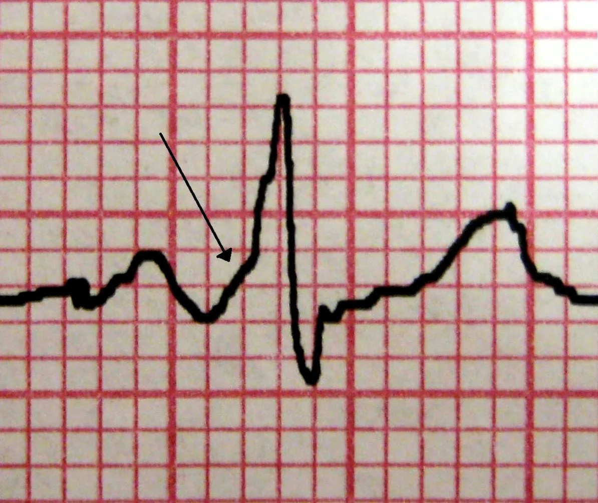
Wolff-Parkinson-White syndrome is the combination of this ECG pattern with symptomatic arrhythmia. The pattern alone, found incidentally, is termed asymptomatic pre-excitation.
| Type | Circuit direction | QRS | Frequency |
|---|---|---|---|
| Orthodromic | Down the AV node, back up the accessory pathway | Narrow - normal ventricular activation | ~95% of AVRT |
| Antidromic | Down the accessory pathway, back up the AV node | Broad and fully pre-excited - easily mistaken for VT | ~5% of AVRT |
A concealed accessory pathway conducts only retrogradely. The resting ECG is completely normal with no delta wave, but orthodromic AVRT can still occur. This is why a normal resting ECG does not exclude an accessory pathway.
Atrial tachycardia
An ectopic atrial focus fires faster than the sinus node. The P wave morphology differs from sinus, there is an isoelectric baseline between P waves (unlike flutter), and the AV node is not part of the circuit. Adenosine therefore usually reveals the atrial activity rather than terminating the rhythm. Multifocal atrial tachycardia, with three or more P wave morphologies and an irregular rhythm, is characteristic of severe COPD.
Clinical features
Symptoms
- Palpitations of abrupt onset and offset - the single most useful historical feature. Patients often describe a switch being flicked, and can name the moment it started and stopped. This contrasts with sinus tachycardia, which warms up and cools down.
- Rapid regular pounding, often described as a rate too fast to count
- Neck pulsation or a pounding in the throat - the frog sign, caused by the right atrium contracting against a closed tricuspid valve. It is more typical of AVNRT than AVRT.
- Chest discomfort and breathlessness
- Anxiety and a sense of impending doom
- Dizziness and presyncope - true syncope is less common and should prompt consideration of a very fast rate or an alternative diagnosis
- Polyuria after the episode - atrial stretch releases atrial natriuretic peptide. A characteristic and frequently examined detail.
- Triggers - caffeine, alcohol, stimulants, stress, exertion, or nothing at all
Examination
During an episode: a regular tachycardia at 140-250/min, often with a normal or slightly reduced blood pressure. Regular cannon a waves may be visible in the JVP because the atria contract against closed AV valves with every beat - unlike the intermittent, irregular cannon waves of ventricular tachycardia.
Between episodes the examination is normal. Examine specifically for structural heart disease, since its presence changes the differential of a broad complex tachycardia considerably and influences drug choice.
Investigations
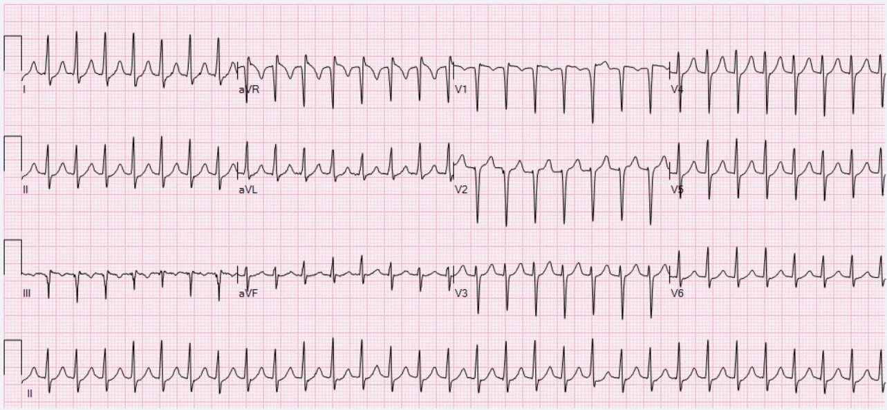
ECG during the episode
Obtain a 12-lead ECG during the tachycardia if at all possible, and keep it - it is the single most valuable document for later diagnosis and for the electrophysiologist.
- Regular narrow complex tachycardia at 140-250/min
- P waves absent, hidden within the QRS, or inverted immediately after it
- Pseudo-R wave in V1 or pseudo-S wave in II, III and aVF - a retrograde P wave distorting the end of the QRS, suggesting AVNRT
- A clearly visible retrograde P wave separated from the QRS (RP interval over 70 ms) - suggests AVRT, since the impulse must travel through ventricular muscle and back up the pathway, which takes longer
- Rate-related ST depression - common, and usually not indicative of coronary disease in a young patient
- QRS alternans - beat-to-beat variation in QRS amplitude, more suggestive of AVRT
Resting ECG between episodes
- Delta wave with a short PR - Wolff-Parkinson-White syndrome
- Completely normal - the usual finding in AVNRT and in concealed accessory pathways
- Look also for evidence of structural heart disease, prior infarction and ventricular hypertrophy
Other investigations
- U&Es, magnesium, calcium, FBC and thyroid function - looking for precipitants
- Ambulatory monitoring - a Holter, patch or event recorder matched to symptom frequency, if no ECG has been captured
- Echocardiogram - to exclude structural heart disease, particularly Ebstein anomaly, which is associated with right-sided accessory pathways
- Exercise testing - abrupt loss of the delta wave at higher heart rates suggests a pathway with a long refractory period and therefore a lower risk of dangerous conduction in atrial fibrillation
- Electrophysiological study - maps the circuit precisely and permits ablation in the same procedure
Acute management
Is the patient stable?
Step 1: vagal manoeuvres
Increasing vagal tone slows conduction through the AV node and can break the circuit. Vagal manoeuvres terminate around 20-25% of episodes with the standard technique, rising to over 40% with the modified Valsalva.2
Carotid sinus massage is an alternative: firm massage over one carotid sinus for 5-10 seconds, with continuous ECG monitoring. Avoid it if there is a carotid bruit, previous stroke or TIA, or known carotid disease, because of the risk of dislodging plaque. Never massage both sides at once.
Step 2: adenosine
Adenosine causes transient, complete AV nodal block lasting a few seconds. In AVNRT and orthodromic AVRT the AV node is part of the circuit, so the arrhythmia terminates abruptly.
- 6 mg as a rapid intravenous bolus into a large proximal vein (antecubital fossa or larger), immediately followed by a 20 ml saline flush with the arm elevated
- If no response after 1-2 minutes, 12 mg
- If still no response, a further 12 mg
- Record a continuous rhythm strip throughout - the response is diagnostically valuable even when the rhythm does not terminate
The half-life is under 10 seconds, which is why the rapid bolus and flush matter enormously: given slowly, the drug is metabolised before it reaches the heart and simply will not work.
Step 3: further options
If vagal manoeuvres and adenosine fail in a stable patient, options include verapamil or diltiazem intravenously (avoiding these in heart failure, and never combining verapamil with an intravenous beta-blocker), an intravenous beta-blocker, or synchronised DC cardioversion. Seek expert help before moving beyond adenosine.
Wolff-Parkinson-White and pre-excited AF
Long-term management of an accessory pathway
- Catheter ablation of the accessory pathway is the definitive treatment, with success rates above 95% and a low complication rate. It is offered to all symptomatic patients.
- Asymptomatic pre-excitation is usually assessed with risk stratification. Ablation is considered in high-risk occupations (pilots, drivers, competitive athletes) and where electrophysiological study shows a pathway capable of rapid conduction.
- Drug therapy where ablation is declined or unsuitable: flecainide or propafenone in a structurally normal heart, or sotalol or amiodarone otherwise
- Avoid AV nodal blocking drugs long term in patients with anterograde pre-excitation
- Driving - the DVLA requires cessation of driving until the arrhythmia is controlled; the rules differ for Group 1 and Group 2 licences.3
Long-term management of AVNRT
- Avoid precipitants - caffeine, alcohol, stimulants, and over-the-counter decongestants. This alone is sufficient for many patients with infrequent episodes.
- Teach the modified Valsalva manoeuvre so the patient can terminate episodes at home
- Drug prophylaxis - a beta-blocker or a rate-limiting calcium channel blocker for frequent or troublesome episodes; flecainide in a structurally normal heart
- Catheter ablation of the slow pathway - curative in over 95% of cases. The main risk is complete heart block requiring a permanent pacemaker, which occurs in under 1% because the slow pathway lies away from the compact AV node. Ablation is now offered early, and is often preferred by younger patients over lifelong medication.
- Reassurance - AVNRT in a structurally normal heart is not life-threatening, and explaining this reduces the considerable anxiety that recurrent episodes cause
Differential diagnosis
| Diagnosis | Clues |
|---|---|
| AVNRT | Rate 140-250, abrupt onset, pseudo-R in V1, neck pounding, terminates with adenosine |
| AVRT | Delta wave on the resting ECG, visible retrograde P wave after the QRS, QRS alternans |
| Atrial flutter with 2:1 block | Rate close to exactly 150, sawtooth waves revealed by adenosine, no isoelectric baseline |
| Atrial tachycardia | Abnormal P wave morphology with an isoelectric baseline, persists after adenosine |
| Sinus tachycardia | Normal P waves, gradual onset and offset, rate rarely above 160 in adults, and a clinical reason for it |
| Ventricular tachycardia | Broad QRS - but consider SVT with aberrancy or antidromic AVRT. Assume VT until proven otherwise. |
Red flags
Prognosis
AVNRT and orthodromic AVRT in a structurally normal heart carry an excellent prognosis. They are not life-shortening, and the main impact is on quality of life through recurrent, unpredictable and frightening episodes. Catheter ablation is curative in over 95% of cases, and most patients require no further treatment.
Wolff-Parkinson-White syndrome is the important exception. The risk of sudden cardiac death, from pre-excited atrial fibrillation degenerating into ventricular fibrillation, is small but real - approximately 0.1% per year in symptomatic patients. Risk is higher where the accessory pathway has a short refractory period, where there are multiple pathways, and where atrial fibrillation is documented. Successful ablation removes this risk, which is the principal argument for offering it to symptomatic patients.
Atrial tachycardia carries the prognosis of its underlying cause, which is more often significant - structural heart disease, chronic lung disease or digoxin toxicity - than in the AV nodal-dependent tachycardias.
References
- Resuscitation Council UK. Adult advanced life support guidelines: peri-arrest arrhythmias. Available here
- Appelboam A, Reuben A, Mann C et al. Postural modification to the standard Valsalva manoeuvre for emergency treatment of supraventricular tachycardias (REVERT): a randomised controlled trial. The Lancet. 2015. Available here
- DVLA. Assessing fitness to drive: a guide for medical professionals. Available here
- Brugada J, Katritsis DG, Arbelo E et al. 2019 ESC Guidelines for the management of patients with supraventricular tachycardia. European Heart Journal. 2020. Available here
- BNF. Adenosine - indications, cautions and contraindications. Available here
- BNF. Verapamil hydrochloride - indications and cautions. Available here
- NICE Clinical Knowledge Summaries. Palpitations. Available here
- Page RL, Joglar JA, Caldwell MA et al. 2015 ACC/AHA/HRS Guideline for the Management of Adult Patients With Supraventricular Tachycardia. Circulation. 2016. Available here
This article is written for revision and education. It is not clinical guidance and must not be used to make decisions about the care of a patient. Always check current NICE guidance and local protocols.