
Myocarditis: Diagnosis and Management
Key points
- Myocarditis: inflammation of the myocardium, most often caused by viral infection or the immune response to it.
- Classic presentation: a young patient with chest pain and a raised troponin, typically 1-2 weeks after a viral prodrome, with unobstructed coronary arteries.
- Presentations: chest pain mimicking ACS, new heart failure, arrhythmia, or sudden cardiac death. Severity ranges from subclinical to fulminant.
- Investigations: raised troponin, raised inflammatory markers, non-specific ECG changes, and echocardiography for ventricular function.
- Cardiac MRI: the key non-invasive test. Subepicardial or mid-wall late gadolinium enhancement in a non-coronary distribution, with oedema on T2 imaging.
- Excluding ACS: coronary angiography or CT coronary angiography is usually required, since chest pain with a raised troponin is acute coronary syndrome until proven otherwise.
- Management: supportive. Treat heart failure and arrhythmia conventionally, and avoid NSAIDs where there is ventricular dysfunction.
- Exercise restriction: abstain from competitive and strenuous exercise for 3-6 months, because exertion during active inflammation increases the risk of ventricular arrhythmia and sudden death.
Introduction
Myocarditis is inflammation of the myocardium, with or without associated necrosis of myocytes. It is a diagnosis that spans an unusually wide range of severity: many cases are subclinical and resolve unnoticed, some present dramatically as cardiogenic shock, and a proportion of sudden cardiac deaths in young people are attributed to it at post-mortem.
The true incidence is unknown precisely because so many cases are never diagnosed. Post-mortem studies suggest myocarditis accounts for around 10-20% of sudden cardiac deaths in people under 40, and it is a recognised cause of dilated cardiomyopathy in patients who never had an obvious acute illness.
The clinical importance for a student lies in a specific and recurring scenario: a young patient with chest pain and a raised troponin. The instinct - correctly - is to treat this as acute coronary syndrome. The skill lies in recognising the features that make myocarditis more likely, so that the patient is investigated appropriately once coronary disease has been excluded, rather than being labelled with a myocardial infarction they never had.
Where the pericardium is also involved - which is common, since the same pathogens affect both - the condition is termed myopericarditis (predominantly pericarditis with some myocardial involvement) or perimyocarditis (predominantly myocarditis with pericardial involvement).
Aetiology
Infective
Viruses are the commonest cause in the developed world. Damage occurs both from direct viral injury to myocytes and, often more importantly, from the immune response that follows - which is why symptoms typically appear one to two weeks after the initial illness.
- Enteroviruses - Coxsackie B in particular, historically the classic cause
- Adenovirus
- Parvovirus B19 and human herpesvirus 6 - now among the most frequently identified viruses on biopsy
- Influenza, SARS-CoV-2, Epstein-Barr virus, cytomegalovirus
- HIV - both the virus itself and opportunistic infection
- Hepatitis C
- Bacterial - diphtheria (through exotoxin), Streptococcus, Staphylococcus, and Lyme disease (Borrelia burgdorferi), which characteristically causes AV block
- Protozoal - Chagas disease (Trypanosoma cruzi), the leading cause worldwide and important in patients from Latin America; also toxoplasmosis
- Fungal - in immunocompromised patients
Immune-mediated
- Systemic autoimmune disease - systemic lupus erythematosus, rheumatoid arthritis, systemic sclerosis, dermatomyositis
- Sarcoidosis - cardiac sarcoidosis characteristically causes heart block and ventricular arrhythmia, and is important because it is treatable with corticosteroids
- Giant cell myocarditis - rare, rapidly progressive, with a very poor prognosis without immunosuppression or transplantation
- Eosinophilic myocarditis - associated with hypereosinophilic syndrome, eosinophilic granulomatosis with polyangiitis, parasitic infection and drug hypersensitivity
- Rheumatic fever - as part of a pancarditis after streptococcal infection
- Post-transplant rejection
Toxic and drug-induced
- Immune checkpoint inhibitors - pembrolizumab, nivolumab, ipilimumab. Checkpoint inhibitor myocarditis is uncommon but has a high mortality, and is an increasingly important cause as these drugs are used more widely.
- Anthracycline chemotherapy - doxorubicin, causing dose-dependent cardiotoxicity
- Clozapine - a recognised cause, particularly in the first weeks of treatment, and one reason for the monitoring schedule
- Cocaine, amphetamines and alcohol
- Hypersensitivity reactions - antibiotics (penicillins, sulfonamides), clozapine, and antipsychotics
- Vaccine-associated myocarditis - reported after mRNA COVID-19 vaccination, predominantly in young males after a second dose. It is rare, typically mild and self-limiting, and the risk of myocarditis from COVID-19 infection itself is substantially higher.
Clinical features
There is no single presentation. Myocarditis is best thought of as having four clinical phenotypes, which may overlap.
| Phenotype | Features |
|---|---|
| Chest pain (ACS-like) | The commonest presentation in young patients. Chest pain with a raised troponin and ECG changes, but unobstructed coronaries on angiography. |
| Heart failure | New-onset breathlessness, orthopnoea, fatigue and oedema over days to weeks, with impaired ventricular function |
| Arrhythmia | Palpitations, syncope, ventricular arrhythmia or heart block. AV block should raise suspicion of Lyme disease, sarcoidosis or giant cell myocarditis. |
| Fulminant myocarditis | Rapid deterioration with cardiogenic shock, severe ventricular dysfunction and often multi-organ failure. Requires intensive care and mechanical support, but paradoxically has a good long-term prognosis in survivors. |
History
- A viral prodrome 1-2 weeks earlier - fever, myalgia, coryza, sore throat, or gastroenteritis. Present in around half of cases, and its absence does not exclude the diagnosis.
- Chest pain - often sharp and pleuritic if the pericardium is involved, but may be indistinguishable from ischaemic pain
- Breathlessness and exercise intolerance - and specifically a change from a previously good exercise tolerance in a young person
- Palpitations, dizziness or syncope
- Fatigue - often profound and disproportionate
- Systemic symptoms - fever, myalgia, arthralgia and rash, suggesting a systemic inflammatory or infective cause
- Drug history - checkpoint inhibitors, clozapine, recent chemotherapy, recreational drugs
- Travel and exposure history - Latin America for Chagas, tick exposure for Lyme disease
Examination
- Often normal, particularly in mild disease
- Fever and tachycardia - a tachycardia disproportionate to the fever is a classic though non-specific finding
- Signs of heart failure - raised JVP, third heart sound, basal crackles, peripheral oedema
- A pericardial rub - if there is pericardial involvement
- Soft first heart sound, and a new mitral regurgitation murmur from ventricular dilatation
- Hypotension and poor perfusion - in fulminant disease
- Arrhythmia or bradycardia - an irregular pulse or a slow rate suggesting heart block
Investigations
Initial tests
- ECG - abnormal in most cases but non-specific. Sinus tachycardia, ST elevation (which may be widespread and saddle-shaped if there is pericarditis, or regional and misleading), T wave inversion, low voltage complexes, AV block, and ventricular or atrial arrhythmia. A new AV block in a young patient should raise suspicion of sarcoidosis, giant cell myocarditis or Lyme disease.
- Troponin - characteristically raised, often out of proportion to the ECG changes, and typically showing a plateau rather than the rapid rise and fall of infarction. A normal troponin does not exclude myocarditis.
- Inflammatory markers - CRP and ESR are usually raised
- FBC - lymphocytosis in viral illness, and eosinophilia pointing to eosinophilic or hypersensitivity myocarditis
- BNP or NT-proBNP - raised where there is ventricular dysfunction
- U&Es, LFTs and thyroid function
- Chest X-ray - may show cardiomegaly and pulmonary oedema, or be entirely normal
Echocardiography
Performed in all cases. Findings include global or regional ventricular dysfunction, wall motion abnormalities that do not follow a coronary territory, chamber dilatation, a pericardial effusion, and functional mitral regurgitation. Transient wall thickening from oedema may be seen. A normal echocardiogram is common in mild disease and does not exclude the diagnosis.
Excluding coronary disease
Cardiac MRI
Cardiac magnetic resonance imaging is the key non-invasive diagnostic test and has largely replaced biopsy in routine practice. The updated Lake Louise criteria require evidence of both myocardial oedema and non-ischaemic myocardial injury.1
- Late gadolinium enhancement in a subepicardial or mid-wall distribution, characteristically in the inferolateral wall. This pattern is non-coronary - infarction produces subendocardial or transmural enhancement following a vascular territory, and this distinction is the single most useful feature.
- Myocardial oedema on T2-weighted imaging and raised T2 mapping values
- Raised native T1 and extracellular volume, reflecting inflammation and interstitial expansion
- Pericardial effusion or pericardial enhancement where there is myopericarditis
- MRI is most sensitive when performed within 2-3 weeks of symptom onset
Endomyocardial biopsy
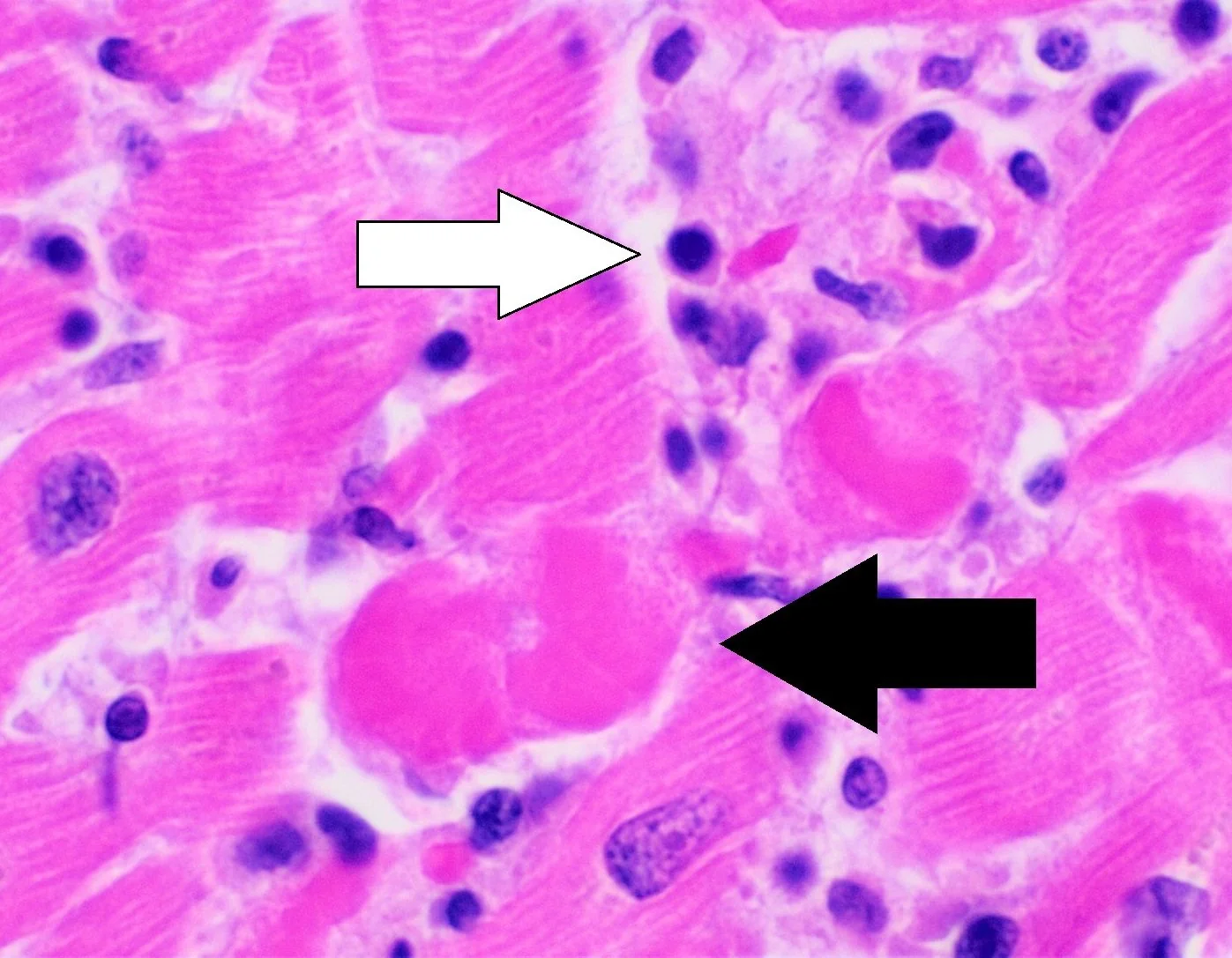
Biopsy remains the gold standard but is reserved for specific situations because of its invasiveness, its sampling error (inflammation is patchy) and the small risk of perforation. Indications include fulminant or rapidly deteriorating disease, unexplained heart failure with ventricular arrhythmia or high-grade AV block, and suspicion of a specific treatable diagnosis such as giant cell myocarditis, cardiac sarcoidosis or eosinophilic myocarditis, where a positive result changes management by justifying immunosuppression.
Aetiological testing
- Viral serology - of limited value in practice, since a positive result rarely alters management
- Lyme serology - where there is AV block or a relevant exposure history
- Autoimmune screen - ANA, ANCA, rheumatoid factor
- Serum ACE and imaging for sarcoidosis - including FDG-PET, which is more sensitive than MRI for active cardiac sarcoid
- HIV testing
- Chagas serology - in patients from endemic areas
Differential diagnosis
| Condition | Distinguishing features |
|---|---|
| Acute coronary syndrome | Older patient, cardiovascular risk factors, regional ECG changes, a rapid troponin rise and fall, and obstructive disease on angiography |
| Acute pericarditis | Sharp positional pleuritic pain relieved by sitting forward, widespread saddle ST elevation with PR depression, a rub, and a normal or only mildly raised troponin |
| Takotsubo cardiomyopathy | Emotional or physical trigger, typically a post-menopausal woman, apical ballooning on echocardiography, and a troponin rise modest relative to the extent of wall motion abnormality |
| Pulmonary embolism | Pleuritic pain, hypoxia, VTE risk factors, right heart strain |
| Dilated cardiomyopathy | More insidious onset without an inflammatory phase; myocarditis may in fact be its precursor |
| Sepsis with myocardial depression | Systemic infection with a clear source and a septic physiology |
| Spontaneous coronary artery dissection | Young women, peripartum period, angiographic appearance of dissection |
Management
There is no specific treatment for viral myocarditis. Management is supportive, directed at the complications, and at avoiding harm during the acute inflammatory phase.
General measures
- Admission and cardiac monitoring - all patients with a raised troponin, arrhythmia, ventricular dysfunction or ongoing symptoms should be monitored, since arrhythmia is unpredictable in the acute phase
- Rest and restriction from exercise - see below; this is the most important piece of advice given
- Treat the underlying cause where one exists - antibiotics for Lyme disease, corticosteroids for cardiac sarcoidosis, withdrawal of a causative drug, immunosuppression for giant cell or eosinophilic myocarditis
- Avoid NSAIDs where there is ventricular dysfunction - they promote sodium and water retention, worsen renal function in heart failure, and were associated with harm in animal models of myocarditis. Where there is significant pericardial involvement and normal ventricular function, an NSAID with colchicine may be used with caution.
Treating heart failure
Standard therapy applies: an ACE inhibitor or ARB, a beta-blocker once the patient is not acutely decompensated, a mineralocorticoid receptor antagonist and an SGLT2 inhibitor, with diuretics for congestion. Anticoagulation is considered where there is severe ventricular dysfunction or a demonstrated mural thrombus.
Treating arrhythmia
Managed conventionally. A temporary pacing wire may be needed for high-grade AV block, which is frequently transient - so permanent pacing decisions are usually deferred, except in sarcoidosis and giant cell myocarditis where the block rarely recovers. Decisions about an ICD are generally postponed until the inflammation has settled and ventricular function has been reassessed, typically at 3-6 months, since function often improves substantially. A wearable cardioverter defibrillator may bridge that interval in high-risk patients.
Fulminant myocarditis
Requires management in a cardiac intensive care unit with inotropic and vasopressor support, and consideration of mechanical circulatory support - an intra-aortic balloon pump, a ventricular assist device or veno-arterial ECMO - as a bridge to recovery or to transplantation. These patients should be discussed early with a transplant centre.
Complications
- Dilated cardiomyopathy - the most important long-term complication. Around 20-30% of patients develop persistent ventricular dysfunction, and myocarditis is thought to account for a substantial minority of cases of dilated cardiomyopathy.
- Chronic heart failure
- Ventricular arrhythmia and sudden cardiac death - the risk is highest during the acute inflammatory phase but persists where myocardial scar remains, and the extent of late gadolinium enhancement on MRI predicts it
- Complete heart block - usually transient, but persistent in sarcoidosis and giant cell myocarditis
- Cardiogenic shock - in fulminant disease
- Mural thrombus and systemic embolism - from a poorly contracting, dilated ventricle
- Pericardial effusion and tamponade - where there is significant pericardial involvement
- Recurrence - uncommon but recognised
Red flags
Prognosis
The majority of patients with acute viral myocarditis and preserved ventricular function recover completely, and the outlook for this group - which includes most young patients presenting with chest pain and a raised troponin - is excellent.
Roughly 20-30% develop persistent left ventricular dysfunction, and myocarditis is an important antecedent of dilated cardiomyopathy. The strongest adverse prognostic markers are a reduced ejection fraction at presentation, NYHA class III-IV symptoms, the extent and persistence of late gadolinium enhancement on cardiac MRI, and sustained ventricular arrhythmia. Because of this, follow-up echocardiography and repeat MRI at several months are routine.
Fulminant myocarditis carries a high in-hospital mortality but, counterintuitively, those who survive the acute illness with mechanical support have a better long-term prognosis than patients with a more indolent presentation - the intense inflammatory response tends to resolve completely rather than smouldering on into chronic dysfunction.
Giant cell myocarditis is the exception in every respect: it is rapidly progressive, and median survival without immunosuppression or transplantation is measured in months. It is the principal reason biopsy retains a role, since the diagnosis fundamentally changes management. Cardiac sarcoidosis likewise carries a distinct prognosis and responds to corticosteroids, making it a diagnosis worth actively pursuing rather than assuming viral aetiology in a patient with heart block and ventricular arrhythmia.
References
- Ferreira VM, Schulz-Menger J, Holmvang G et al. Cardiovascular magnetic resonance in nonischemic myocardial inflammation: expert recommendations (updated Lake Louise criteria). Journal of the American College of Cardiology. 2018. Available here
- Caforio ALP, Pankuweit S, Arbustini E et al. Current state of knowledge on aetiology, diagnosis, management and therapy of myocarditis: ESC Working Group position statement. European Heart Journal. 2013. Available here
- NICE NG106. Chronic heart failure in adults: diagnosis and management. 2018. Available here
- NICE NG185. Acute coronary syndromes. 2020, updated 2025. Available here
- NICE NG95. Lyme disease. 2018. Available here
- Zeppenfeld K, Tfelt-Hansen J, de Riva M et al. 2022 ESC Guidelines for the management of patients with ventricular arrhythmias and the prevention of sudden cardiac death. European Heart Journal. 2022. Available here
- MHRA Drug Safety Update. Clozapine: monitoring for myocarditis and cardiomyopathy. Available here
- Lyon AR, Lopez-Fernandez T, Couch LS et al. 2022 ESC Guidelines on cardio-oncology. European Heart Journal. 2022. Available here
This article is written for revision and education. It is not clinical guidance and must not be used to make decisions about the care of a patient. Always check current NICE guidance and local protocols.