
Carotid Artery Disease
Key points
- Carotid artery disease: atherosclerotic stenosis at the carotid bifurcation and the origin of the internal carotid artery, responsible for roughly 15 to 20% of ischaemic strokes.
- Mechanism: artery-to-artery thromboembolism from an ulcerated plaque, rather than haemodynamic hypoperfusion, causes the great majority of events.
- Amaurosis fugax: painless transient monocular visual loss, classically described as a curtain descending, from embolism to the retinal circulation via the ophthalmic artery.
- Symptomatic disease: stenosis with an ipsilateral TIA, stroke or retinal event in the preceding six months. Asymptomatic disease is managed very differently.
- Carotid bruit: an unreliable sign. It may be absent in a near-occlusion, and present with a haemodynamically insignificant stenosis.
- First-line imaging: urgent carotid duplex ultrasound, confirmed by CT or MR angiography before any intervention.
- Surgery: carotid endarterectomy for symptomatic stenosis of 50% or more by NASCET criteria, ideally within seven days of the index event.
- Why speed matters: the risk of recurrent stroke after a TIA is highest in the first few days, so the benefit of endarterectomy falls sharply with delay.
Introduction and pathophysiology
Atherosclerotic plaque forms preferentially at the carotid bulb, the dilated segment at the origin of the internal carotid artery, where flow separates and shear stress is low and oscillatory. Plaque here is responsible for around 15 to 20% of all ischaemic strokes, and is one of the few causes of stroke that can be treated surgically.
The key conceptual point is that carotid stenosis usually causes stroke by embolism, not by starvation of flow. An ulcerated or fissured plaque exposes thrombogenic material to the bloodstream, platelet-fibrin thrombus forms on its surface, and fragments break off and travel distally into the middle cerebral or ophthalmic territory.
- Artery-to-artery thromboembolism - the dominant mechanism. It explains why the symptoms are focal and abrupt, and why an unstable plaque of moderate stenosis can be more dangerous than a smooth tight one.
- Plaque rupture with acute thrombosis and occlusion
- Haemodynamic hypoperfusion - relatively rare, requiring a tight stenosis plus an incomplete circle of Willis, and typically producing watershed infarction between vascular territories
- Carotid dissection - a separate entity, more common in younger patients, presenting with neck pain, a partial Horner syndrome and stroke, often after minor trauma or neck manipulation
The most important classification in the whole topic is symptomatic versus asymptomatic. Symptomatic disease means an ipsilateral TIA, stroke or retinal artery event within the previous six months. It carries a high early risk of recurrence and a clear benefit from surgery. Asymptomatic disease carries a much lower annual risk on modern medical therapy, and the case for intervention is correspondingly weaker.
Risk factors
- Increasing age and male sex
- Smoking
- Hypertension - the strongest modifiable risk factor for stroke overall
- Diabetes mellitus
- Hyperlipidaemia
- Atherosclerosis elsewhere - coronary artery disease, peripheral arterial disease, abdominal aortic aneurysm
- Chronic kidney disease
- Previous neck irradiation, which produces a diffuse fibrotic stenosis that is technically difficult to operate on
- Family history of stroke or premature vascular disease
Clinical features
Territory-specific symptoms
| Carotid (anterior circulation) | Vertebrobasilar (posterior circulation) |
|---|---|
| Unilateral weakness or sensory loss of the face, arm or leg, contralateral to the diseased artery | Bilateral or alternating motor or sensory symptoms |
| Dysphasia, if the dominant hemisphere is affected | Dysarthria and dysphagia |
| Amaurosis fugax - transient painless monocular visual loss, ipsilateral to the diseased artery | Homonymous hemianopia or cortical blindness |
| Homonymous hemianopia from middle cerebral territory involvement | Vertigo, ataxia, diplopia, and crossed signs |
| Higher cortical dysfunction - neglect, apraxia | Drop attacks and loss of consciousness |
Amaurosis fugax deserves particular attention because it is the symptom most specific to carotid disease. The ophthalmic artery is the first branch of the internal carotid, so an embolus travelling up the carotid may lodge in the retinal circulation. The patient describes painless loss of vision in one eye, often like a curtain or shade coming down, lasting seconds to minutes and then clearing completely. On fundoscopy a bright refractile Hollenhorst plaque may occasionally be seen at a retinal arteriolar bifurcation.
The crucial anatomical point is the crossed pattern: because the ophthalmic artery arises before the internal carotid supplies the hemisphere, carotid disease causes ipsilateral visual symptoms and contralateral limb symptoms. A patient with right-sided amaurosis fugax and left-sided weakness has right carotid disease.
Examination
- Full neurological examination, with an NIHSS score if there is a deficit
- Carotid auscultation for a bruit, remembering that it is a poor test - sensitivity and specificity are both around 50 to 60%, and a bruit may disappear as a stenosis becomes critical because flow across it falls
- Blood pressure in both arms, and cardiovascular examination for atrial fibrillation, murmurs and heart failure
- Fundoscopy for retinal emboli and hypertensive or diabetic changes
- Peripheral pulses and abdominal palpation for coexisting arterial disease and aneurysm
- Look for signs of dissection - neck pain, a partial Horner syndrome with ptosis and miosis but preserved sweating, and a history of trauma or neck manipulation
Investigations
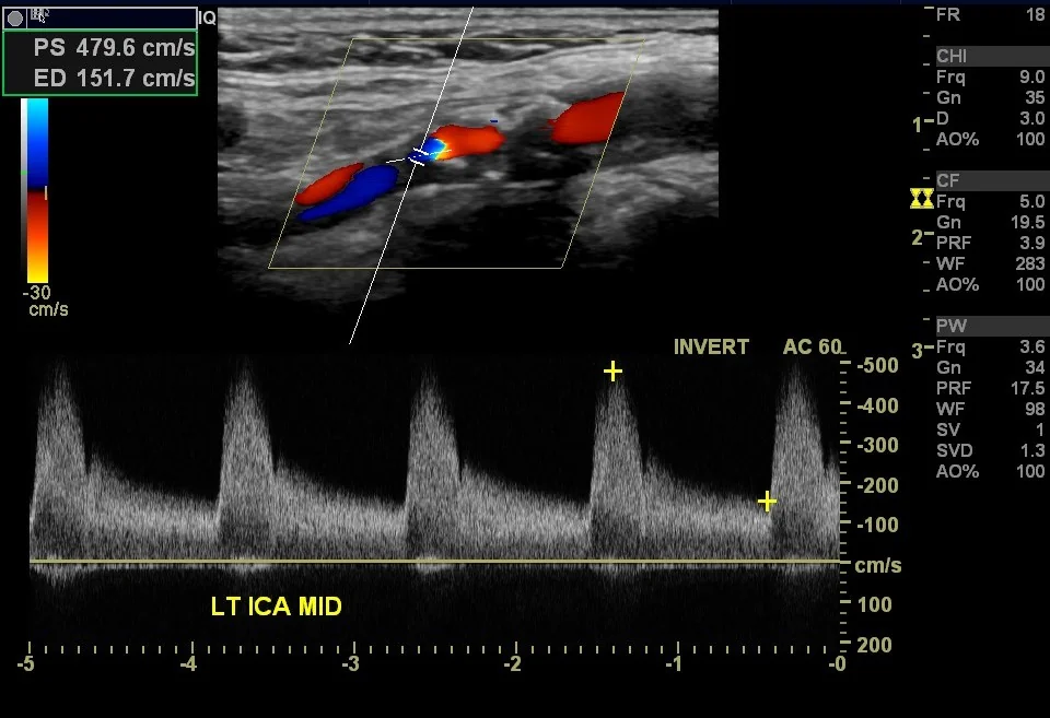
- Carotid duplex ultrasound - first line, non-invasive and quick. It grades stenosis by peak systolic velocity across the lesion as well as by direct imaging, and assesses plaque morphology. In a patient with a TIA it should be performed urgently, within days.
- CT angiography or MR angiography - used to confirm the duplex finding and to define the anatomy before intervention, since operating on the basis of duplex alone risks acting on an overestimate. They also distinguish a near-occlusion from a complete occlusion, which is a crucial distinction because an occluded artery is not operated on.
- Brain imaging - CT head immediately in suspected stroke to exclude haemorrhage before thrombolysis; diffusion-weighted MRI is preferred in TIA, since it identifies infarction in a substantial proportion of patients whose symptoms have resolved
- ECG and prolonged cardiac monitoring - to detect atrial fibrillation, which is a competing and commoner cause of embolic stroke and changes treatment from antiplatelet to anticoagulant
- Echocardiography where a cardiac source is suspected
- Bloods - FBC, U&Es, glucose and HbA1c, lipid profile, clotting, ESR (to consider giant cell arteritis in visual loss), and thrombophilia or vasculitis screen in the young
Management
Immediate management of a TIA or minor stroke
- Aspirin 300 mg immediately unless contraindicated, and continued until assessed
- Specialist assessment within 24 hours for anyone with a suspected TIA in the preceding week. NICE explicitly advises against using the ABCD2 score to stratify who is seen urgently - all suspected TIAs are assessed urgently.1
- Brain imaging, and urgent carotid imaging in anyone who would be a candidate for carotid intervention
- Exclude mimics - hypoglycaemia, seizure with Todd paresis, migraine aura, and giant cell arteritis in transient visual loss
- Advise not to drive until assessed. Group 1 licence holders must not drive for at least one month after a TIA or stroke and need not notify the DVLA if recovery is complete; Group 2 holders must notify and are disqualified for a year. Check current DVLA guidance.2
Medical therapy for all patients
Best medical therapy has improved considerably, and it is the reason the case for operating on asymptomatic stenosis has weakened over the last two decades.
- Antiplatelet therapy - clopidogrel 75 mg daily is the usual long-term choice after a TIA or ischaemic stroke. NICE supports a short course of dual antiplatelet therapy with clopidogrel and aspirin for high-risk TIA and minor stroke, followed by clopidogrel alone.
- Anticoagulation instead of antiplatelet therapy if atrial fibrillation is found - the two mechanisms are competing explanations and it matters which is acted on
- A high-intensity statin, atorvastatin 80 mg daily
- Blood pressure control, introduced carefully in the days after an acute stroke but pursued firmly thereafter
- Smoking cessation, diabetes optimisation, weight and alcohol management, and exercise
Carotid endarterectomy
The plaque is removed through an incision along the anterior border of sternocleidomastoid, with the artery clamped and either a shunt inserted or the operation performed under regional anaesthesia so that cerebral function can be monitored directly. The arteriotomy is usually closed with a patch to reduce restenosis.
| Group | Stenosis | Recommendation |
|---|---|---|
| Symptomatic - ipsilateral TIA, stroke or retinal event within 6 months | 50 to 99% | Offer carotid endarterectomy, ideally within 7 days of the index event, alongside best medical therapy1 |
| Symptomatic | Under 50% | Best medical therapy alone; surgery is not beneficial |
| Symptomatic | Complete occlusion | No intervention - the vessel cannot be reopened safely and the embolic source is gone |
| Asymptomatic | 60 to 99% | Individualised decision in a specialist multidisciplinary setting. Benefit over modern medical therapy is small and depends on a low institutional complication rate and a long life expectancy. |
| Asymptomatic | Under 60% | Best medical therapy alone |
The evidence base is the NASCET and ECST trials, which together showed a clear reduction in ipsilateral stroke with endarterectomy in symptomatic severe stenosis, and a smaller but real benefit in moderate stenosis.3 Pooled reanalyses demonstrated that the benefit is highly time-dependent: the number needed to treat to prevent one stroke is around 5 if surgery is performed within two weeks of symptoms, but rises steeply thereafter, and by 12 weeks there is little benefit at all. This is the reason for the seven-day target.
Carotid artery stenting
An endovascular alternative in which a stent is deployed across the stenosis, usually with an embolic protection device. It avoids a neck incision and cranial nerve injury, and is useful in a hostile neck after radiotherapy or previous surgery, in high lesions that are surgically inaccessible, and in patients with severe cardiac comorbidity.
Randomised trials, including CREST and the ICSS study, found a higher periprocedural stroke risk with stenting and a higher periprocedural myocardial infarction risk with endarterectomy, with similar long-term outcomes. The excess stroke risk with stenting is concentrated in patients over about 70, so endarterectomy remains the default in older patients.
Complications of untreated disease
- Ischaemic stroke, with the associated mortality, disability, and loss of independence
- Recurrent TIA, and the cumulative cognitive effect of multiple small infarcts
- Permanent monocular blindness from central retinal artery occlusion
- Vascular dementia, from accumulated small vessel and embolic damage
- Complete carotid occlusion, which paradoxically removes the embolic source but may cause a watershed infarct at the time it occurs
- Death - stroke remains a leading cause of death and the leading cause of acquired adult disability in the UK
Red flags
Prognosis
The natural history of symptomatic carotid stenosis is front-loaded. The risk of stroke after a TIA is highest in the first 48 hours to two weeks, with several per cent of patients suffering a completed stroke within that window, and it then falls. This is why the whole service model - urgent assessment, urgent imaging, and surgery within seven days - is built around speed rather than around thoroughness.
With endarterectomy plus best medical therapy, the risk of ipsilateral stroke in symptomatic severe stenosis falls substantially compared with medical therapy alone, and the benefit persists for years. In asymptomatic disease the annual stroke risk on modern medical therapy has fallen to around 1% or less, which is why the older trial results supporting surgery are widely regarded as no longer directly applicable and why practice has become more conservative.
The transferable point is that carotid stenosis is a marker of systemic atherosclerosis. Whatever happens to the artery in the neck, these patients remain at substantial risk of myocardial infarction and vascular death, and the statin, the antiplatelet and the blood pressure control matter to their survival at least as much as the operation does.
References
- NICE NG128. Stroke and transient ischaemic attack in over 16s: diagnosis and initial management. 2019, updated 2022. Available here
- DVLA. Assessing fitness to drive: a guide for medical professionals. Available here
- North American Symptomatic Carotid Endarterectomy Trial Collaborators. Beneficial effect of carotid endarterectomy in symptomatic patients with high-grade carotid stenosis. New England Journal of Medicine. 1991. Available here
This article is written for revision and education. It is not clinical guidance and must not be used to make decisions about the care of a patient. Always check current NICE guidance and local protocols.