
Giant Cell Arteritis: Diagnosis and Management
Key points
- Giant cell arteritis (GCA): a large and medium vessel vasculitis affecting the extracranial branches of the carotid artery, almost always in people over 50, with a strong association with polymyalgia rheumatica.
- Classic triad: new headache (often temporal), scalp tenderness, and jaw claudication - pain on chewing that eases with rest.
- Medical emergency: any visual disturbance - amaurosis fugax, diplopia, or sudden visual loss - signals imminent or actual anterior ischaemic optic neuropathy, which can cause permanent blindness within hours.
- Treat before confirming: start high-dose corticosteroids immediately on clinical suspicion - do not wait for biopsy or imaging results, since delay risks irreversible vision loss.
- Investigations: markedly raised ESR/CRP, and either temporal artery ultrasound (showing the 'halo sign') or temporal artery biopsy to confirm the diagnosis after treatment has started.
- Visual symptoms: treat as an emergency - IV methylprednisolone and same-day ophthalmology review.
- Steroid-sparing therapy: tocilizumab (anti-IL-6) reduces relapse and cumulative steroid exposure in patients who need it, alongside a slow steroid taper over 1-2 years.
- Long-term risk: aortic aneurysm and dissection - large vessel involvement warrants imaging surveillance.
Introduction
Giant cell arteritis (GCA), also known as temporal arteritis, is a granulomatous large and medium vessel vasculitis that predominantly affects the extracranial branches of the carotid artery - classically the superficial temporal artery - in people over the age of 50. It is the commonest vasculitis in this age group and is strongly linked to polymyalgia rheumatica, with which it shares an overlapping population and, in many patients, coexists.1
GCA is one of the true medical emergencies in rheumatology, not because it is immediately life-threatening in most cases, but because untreated inflammation of the ophthalmic and posterior ciliary arteries can cause sudden, irreversible blindness. This single fact governs the whole approach to management: treatment starts on clinical suspicion, before any confirmatory test result is back.
Aetiology
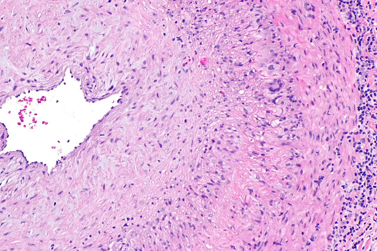
GCA is a granulomatous vasculitis in which T cells and macrophages infiltrate the vessel wall, forming giant cells and driving intimal hyperplasia that narrows or occludes the vessel lumen. It shows a striking predilection for the extracranial branches of the carotid artery, though large vessel involvement of the aorta and its major branches occurs in a significant minority and is increasingly recognised with modern imaging.
- Age over 50 - the disease essentially does not occur below this age
- Increasing incidence with age, peaking in the 70s and 80s
- Female sex (roughly 2-3x)
- Northern European / Scandinavian ancestry
- Polymyalgia rheumatica - around 40-60% of patients with GCA have coexisting PMR features, and a smaller proportion of PMR patients go on to develop GCA
Clinical features
- New headache - typically unilateral, temporal, and a distinct change from the patient's usual pattern
- Scalp tenderness - classically noticed when combing hair or resting the head on a pillow
- Jaw claudication - pain in the jaw or tongue on chewing, which eases with rest; highly specific when present
- Temporal artery abnormality - tenderness, thickening, or reduced/absent pulsation on palpation
- Constitutional features - fever, malaise, weight loss, night sweats, overlapping with PMR
- Symptoms of coexisting PMR - bilateral shoulder and hip girdle pain and stiffness (see the polymyalgia rheumatica article)
Visual symptoms - the emergency
- Amaurosis fugax - transient monocular visual loss, a warning sign that permanent loss may follow imminently
- Diplopia
- Sudden, painless, permanent visual loss - from anterior ischaemic optic neuropathy (AION), caused by occlusion of the posterior ciliary arteries supplying the optic nerve head. Once established, visual loss is usually irreversible, and the fellow eye is at high risk within days if treatment is not started
Large vessel involvement
Aortitis and involvement of the subclavian, axillary or other large arteries can cause limb claudication, asymmetrical blood pressures, absent pulses, and bruits - and predisposes to aortic aneurysm formation over subsequent years.
Differential diagnosis
- Migraine and tension headache - lack the systemic features, raised inflammatory markers and age profile of GCA
- Trigeminal neuralgia - brief, electric, stimulus-triggered pain rather than a constant headache with scalp tenderness
- Other primary vasculitides (Takayasu arteritis - typically under 40, ANCA-associated vasculitis)
- Multiple myeloma or other malignancy - can cause a raised ESR and constitutional symptoms in an older patient; consider if the response to steroids is atypical
- Retinal artery occlusion of another cause - always ask about GCA symptoms in any older patient with sudden visual loss
Investigations
Investigations confirm the diagnosis but must never delay treatment - start corticosteroids first, on the basis of the clinical picture, and investigate in parallel or immediately after.2
- ESR and CRP - typically markedly raised (ESR often above 50 mm/h); characteristic but a normal ESR does not fully exclude GCA if clinical suspicion is high
- FBC - normocytic anaemia and thrombocytosis are common
- LFTs - a raised alkaline phosphatase is a recognised, if non-specific, association
- Temporal artery ultrasound - now recommended as a first-line imaging investigation where available; shows the 'halo sign', a hypoechoic ring around the arterial lumen caused by wall oedema
- Temporal artery biopsy - remains the traditional gold standard, but skip lesions mean a segment of at least 1 cm should be taken, and a negative biopsy does not exclude the diagnosis if clinical suspicion remains high
- PET-CT or CT/MR angiography - used where large vessel involvement is suspected (limb claudication, asymmetrical pulses/blood pressures, unexplained aortic dilatation)
Management
Start high-dose corticosteroids immediately on clinical suspicion of GCA, without waiting for biopsy or imaging confirmation.3
| Presentation | Treatment |
|---|---|
| No visual symptoms | Oral prednisolone 40-60 mg daily |
| Visual symptoms (amaurosis fugax, diplopia, visual loss) or jaw/tongue claudication | IV methylprednisolone (typically 500 mg-1 g daily for up to 3 days), plus same-day ophthalmology review |
Once symptoms and inflammatory markers have settled, prednisolone is tapered slowly, typically over 1-2 years, guided by symptoms and CRP/ESR rather than a fixed schedule, since relapse during tapering is common.
- Tocilizumab (anti-IL-6 receptor) - NICE-approved as a steroid-sparing agent, shown to reduce relapse rates and cumulative steroid exposure, particularly for relapsing or refractory disease
- Methotrexate - an alternative steroid-sparing option where tocilizumab is unsuitable
- Low-dose aspirin - considered in some patients to reduce the risk of ischaemic complications, though the evidence is mixed and it is not universally recommended
- Bone protection - bisphosphonate, calcium and vitamin D, given the near-certainty of prolonged high-dose steroid exposure
- Gastroprotection with a proton pump inhibitor while on high-dose steroids
Investigating for large vessel involvement
Because large vessel GCA carries a long-term risk of aortic aneurysm and dissection, patients with confirmed disease, particularly those with large vessel features at diagnosis, are usually offered periodic imaging surveillance (chest X-ray or CT) over subsequent years.
Complications
The most feared complication is irreversible visual loss from AION, occurring in a meaningful minority of untreated or late-treated patients. Cerebrovascular events (stroke) can occur from involvement of intracranial-supplying vessels. Long-term, aortic aneurysm formation and dissection are recognised complications of large vessel involvement. Treatment itself carries substantial burden - osteoporosis, diabetes, weight gain, cataracts, infection risk and adrenal suppression from prolonged high-dose corticosteroids, which is precisely why steroid-sparing agents are used where relapse or prolonged treatment is anticipated.
Red flags
Prognosis
With prompt treatment, most cranial symptoms resolve rapidly and further visual loss is prevented, though any vision already lost to AION rarely recovers. Relapse during steroid tapering is common, affecting a substantial proportion of patients, which is why steroid-sparing therapy has become increasingly central to management. Long-term outcome is otherwise generally good, though patients require ongoing monitoring both for disease relapse and for the cumulative effects of corticosteroid treatment.
References
- NICE Clinical Knowledge Summaries. Giant cell arteritis. Available here
- British Society for Rheumatology. Guideline for the management of giant cell arteritis. Rheumatology. 2020. Available here
- Hellmich B, Agueda A, Monti S et al. 2018 update of the EULAR recommendations for the management of large vessel vasculitis. Annals of the Rheumatic Diseases. 2020. Available here
- NICE TA573. Tocilizumab for treating giant cell arteritis. Available here
- Ponte C, Grayson PC, Robson JC et al. 2022 ACR/EULAR classification criteria for giant cell arteritis. Annals of the Rheumatic Diseases. 2022. Available here
This article is written for revision and education. It is not clinical guidance and must not be used to make decisions about the care of a patient. Always check current NICE guidance and local protocols.