
Varicose Veins and Chronic Venous Insufficiency
Key points
- Varicose veins: dilated, tortuous, elongated superficial veins, caused by valvular incompetence allowing reflux down the superficial system.
- The underlying problem: ambulatory venous hypertension - failure of the calf muscle pump and valves to lower venous pressure during walking.
- Anatomy: the great saphenous vein runs medially from in front of the medial malleolus to the saphenofemoral junction; the small saphenous runs behind the lateral malleolus up the posterior calf to the popliteal fossa.
- Primary versus secondary: around 95% are primary. Secondary causes are previous deep vein thrombosis, pelvic masses, pregnancy and arteriovenous malformation.
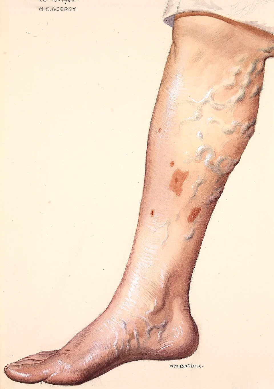
- Skin changes: varicose eczema, haemosiderin staining, lipodermatosclerosis producing the inverted champagne bottle leg, atrophie blanche and ulceration.
- Venous ulcer: shallow, in the gaiter area above the medial malleolus, with sloping edges and a granulating base, and relatively painless compared with an arterial ulcer.
- Before any compression: measure the ABPI. Compressing a leg with arterial disease can cause tissue necrosis.
- NICE order of treatment: endothermal ablation first, then ultrasound-guided foam sclerotherapy if that is unsuitable, then open surgery if neither is possible.
Anatomy and physiology
Venous return from the leg works against gravity, and depends on three things: the calf muscle pump, which squeezes deep veins during walking; one-way valves, which prevent blood falling back down; and perforating veins, which direct flow from the superficial to the deep system and not the reverse.
When these work, the venous pressure at the ankle falls from around 90 mmHg on standing still to roughly 20 to 30 mmHg during walking. When valves fail, blood refluxes on each step and the pressure barely falls. This sustained ambulatory venous hypertension is the single pathological process behind everything in this topic, from aching legs to ulceration.
- Great (long) saphenous vein - arises from the medial end of the dorsal venous arch, passes in front of the medial malleolus, runs up the medial calf and thigh, and drains into the femoral vein at the saphenofemoral junction, about 4 cm inferolateral to the pubic tubercle
- Small (short) saphenous vein - arises from the lateral end of the dorsal venous arch, passes behind the lateral malleolus, runs up the midline of the posterior calf and pierces the deep fascia to join the popliteal vein at the saphenopopliteal junction
- Deep veins - the anterior and posterior tibial, peroneal, popliteal and femoral veins, carrying about 90% of the venous return
- Perforating veins - connect superficial to deep, with valves directing flow inwards. The medial calf perforators are the ones classically implicated in gaiter-area ulceration.
The saphenous veins run superficial to the deep fascia, which is why they can be seen and why they dilate when incompetent, and it is also why they are safe to remove: the deep system carries the great majority of the venous return.
Pathophysiology
- Valvular incompetence develops, either from an intrinsic weakness of the vein wall that allows dilatation until the valve cusps no longer meet, or from destruction of valves by a previous thrombosis
- Reflux occurs down the superficial system on standing and during the relaxation phase of walking
- Ambulatory venous hypertension results, since the calf pump cannot lower the pressure in a column of blood that keeps refilling from above
- Capillary hypertension develops in the skin of the gaiter area, where the pressure is highest, with capillary elongation and increased permeability
- Extravasation of red cells and protein - haemoglobin breaks down to haemosiderin, staining the skin brown, and fibrinogen forms pericapillary fibrin cuffs that impair oxygen diffusion
- Leucocyte trapping and activation in the congested microcirculation releases proteases and free radicals, producing chronic inflammation
- Fibrosis of the dermis and subcutaneous fat (lipodermatosclerosis), and eventually skin breakdown into a venous ulcer
Primary and secondary disease
- Primary (about 95%) - intrinsic vein wall weakness with progressive valvular incompetence and no identifiable precipitant
- Secondary to previous deep vein thrombosis - the post-thrombotic syndrome, where deep valves are destroyed and deep veins may remain obstructed. This produces the most severe venous hypertension and the most refractory ulceration.
- Secondary to obstruction - a pelvic or abdominal mass, pregnancy, or May-Thurner compression of the left common iliac vein by the right common iliac artery
- Secondary to an arteriovenous fistula or malformation, including congenital syndromes such as Klippel-Trenaunay
Risk factors and classification
- Increasing age and female sex
- Pregnancy - progesterone-mediated venous relaxation, increased circulating volume and mechanical compression by the uterus. Varicosities appearing in pregnancy often regress after delivery, so intervention is usually deferred.
- Family history, which is strong and suggests a heritable connective tissue component
- Obesity, particularly in women
- Prolonged standing occupations
- Previous deep vein thrombosis or leg fracture
- Previous varicose vein surgery, since recurrence is common
| Class | Findings |
|---|---|
| C0 | No visible or palpable signs of venous disease |
| C1 | Telangiectasia (thread veins) or reticular veins |
| C2 | Varicose veins |
| C3 | Oedema |
| C4a | Pigmentation (haemosiderin staining) or varicose eczema |
| C4b | Lipodermatosclerosis or atrophie blanche |
| C5 | Healed venous ulcer |
| C6 | Active venous ulcer |
The full CEAP system also records aetiology, anatomy and pathophysiology, but the clinical class is the part used day to day, and C4 and above are the classes that mandate referral to a vascular service.
Clinical features
Symptoms
- Aching, heaviness and throbbing in the legs, worse at the end of the day and on prolonged standing, relieved by elevation - the pattern is the opposite of arterial rest pain, which is relieved by dependency
- Itching, particularly over varicosities and areas of eczema
- Ankle swelling, worse in the evening
- Night cramps and restless legs
- Cosmetic concern, which is a legitimate symptom but is not funded for intervention on the NHS in isolation
- Bleeding from a thin-walled varicosity, which can be surprisingly brisk
Signs of chronic venous insufficiency
- Haemosiderin staining - brown pigmentation of the gaiter area from breakdown of extravasated red cells
- Varicose (venous) eczema - itchy, scaly, erythematous skin, which may become secondarily infected or develop contact sensitisation to topical treatments
- Lipodermatosclerosis - fibrosis and induration of the skin and subcutaneous fat, producing a tight, woody, narrowed lower calf above a swollen ankle. The classic description is an inverted champagne bottle leg.
- Atrophie blanche - smooth white atrophic scarred plaques with surrounding telangiectasia, marking previous ulceration
- Oedema, initially pitting and later firm
- Venous ulceration, most often in the gaiter area above the medial malleolus
| Venous | Arterial | Neuropathic | |
|---|---|---|---|
| Site | Gaiter area, especially above the medial malleolus | Pressure points, tips of toes, heel, lateral malleolus | Under the metatarsal heads and other weight-bearing points |
| Edge | Sloping, shallow, irregular | Punched out, sharply defined, deep | Punched out with surrounding callus |
| Base | Granulating, often with slough and exudate | Pale, necrotic, may expose tendon or bone | Variable, often clean |
| Pain | Aching, relieved by elevation | Severe, worse on elevation and at night, relieved by dependency | Painless - the defining feature |
| Surrounding skin | Pigmentation, eczema, lipodermatosclerosis, oedema | Cool, pale, hairless, shiny, absent pulses | Warm with palpable pulses, callus, deformity, dry skin |
| ABPI | Normal, usually above 0.9 | Reduced, usually below 0.8 | Often normal, but may be falsely high if calcified |
| Treatment | Compression | Revascularisation - compression is contraindicated | Offloading, debridement, glycaemic control |
Examination
- Examine the patient standing, in good light, with both legs fully exposed
- Inspect the distribution of varicosities, and map whether they follow the great or small saphenous territory
- Look for skin changes in the gaiter area, and for ulcers, including behind the malleoli and between the toes
- Palpate the varicosities, the saphenofemoral junction for a saphena varix, and feel for tenderness suggesting thrombophlebitis
- Assess the arterial supply - palpate all pedal pulses and measure the ABPI, because compression cannot be applied safely without it
- Examine the abdomen and pelvis where secondary causes are suspected
- The classical tourniquet (Trendelenburg) test and the tap and cough impulse tests are described in textbooks, but duplex ultrasound has replaced them entirely in practice. Mention them, then say you would request a duplex.
Investigations
- Duplex ultrasound - the investigation of choice and mandatory before any intervention. It demonstrates reflux (conventionally more than 0.5 seconds of reverse flow on release of calf compression), maps which junctions and perforators are incompetent, confirms deep vein patency and excludes post-thrombotic obstruction.
- ABPI - before any compression therapy, and in every leg ulcer. Compression requires an ABPI above about 0.8.
- Bloods - FBC, HbA1c and albumin where an ulcer is not healing, since anaemia, diabetes and malnutrition all impair healing
- Ulcer swab - only if there are clinical signs of infection. All chronic ulcers are colonised, and swabbing a clean ulcer leads to unnecessary antibiotics.
- Biopsy - any ulcer that fails to heal after 12 weeks of adequate compression, or that has a rolled or everted edge, should be biopsied to exclude malignancy
- Cross-sectional imaging of the abdomen and pelvis where an obstructive secondary cause is suspected
- Patch testing where varicose eczema worsens with topical treatment, since contact allergy to dressings and creams is common
Management
Conservative measures
- Weight loss and regular walking, which activates the calf pump
- Leg elevation above the level of the heart when resting, and avoiding prolonged standing
- Emollients for dry skin, and a short course of a topical corticosteroid for active varicose eczema
- Graduated compression stockings - class 1 or 2, applied in the morning before the leg swells, after the ABPI has been checked. NICE notes that compression hosiery should not be offered as a treatment for varicose veins unless interventional treatment is unsuitable.1
- Skin care and avoidance of trauma, since minor injuries in this skin heal badly
Referral to a vascular service
NICE recommends referral for any of the following:1
- Symptomatic primary or recurrent varicose veins
- Lower limb skin changes thought to be caused by chronic venous insufficiency - pigmentation or eczema
- Superficial vein thrombosis with suspected venous incompetence
- A venous leg ulcer - an ulcer below the knee that has not healed within two weeks
- A healed venous leg ulcer
- Bleeding from a varicosity - refer immediately if bleeding is uncontrolled
Interventional treatment
NICE sets out a clear order of preference, and it is worth learning in that order:1
| Order | Treatment | How it works | Notes |
|---|---|---|---|
| First | Endothermal ablation - radiofrequency or endovenous laser | A catheter within the vein delivers heat, causing thermal injury, thrombosis and fibrosis of the vein | Performed under local tumescent anaesthesia as a day case. Fast recovery, low complication rate. |
| Second | Ultrasound-guided foam sclerotherapy | Sclerosant foam is injected under ultrasound guidance, causing endothelial damage and fibrosis | Used if endothermal ablation is unsuitable. Higher recurrence, and rarely visual disturbance or stroke from foam embolisation through a patent foramen ovale. |
| Third | Surgery - saphenofemoral or saphenopopliteal ligation with stripping and phlebectomies | The junction is ligated and the truncal vein stripped, with avulsion of visible varicosities | Requires general anaesthesia and has a longer recovery. Risk of saphenous or sural nerve injury, haematoma and wound infection. |
Compression hosiery alone is offered only when none of these interventions is suitable. Thread veins and reticular veins are treated for cosmetic reasons only and are not funded on the NHS.
Venous leg ulcer
- Measure the ABPI first. Compression is contraindicated below about 0.8 and must be applied cautiously between 0.8 and 0.9.
- Multi-layer graduated compression bandaging is the cornerstone of treatment, aiming for around 40 mmHg at the ankle, applied by a trained practitioner and changed weekly
- Simple non-adherent dressings. There is no good evidence that any particular dressing improves healing over a simple one, and expensive dressings are frequently used in place of adequate compression.
- Manage the surrounding skin with emollients and treat varicose eczema
- Antibiotics only for clinical infection, not for colonisation or a positive swab
- Early endovenous ablation of superficial reflux. The EVRA trial showed that early intervention in addition to compression heals ulcers significantly faster than compression alone with deferred intervention, so referral should not wait for the ulcer to heal.2
- Compression hosiery long term after healing, because recurrence without it is high
- Pain control, nutritional assessment and treatment of oedema, and consideration of pentoxifylline as an adjunct in refractory cases
Complications
Of the disease
- Venous ulceration, the endpoint of chronic venous hypertension and the commonest cause of leg ulceration in the UK
- Superficial vein thrombosis (thrombophlebitis) - a tender, hard, red cord along a varicosity. It is not benign: it extends into the deep system in a meaningful minority, so consider duplex and anticoagulation where it is extensive or close to the saphenofemoral junction.
- Deep vein thrombosis and pulmonary embolism
- Haemorrhage from a thin-walled varicosity, which can be substantial and is treated by lying the patient flat, elevating the leg and applying direct pressure
- Varicose eczema, contact dermatitis and cellulitis
- Lipodermatosclerosis and permanent skin changes
- Marjolin ulcer - squamous cell carcinoma arising in a chronic ulcer, which is why a non-healing ulcer must be biopsied
Of treatment
- Recurrence, which occurs after every modality and is commoner after foam sclerotherapy than after ablation or surgery
- Nerve injury - the saphenous nerve accompanies the great saphenous vein below the knee and the sural nerve accompanies the small saphenous, so numbness is a recognised complication
- Endovenous heat-induced thrombosis extending into the deep system after ablation
- Skin burns, pigmentation and matting after thermal ablation or sclerotherapy
- Haematoma, wound infection and lymphatic injury after open surgery
- Rarely, deep vein thrombosis, pulmonary embolism, and neurological events after foam sclerotherapy
Red flags
Prognosis
Uncomplicated varicose veins are a chronic but benign condition, and most patients do not progress to skin changes or ulceration. Where they do, the progression is slow and predictable, which is why NICE sets referral thresholds at the point where skin changes appear rather than waiting for an ulcer.
Venous ulceration is the outcome that carries the real burden. It affects around 1% of the UK adult population at some point, consumes a substantial share of community nursing time, and heals in around 70% of patients within six months with adequate compression. Recurrence is the central problem: without long-term compression hosiery and correction of superficial reflux, up to two thirds recur within five years.
Two practical conclusions follow. First, most treatment failure is failure of compression rather than failure of the ulcer to respond - either the bandaging is inadequate, or the patient cannot tolerate it and no one has asked why. Second, patients with an active or healed venous ulcer should be referred for assessment of superficial reflux, because correcting it both speeds healing and reduces recurrence.
References
- NICE CG168. Varicose veins: diagnosis and management. 2013, updated 2024. Available here
- Gohel MS, Heatley F, Liu X et al. A randomized trial of early endovenous ablation in venous ulceration (EVRA). New England Journal of Medicine. 2018. Available here
This article is written for revision and education. It is not clinical guidance and must not be used to make decisions about the care of a patient. Always check current NICE guidance and local protocols.