
Breast Cancer
Key points
- Epidemiology: the commonest cancer in UK women, with around 56,000 new cases a year and a lifetime risk of about one in seven. Around 400 men are diagnosed annually.
- Dominant risk factor: cumulative unopposed oestrogen exposure - early menarche, late menopause, nulliparity, late first pregnancy and combined HRT - together with increasing age.
- Triple assessment: clinical examination, imaging (ultrasound under 40, mammography plus ultrasound over 40) and core biopsy, each scored 1 to 5.
- Commonest histology: invasive ductal carcinoma of no special type, about 70 to 80%. Lobular carcinoma is diffuse, more often bilateral and frequently invisible on mammography.
- Receptor status: every tumour is tested for oestrogen receptor, progesterone receptor and HER2. This determines endocrine therapy and anti-HER2 therapy and carries prognostic weight.
- Screening: the NHS Breast Screening Programme invites women aged 50 to 70 for mammography every three years.
- Surgery: wide local excision with radiotherapy gives equivalent survival to mastectomy in suitable tumours. The axilla is staged by sentinel lymph node biopsy.
- Endocrine therapy: tamoxifen before the menopause, an aromatase inhibitor after it, for at least five years in oestrogen receptor positive disease.
Introduction
Breast cancer is the commonest cancer in the UK and the commonest cancer in women, accounting for around 15% of all new cancer diagnoses. Roughly 56,000 women and 400 men are diagnosed each year, and the lifetime risk for a woman is approximately one in seven.1
It is also one of the clearest illustrations of what organised cancer care achieves. Mortality has fallen by around 40% since the late 1980s, despite rising incidence, because of the combined effect of population screening, prompt referral pathways, multidisciplinary team working, and systemic therapies matched to tumour biology.
For finals, the topic divides into four things you must be able to do: identify who needs urgent referral, describe triple assessment, explain what receptor status means for treatment, and outline the surgical and adjuvant options. The pathology detail is secondary to those.
Risk factors
The unifying theme of most modifiable risk is lifetime exposure of breast epithelium to oestrogen.
- Increasing age - the single strongest risk factor. Around 80% of cases occur in women over 50.
- Female sex - though male breast cancer exists and is more likely to be associated with a BRCA2 mutation
- Reproductive history - early menarche (before 12), late menopause (after 55), nulliparity, and first pregnancy after the age of 30
- Hormone replacement therapy - combined oestrogen and progestogen HRT increases risk, with the excess related to duration of use and declining after stopping. Oestrogen-only HRT carries a smaller increase.
- Combined oral contraception - a small increase in risk while taking it, which returns to baseline within about ten years of stopping
- Obesity after the menopause - adipose tissue aromatase converts androgens to oestrogen, so the fat becomes the principal oestrogen source
- Alcohol, with a dose-dependent increase, and smoking
- Previous chest irradiation, particularly mantle radiotherapy for Hodgkin lymphoma in adolescence
- Dense breast tissue on mammography, and previous atypical ductal or lobular hyperplasia or lobular carcinoma in situ
- Previous breast cancer, which raises the risk in the contralateral breast
Family history and genetics
Around 5 to 10% of breast cancers are attributable to a high-penetrance inherited mutation.
- BRCA1 - autosomal dominant, on chromosome 17. Lifetime breast cancer risk around 60 to 70% and ovarian cancer risk around 40%. Tumours are more often triple negative and higher grade.
- BRCA2 - on chromosome 13. Lifetime breast cancer risk of similar magnitude, lower ovarian risk, and an increased risk of male breast cancer, prostate and pancreatic cancer.
- TP53 (Li-Fraumeni syndrome), PTEN (Cowden syndrome) and STK11 (Peutz-Jeghers syndrome) - rarer high-risk syndromes
- Referral for genetic assessment is guided by NICE CG164, based on the number of affected relatives, their age at diagnosis, bilateral disease, male breast cancer, and associated ovarian cancer2
- Management of a confirmed high-risk mutation includes annual MRI surveillance from a younger age, risk-reducing mastectomy and salpingo-oophorectomy, and chemoprevention with tamoxifen or anastrozole
Protective factors are breastfeeding, an early first pregnancy, higher parity and regular physical activity - all of which reduce cumulative ovulatory cycles or oestrogen exposure.
Pathology
In situ disease
- Ductal carcinoma in situ (DCIS) - malignant cells confined within the ducts by an intact basement membrane. It is usually impalpable and detected on screening as microcalcification, and it accounts for around 20% of screen-detected cancers. It is a genuine precursor of invasive cancer and is treated with excision, and radiotherapy after breast-conserving surgery.
- Lobular carcinoma in situ (LCIS) - usually an incidental finding on a biopsy taken for another reason. It is best regarded as a risk marker rather than a precursor, indicating an increased risk of invasive cancer in either breast, and is generally managed with surveillance rather than excision.
Invasive disease
| Type | Share | Features |
|---|---|---|
| Invasive ductal carcinoma of no special type | 70 to 80% | The default type. Forms a discrete mass with a desmoplastic reaction, so it feels hard and is visible on mammography. |
| Invasive lobular carcinoma | 10 to 15% | Cells infiltrate in single files without forming a mass, so it presents as diffuse thickening rather than a lump, is frequently occult on mammography, and is more often multifocal and bilateral. MRI is often needed to define its extent. |
| Tubular, mucinous, cribriform and papillary | Each a few per cent | Special types with a generally better prognosis |
| Inflammatory breast cancer | 1 to 5% | Tumour emboli obstruct dermal lymphatics, producing a red, hot, swollen, oedematous breast with peau d'orange. Frequently misdiagnosed as mastitis. Aggressive, and treated with neoadjuvant chemotherapy first. |
| Paget disease of the nipple | 1 to 3% | Eczematous, itchy, ulcerated change of the nipple that spreads to the areola, caused by intraepidermal spread of malignant cells. An underlying in situ or invasive carcinoma is present in most cases. |
Receptor status
Every invasive tumour is tested for three receptors, and the result dictates systemic treatment more than the histological type does.
- Oestrogen receptor (ER) - positive in around 70 to 80%. ER-positive disease is treated with endocrine therapy and has a better prognosis, though it retains a risk of late recurrence beyond ten years.
- Progesterone receptor (PR) - usually reported alongside ER and adds prognostic information
- HER2 - overexpressed in around 15 to 20%. Historically an adverse prognostic feature, but the availability of trastuzumab and other HER2-directed agents has transformed the outlook.
- Triple negative - ER, PR and HER2 all negative, around 15%. Commoner in younger women, in Black women and in BRCA1 carriers. There is no endocrine or anti-HER2 option, so chemotherapy is the mainstay, and prognosis is worse.
Clinical features
- A painless, hard, irregular, poorly mobile lump - the classic presentation. Around 90% of breast lumps are benign, but pain is a poor discriminator in either direction.
- Skin tethering or dimpling, produced by malignant infiltration shortening the suspensory ligaments of Cooper. Ask the patient to raise their arms or press their hands on their hips to make it visible.
- Peau d'orange - dermal lymphatic obstruction pulling the skin inwards at each hair follicle, giving an orange-peel texture
- Nipple retraction or inversion, particularly if new and unilateral, and nipple distortion or deviation
- Blood-stained or single-duct nipple discharge
- Eczematous nipple change - Paget disease
- Skin ulceration or a fungating mass in advanced disease
- Axillary or supraclavicular lymphadenopathy
- Symptoms of metastatic disease - bone pain and pathological fracture, hypercalcaemia, jaundice and hepatomegaly, breathlessness and pleural effusion, headache and seizures
The classic metastatic sites are bone, lung, liver and brain, with bone the commonest, and lobular carcinoma showing an unusual tendency to metastasise to the peritoneum, retroperitoneum and gastrointestinal tract.
Triple assessment
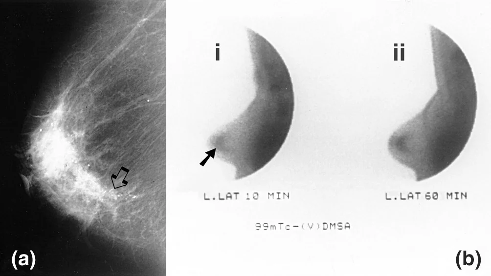
Every patient referred with a suspicious breast symptom undergoes triple assessment, ideally in a one-stop clinic. Each component is independently scored from 1 (normal) to 5 (malignant), prefixed by the modality.
| Component | Prefix | What is done |
|---|---|---|
| Clinical examination | P (palpation) | Full history and examination of both breasts, axillae and supraclavicular fossae, with the findings scored P1 to P5 |
| Imaging | M (mammography) or U (ultrasound) | Ultrasound is first line under 40, because dense young breast tissue makes mammography insensitive. Mammography plus ultrasound over 40. MRI is added for lobular cancers, dense breasts, implants and when planning breast conservation in extensive disease. |
| Pathology | B (core biopsy) or C (cytology) | Core biopsy is preferred because it distinguishes invasive from in situ disease and provides tissue for receptor testing. Fine needle aspiration cytology cannot do either and is now largely reserved for cyst aspiration and node sampling. |
Discordance between the three components is itself a finding. A benign biopsy of a clinically and radiologically suspicious lesion should not be accepted at face value: it should be discussed at the multidisciplinary meeting and the lesion re-sampled or excised.
Staging investigations
- Axillary ultrasound with biopsy of any abnormal node at the time of diagnosis
- Bloods - FBC, U&Es, LFTs, bone profile including calcium
- CT of the chest, abdomen and pelvis, and a bone scan, for patients with clinically node-positive or higher-stage disease, or symptoms suggesting metastasis. Routine staging imaging is not indicated in early node-negative disease.
- Receptor testing on the core biopsy - ER, PR and HER2
- Genetic testing where the age at diagnosis, family history or triple-negative phenotype meets referral criteria
Screening
The NHS Breast Screening Programme invites women aged 50 to 70 for two-view mammography every three years, with self-referral available beyond 70. Around 1 in 100 women screened is found to have cancer, and screening detects cancers at an earlier stage, including impalpable DCIS.
Management
Every case is discussed at a multidisciplinary team meeting comprising surgeons, oncologists, radiologists, pathologists and breast care nurses. Treatment combines local control of the breast and axilla with systemic therapy directed by receptor status.
Surgery to the breast
- Wide local excision (breast-conserving surgery) with adjuvant radiotherapy gives equivalent overall survival to mastectomy for suitable tumours. Suitability depends on the tumour-to-breast size ratio and on the disease being unifocal or accessible in one specimen.
- Mastectomy for large tumours relative to breast size, multifocal disease, extensive DCIS, previous breast radiotherapy, inflammatory cancer, or patient preference including risk-reducing surgery in BRCA carriers
- Reconstruction should be discussed with everyone undergoing mastectomy, whether immediate or delayed, using an implant, an autologous flap such as a deep inferior epigastric perforator (DIEP) or latissimus dorsi flap, or a combination
- Neoadjuvant chemotherapy may shrink a large tumour enough to permit breast conservation, and gives information about how the tumour responds to treatment
Surgery to the axilla
- Pre-operative axillary ultrasound with biopsy of suspicious nodes in everyone
- Sentinel lymph node biopsy where the axilla is clinically and radiologically normal. Blue dye and a radioisotope identify the first draining node or nodes, which are removed and examined. If negative, the rest of the axilla is spared.
- Axillary node clearance where nodal disease is confirmed pre-operatively or on sentinel node biopsy, though axillary radiotherapy is increasingly used instead in selected patients with limited nodal involvement
- Axillary node clearance carries a substantial risk of lymphoedema, in the region of 20%, along with shoulder stiffness, seroma and intercostobrachial nerve injury causing numbness of the inner upper arm. Avoiding unnecessary clearance is the whole point of sentinel node biopsy.
Radiotherapy
- Whole breast radiotherapy after every wide local excision, which reduces local recurrence substantially
- Chest wall radiotherapy after mastectomy where there is node-positive disease, a large tumour or involved margins
- Axillary or supraclavicular fossa radiotherapy where indicated by nodal disease
- Side effects include skin erythema and fatigue acutely, and later fibrosis, lymphoedema, rib fracture and a small increase in cardiac and second-malignancy risk
Endocrine therapy
| Agent | Mechanism | Used in | Key adverse effects |
|---|---|---|---|
| Tamoxifen | Selective oestrogen receptor modulator - antagonist in breast tissue, agonist in endometrium and bone | Pre-menopausal women | Endometrial cancer, venous thromboembolism, hot flushes, vaginal discharge. Report any postmenopausal bleeding urgently. |
| Anastrozole, letrozole, exemestane | Aromatase inhibitors - block peripheral conversion of androgens to oestrogen. Ineffective while the ovaries are producing oestrogen. | Post-menopausal women | Osteoporosis (baseline and monitoring DEXA required), arthralgia and myalgia, hot flushes, dyslipidaemia |
| Goserelin and other GnRH agonists | Ovarian suppression | Pre-menopausal women at higher risk, often combined with an aromatase inhibitor | Menopausal symptoms, bone loss |
| Fulvestrant | Selective oestrogen receptor degrader | Advanced or metastatic disease | Injection site reactions, hot flushes |
Treatment is given for at least five years, extended to ten in higher-risk disease, and reduces both recurrence and mortality. Adherence is the practical problem: a substantial minority stop early because of side effects, so these should be asked about and actively managed rather than assumed to be tolerable.5
Chemotherapy and targeted therapy
- Adjuvant chemotherapy - typically an anthracycline and a taxane, offered where the risk of recurrence justifies it. Genomic assays such as Oncotype DX are used in intermediate-risk ER-positive, node-negative disease to identify women who can safely avoid chemotherapy.
- Neoadjuvant chemotherapy - given before surgery in large, inflammatory, or HER2-positive and triple-negative tumours, to downstage the tumour and assess response
- Trastuzumab and other HER2-directed agents such as pertuzumab and trastuzumab emtansine, for HER2-positive disease. Trastuzumab is cardiotoxic, so left ventricular ejection fraction must be measured at baseline and monitored during treatment, and it should not be given with an anthracycline concurrently.
- Bisphosphonates - adjuvant zoledronic acid reduces bone recurrence and improves survival in postmenopausal women
- PARP inhibitors such as olaparib in BRCA-mutated disease, and immune checkpoint inhibitors in triple-negative disease, are increasingly used
- Metastatic disease is treated with the intent of prolonging life and controlling symptoms, with endocrine therapy, CDK4/6 inhibitors, chemotherapy, targeted agents, bone-directed therapy and palliative radiotherapy
Complications
- Lymphoedema of the arm after axillary clearance or radiotherapy - chronic, distressing and largely irreversible, managed with compression, exercise and specialist lymphoedema services
- Local and regional recurrence, and distant metastasis, which may occur many years later in ER-positive disease
- Seroma, wound infection and flap or implant failure after surgery, and capsular contracture after implant reconstruction
- Intercostobrachial nerve injury causing numbness or neuropathic pain in the inner upper arm, and shoulder stiffness
- Treatment toxicity - endometrial cancer and venous thromboembolism with tamoxifen, osteoporosis and arthralgia with aromatase inhibitors, cardiotoxicity with trastuzumab and anthracyclines, neuropathy with taxanes
- Premature menopause and infertility after chemotherapy, which must be discussed and fertility preservation offered before treatment starts
- Psychological morbidity - anxiety, depression, altered body image and effects on relationships and sexuality, which are common and under-treated
- Complications of metastatic disease - pathological fracture, metastatic spinal cord compression, hypercalcaemia, malignant pleural effusion and brain metastases
Red flags
Prognosis
Overall five-year survival in the UK is around 85%, and around 76% at ten years, but the aggregate figure conceals an enormous spread. Stage at diagnosis dominates: five-year survival is close to 100% for stage 1 disease and around 25 to 30% for stage 4.
The Nottingham Prognostic Index remains a useful bedside summary and is worth being able to state: (0.2 x tumour size in cm) + histological grade (1 to 3) + nodal status score (1 for no nodes, 2 for one to three nodes, 3 for four or more). A score below 3.4 indicates a good prognosis, 3.4 to 5.4 moderate, and above 5.4 poor. Contemporary practice supplements it with online tools such as PREDICT, which incorporate receptor status and estimate the benefit of each treatment option.
Two nuances are worth carrying. ER-positive disease has a better short-term prognosis but a persistent risk of late recurrence beyond ten years, which is why extended endocrine therapy is offered and why discharge from follow-up does not mean the risk is zero. Triple-negative and HER2-positive disease recur earlier if they recur at all, so a patient who is disease-free at five years has largely passed the period of greatest risk. Explaining that difference is often more useful to a patient than quoting a single survival percentage.
References
- Cancer Research UK. Breast cancer statistics. Available here
- NICE CG164. Familial breast cancer: classification, care and managing breast cancer and related risks in people with a family history of breast cancer. 2013, updated 2019. Available here
- NICE NG12. Suspected cancer: recognition and referral. 2015, updated 2023. Available here
- Independent UK Panel on Breast Cancer Screening. The benefits and harms of breast cancer screening: an independent review. The Lancet. 2012. Available here
- NICE NG101. Early and locally advanced breast cancer: diagnosis and management. 2018, updated 2023. Available here
This article is written for revision and education. It is not clinical guidance and must not be used to make decisions about the care of a patient. Always check current NICE guidance and local protocols.