
Systemic Lupus Erythematosus: Diagnosis and Management
Key points
- SLE: a chronic multisystem autoimmune disease caused by loss of tolerance to nuclear antigens, with immune complex deposition driving inflammation in skin, joints, kidneys, serosal surfaces, blood and the nervous system.
- Epidemiology: predominantly affects women of childbearing age (roughly 9:1), and is commoner and more severe in people of Black African-Caribbean and Asian ancestry.
- Screening antibody: ANA is positive in over 95% of SLE - a negative ANA makes the diagnosis very unlikely - but it is not specific.
- Specific antibody: anti-dsDNA is far more specific and its titre tracks disease activity, particularly of lupus nephritis.
- Complement: C3 and C4 fall during an active flare as they are consumed by immune complexes - low complement plus rising anti-dsDNA suggests active disease.
- Background therapy: hydroxychloroquine is given to almost everyone with SLE - it reduces flares and disease damage over time, and requires annual retinal screening.
- Renal involvement: lupus nephritis is a major determinant of long-term outcome - screen with urinalysis and urine protein:creatinine ratio at every review, and biopsy if abnormal.
- Flare versus infection: CRP is often normal in a lupus flare (unlike ESR, which rises) - a high CRP in an unwell lupus patient should raise suspicion of infection.
Introduction
Systemic lupus erythematosus (SLE) is a chronic autoimmune disease in which the immune system loses tolerance to nuclear self-antigens, producing autoantibodies and immune complexes that deposit in tissues throughout the body and drive inflammation wherever they lodge. Because almost any organ can be affected, SLE is notorious for its protean presentation - it is often called 'the great imitator'.1
It predominantly affects women of childbearing age, with a female-to-male ratio of around 9:1, and is both commoner and tends to run a more severe course, particularly with renal involvement, in people of Black African-Caribbean and Asian ancestry.
It is examined heavily because it forces you to hold a wide differential in mind at once - a young woman with joint pain, a rash, and an abnormal urine dip is a classic exam vignette - and because its serology (ANA, anti-dsDNA, complement) is genuinely useful for both diagnosis and monitoring, unlike some autoantibody panels that are tested more by habit than utility.
Aetiology and pathophysiology
SLE arises from a combination of genetic susceptibility and environmental triggers that together break tolerance to nuclear antigens (particularly double-stranded DNA and nuclear proteins). Impaired clearance of apoptotic cell debris exposes the immune system to nuclear material, driving autoantibody production. The resulting immune complexes deposit in tissues - classically the glomerular basement membrane - where they fix complement and recruit inflammatory cells, causing tissue damage. This immune-complex (type III hypersensitivity) mechanism explains why complement levels fall during a flare, as C3 and C4 are consumed.
- Genetic factors - complement deficiencies (C1q, C2, C4), certain HLA alleles, and a strong familial tendency
- UV light - a well-recognised trigger of both skin flares and systemic disease activity
- Epstein-Barr virus infection has been linked to disease onset
- Hormonal factors - oestrogen is thought to contribute to the strong female predominance
- Drugs - certain drugs cause a drug-induced lupus syndrome (see below)
Clinical features
SLE can affect essentially any organ system, often with different combinations of features in different patients and at different times in the same patient.
Constitutional and mucocutaneous
- Fatigue, low-grade fever, weight loss - common and often the earliest symptoms
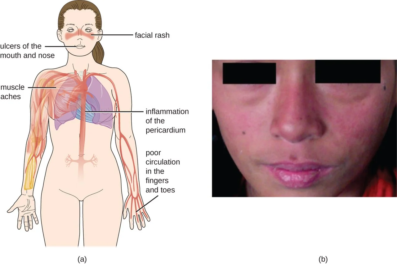
- Malar ('butterfly') rash - erythema over the cheeks and nasal bridge, characteristically sparing the nasolabial folds, often triggered or worsened by sunlight
- Discoid lupus - scarring, plaque-like lesions that can occur with or without systemic disease
- Photosensitivity
- Oral and nasal ulcers, typically painless
- Non-scarring alopecia
Musculoskeletal
Arthralgia and a non-erosive, symmetrical small-joint arthritis are very common. A minority develop Jaccoud's arthropathy - reversible joint deformity from ligamentous laxity rather than erosive bone destruction, which distinguishes it radiographically from rheumatoid arthritis.
Renal
Lupus nephritis develops in up to half of patients and is a major determinant of long-term morbidity. It presents with proteinuria, haematuria and, in more severe classes, hypertension and declining renal function. It is often asymptomatic in its early stages, which is why routine urine screening at every review is essential.
Serosal and cardiopulmonary
- Pleuritis and pleural effusion - the commonest pulmonary manifestation
- Pericarditis - the commonest cardiac manifestation, presenting with pleuritic chest pain
- Libman-Sacks endocarditis - sterile verrucous vegetations, usually on the mitral valve
- Accelerated atherosclerosis - a major contributor to long-term mortality
- Myocarditis and pulmonary hypertension - less common but serious
Neuropsychiatric
A wide range of presentations, from headache and mood disturbance to seizures, psychosis, and cerebrovascular events (the latter often related to coexisting antiphospholipid syndrome). Distinguishing neuropsychiatric lupus from corticosteroid-induced psychosis, or from an unrelated cause, can be genuinely difficult and usually needs specialist input.
Haematological
- Normocytic anaemia of chronic disease, or autoimmune haemolytic anaemia (Coombs-positive)
- Leucopenia, particularly lymphopenia
- Thrombocytopenia
Investigations
Autoantibodies
| Antibody | Sensitivity/specificity | Notes |
|---|---|---|
| ANA | Sensitive (>95%), not specific | Screening test - a negative result makes SLE very unlikely |
| Anti-dsDNA | Specific, moderate sensitivity | Titre correlates with disease activity, particularly lupus nephritis |
| Anti-Sm | Highly specific, low sensitivity | Confirms the diagnosis when positive, but rarely positive |
| Anti-Ro (SSA) / Anti-La (SSB) | Not specific to SLE (also Sjögren syndrome) | Associated with photosensitivity, neonatal lupus and congenital heart block |
| Antiphospholipid antibodies | Not specific to SLE | Anticardiolipin, lupus anticoagulant, anti-beta2-glycoprotein I - screen for secondary APS |
Other laboratory tests
- FBC - cytopenias affecting any or all lineages
- Complement C3 and C4 - fall during active disease as they are consumed by immune complex formation, and are a useful marker of a flare
- ESR and CRP - ESR typically rises with disease activity, but CRP is often normal in an uncomplicated flare; a raised CRP in an unwell lupus patient should prompt a careful search for infection or serositis
- U&Es, urinalysis and urine protein:creatinine ratio - screen for renal involvement at every review
- Renal biopsy - performed if proteinuria, haematuria or declining renal function suggest nephritis, to classify the histological class (I-VI) and guide treatment intensity
- Direct antiglobulin (Coombs) test - if haemolytic anaemia is suspected
Classification criteria
The 2019 EULAR/ACR classification criteria require a positive ANA as an entry criterion, followed by a weighted scoring system across clinical domains (constitutional, haematological, neuropsychiatric, mucocutaneous, serosal, musculoskeletal, renal) and immunological domains (antiphospholipid antibodies, complement, SLE-specific antibodies), with a threshold score confirming classification.2 In practice, diagnosis is clinical, supported by this framework rather than dictated by it.
Differential diagnosis
- Rheumatoid arthritis - overlapping arthralgia, but SLE arthritis is typically non-erosive
- Other connective tissue diseases - mixed connective tissue disease, Sjögren syndrome (often overlaps directly with SLE), systemic sclerosis
- Fibromyalgia - can coexist with SLE and cause diagnostic confusion when fatigue and diffuse pain dominate
- Viral infection (parvovirus B19, acute HIV) - can mimic several features, including arthralgia, rash and cytopenias
- Drug-induced lupus - see above
- Vasculitis - overlapping organ involvement, distinguished by antibody profile and biopsy where needed
Management
Management is stratified by severity and organ involvement, with the goal of controlling disease activity while minimising cumulative treatment toxicity - particularly from long-term corticosteroids.3
General measures
- Sun protection - high-factor sunscreen and avoidance of UV exposure, given its role as a trigger
- Smoking cessation - smoking worsens disease activity and reduces the effectiveness of hydroxychloroquine
- Cardiovascular risk management - blood pressure, lipids and glucose, given the markedly increased cardiovascular risk
- Vaccination - kept up to date, with live vaccines avoided in significantly immunosuppressed patients
Background and mild disease
Hydroxychloroquine is recommended for essentially all patients with SLE unless contraindicated. It reduces the frequency of flares, protects against long-term organ damage accrual, and has a favourable safety profile - its main risk is a dose-dependent retinopathy, requiring baseline and then annual ophthalmological screening.
NSAIDs help musculoskeletal pain and serositis. Topical corticosteroids are used for cutaneous disease.
Moderate to severe disease
Corticosteroids (oral or intravenous, depending on severity) are used for flares, at the lowest effective dose and tapered as quickly as disease activity allows, given the cumulative harms of long-term steroid use.
- Mycophenolate mofetil and azathioprine - used for organ-threatening disease, including lupus nephritis, and as steroid-sparing agents
- Methotrexate - useful for arthritis and cutaneous disease
- Cyclophosphamide - reserved for severe, organ- or life-threatening disease (severe nephritis, CNS lupus), given its toxicity
Biologic therapy
- Belimumab - a monoclonal antibody against B-lymphocyte stimulator (BAFF/BLyS), used as an add-on for active disease despite standard therapy
- Anifrolumab - targets the type I interferon receptor, reflecting the central role of interferon signalling in SLE pathogenesis
- Rituximab - anti-CD20, used off-label for severe or refractory disease, particularly haematological and renal involvement
Lupus nephritis
Treatment intensity is guided by biopsy class. Induction therapy for proliferative nephritis (class III/IV) typically uses mycophenolate mofetil or cyclophosphamide with corticosteroids, followed by a less intensive maintenance regimen. An ACE inhibitor or ARB is added for its antiproteinuric and renoprotective effect.
Complications
Long-term outcomes are shaped by three main threats: progressive renal impairment from lupus nephritis, accelerated cardiovascular disease from chronic inflammation (now a leading cause of death in SLE), and complications of treatment itself, particularly infection from immunosuppression and osteoporosis or avascular necrosis from cumulative corticosteroid exposure. Secondary antiphospholipid syndrome adds a further burden of thrombotic risk.
Red flags
Prognosis
Survival in SLE has improved substantially over recent decades, with modern treatment achieving 10-year survival well above 90% in most cohorts, though outcomes remain worse with significant renal or neuropsychiatric involvement and in some ethnic groups with more severe disease patterns. The disease typically follows a relapsing-remitting course, and long-term management is focused as much on minimising cumulative organ damage and treatment toxicity as on treating individual flares.
References
- NICE Clinical Knowledge Summaries. Systemic lupus erythematosus. Available here
- Aringer M, Costenbader K, Daikh D et al. 2019 European League Against Rheumatism/American College of Rheumatology classification criteria for systemic lupus erythematosus. Annals of the Rheumatic Diseases. 2019. Available here
- Fanouriakis A, Kostopoulou M, Andersen J et al. EULAR recommendations for the management of systemic lupus erythematosus. Annals of the Rheumatic Diseases. 2024. Available here
- Fanouriakis A, Kostopoulou M, Cheema K et al. EULAR recommendations for the management of lupus nephritis. Annals of the Rheumatic Diseases. 2020. Available here
- Kaul A, Gordon C, Crow MK et al. Systemic lupus erythematosus. Nature Reviews Disease Primers. 2016. Available here
This article is written for revision and education. It is not clinical guidance and must not be used to make decisions about the care of a patient. Always check current NICE guidance and local protocols.