
Cardiovascular Disease: Risk Assessment and Prevention
Key points
- Cardiovascular disease: an umbrella term for conditions caused by atherosclerosis of the coronary, cerebral and peripheral arteries.
- Pathophysiology: endothelial injury allows LDL to enter the arterial wall, where it is taken up by macrophages to form a plaque that narrows the lumen or ruptures.
- Presentation: silent for decades, then presents as angina, myocardial infarction, stroke, TIA or intermittent claudication depending on the arterial territory affected.
- Risk factors: modifiable (smoking, hypertension, diabetes, dyslipidaemia, obesity, inactivity) and non-modifiable (age, sex, ethnicity, family history).
- Risk assessment: QRISK3 estimates the 10-year risk of a first myocardial infarction or stroke in people aged 25-84 without established cardiovascular disease.
- Primary prevention: lifestyle change for everyone; offer atorvastatin 20 mg if the QRISK3 score is 10% or more.
- Secondary prevention: atorvastatin 80 mg, antiplatelet therapy and aggressive risk factor control in anyone with established disease.
- Monitoring: recheck the lipid profile at 2-3 months, aiming for a reduction in non-HDL cholesterol of more than 40%.
Introduction
Cardiovascular disease (CVD) is an umbrella term for the conditions that result from atherosclerosis affecting the arterial tree. It includes coronary artery disease, cerebrovascular disease and peripheral arterial disease, and it remains responsible for around a quarter of all deaths in the UK.1
The important idea for exams is that these are not separate diseases. They share one pathological process, one set of risk factors and largely one preventive strategy, which is why a patient presenting with intermittent claudication needs their coronary and cerebrovascular risk addressed at the same time.
Much of the clinical work in CVD happens before any symptoms appear. Identifying who is at risk, quantifying that risk, and intervening early is a core primary care skill and a common OSCE and written exam topic.
Aetiology
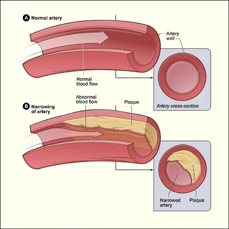
Atherosclerosis is a chronic inflammatory process affecting medium and large arteries. It develops over decades and is well established long before it causes symptoms.2
Plaque development
- Endothelial dysfunction. Shear stress, smoking, hyperglycaemia and hypertension damage the endothelium, making it more permeable and more adhesive to leucocytes.
- Lipid entry and oxidation. LDL cholesterol passes into the intima and is oxidised, which is pro-inflammatory.
- Foam cell formation. Monocytes migrate in, become macrophages and engulf oxidised LDL, becoming lipid-laden foam cells. Aggregated foam cells form the fatty streak, visible even in adolescence.
- Fibrous plaque. Smooth muscle cells migrate from the media and lay down a collagen cap over a necrotic lipid core, producing a mature atheromatous plaque.
Why plaques cause events
A plaque causes problems in two ways. A large, stable plaque progressively narrows the lumen and limits flow when demand rises, producing stable symptoms such as angina or claudication. A smaller plaque with a thin, inflamed cap may rupture or erode, exposing thrombogenic material and triggering acute thrombosis. This is the mechanism behind myocardial infarction and most ischaemic strokes.
This explains an initially counterintuitive point: the plaque that causes a myocardial infarction is often not the most stenotic one. Plaque stability matters as much as plaque size, which is why statins reduce events out of proportion to the change they produce in luminal diameter.
Risk factors
Risk factors are conventionally split into those that can and cannot be modified. In practice the distinction matters because it determines where the consultation goes.3
| Modifiable | Non-modifiable |
|---|---|
| Smoking | Increasing age |
| Hypertension | Male sex |
| Diabetes mellitus | South Asian ethnicity |
| Dyslipidaemia (raised LDL, low HDL) | Family history of premature CVD |
| Obesity and physical inactivity | Family history of hyperlipidaemia |
| Excess alcohol and poor diet | |
| Chronic kidney disease |
A family history of premature cardiovascular disease means a first-degree relative affected before the age of 55 in men or 65 in women. This is worth asking about explicitly, because it both raises the calculated risk and flags the possibility of an inherited lipid disorder.
Several conditions raise cardiovascular risk more than their prevalence suggests and are easy to forget: chronic kidney disease, rheumatoid arthritis and other chronic inflammatory conditions, systemic lupus erythematosus, severe mental illness, and HIV. Atypical antipsychotics and long-term corticosteroids also contribute.
Clinical features
Atherosclerosis is asymptomatic until a vessel is critically narrowed or a plaque ruptures. How it presents depends entirely on which arterial bed is involved.
| Territory | Stable presentation | Acute presentation |
|---|---|---|
| Coronary | Stable angina | Acute coronary syndrome |
| Cerebral | Vascular cognitive impairment | Stroke or transient ischaemic attack |
| Peripheral (limbs) | Intermittent claudication | Acute limb ischaemia |
| Renal | Renovascular hypertension | Flash pulmonary oedema |
| Mesenteric | Mesenteric angina (post-prandial pain, weight loss) | Acute mesenteric ischaemia |
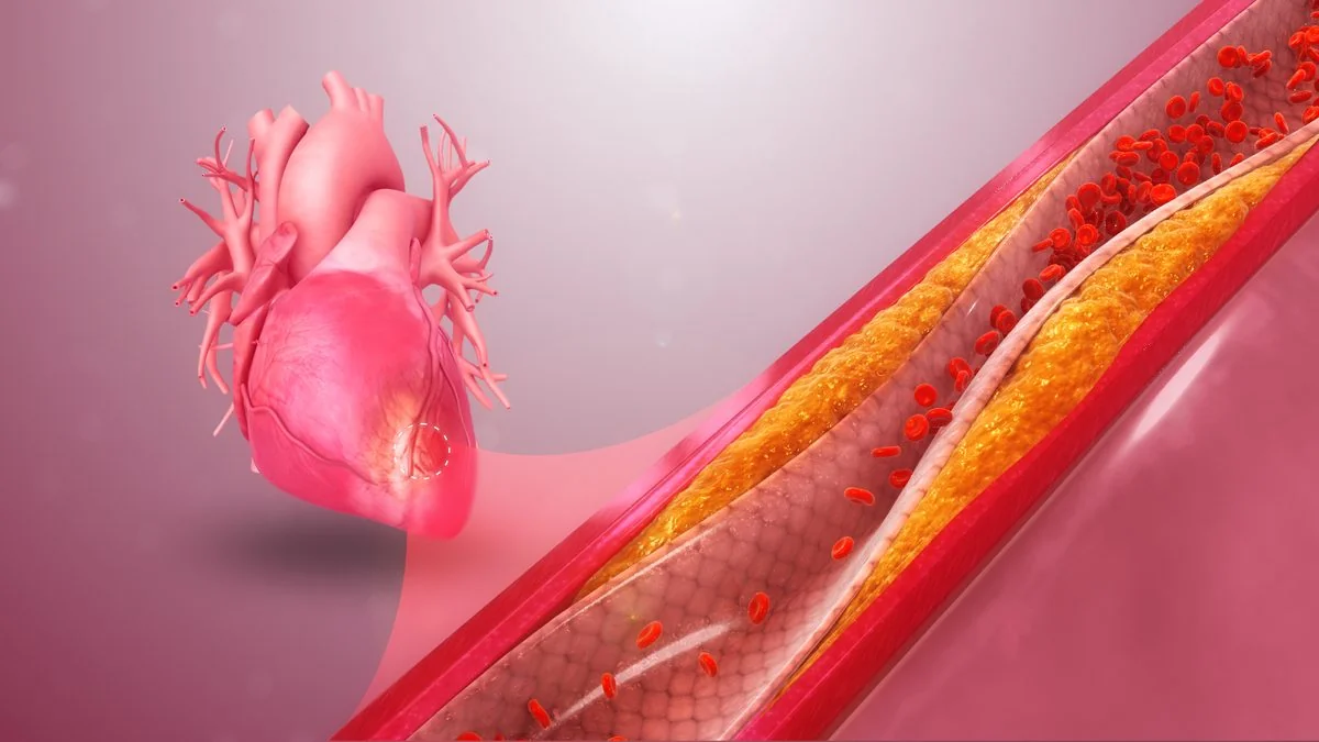
Because the process is diffuse, disease in one territory should prompt you to look for it in the others. A patient with claudication has a substantially raised risk of myocardial infarction and stroke, and their long-term prognosis is usually determined by their coronary disease rather than their legs.
Clinical examination
Examination in an asymptomatic patient is often normal, but it is used to look for evidence of established disease, for signs of severe dyslipidaemia, and for contributing risk factors.
- General: BMI and waist circumference; tar staining; xanthelasma, corneal arcus and tendon xanthomata (the last strongly suggests familial hypercholesterolaemia)
- Cardiovascular: blood pressure in both arms, heart rate and rhythm, murmurs, signs of heart failure
- Peripheral vessels: all peripheral pulses, capillary refill, carotid and abdominal bruits, and an abdominal examination for an aortic aneurysm
- Feet: ulceration, hair loss, and in diabetes a full diabetic foot check including monofilament sensation
The ankle-brachial pressure index is a useful bedside measure where peripheral arterial disease is suspected. A value below 0.9 supports the diagnosis; a value above 1.3 suggests incompressible, calcified vessels, which is common in longstanding diabetes and makes the test unreliable.
Investigations
Formal risk assessment
QRISK3 is the tool recommended in the UK. It estimates the percentage risk of a first heart attack or stroke over the next 10 years, and it is validated for people aged 25 to 84 who do not already have cardiovascular disease.4
QRISK3 incorporates age, sex, ethnicity, postcode-based deprivation, smoking, diabetes, family history, cholesterol ratio, blood pressure and BMI, along with several conditions that older tools missed, including chronic kidney disease, atrial fibrillation, migraine, corticosteroid use, severe mental illness and systemic lupus erythematosus.
Baseline investigations
- Full lipid profile including total cholesterol, HDL, non-HDL and triglycerides. A fasting sample is not required.
- HbA1c to identify undiagnosed diabetes or prediabetes
- Renal function and eGFR, plus a urine albumin:creatinine ratio
- Liver function tests before starting a statin, and TFTs, since hypothyroidism causes a secondary dyslipidaemia that should be corrected first
- Blood pressure, confirmed with ambulatory or home readings before diagnosing hypertension
- ECG if there are symptoms or examination findings suggesting established disease
Management
Management is divided into primary prevention, in people without established disease, and secondary prevention, in people who have already had an event. Lifestyle advice is common to both and should never be omitted because a drug has been started.
Lifestyle measures
- Smoking cessation - the single most effective intervention. Offer behavioural support plus pharmacotherapy rather than advice alone.
- Diet - a Mediterranean-style pattern: more vegetables, fruit, wholegrains, oily fish and unsaturated fats; less saturated fat, salt and refined sugar
- Physical activity - at least 150 minutes of moderate intensity activity per week, plus muscle-strengthening on two days
- Weight - aim for a BMI under 25 kg/m², with a lower threshold in people of South Asian ethnicity
- Alcohol - no more than 14 units per week, spread over three or more days
Primary prevention
Offer atorvastatin 20 mg once daily to people with a QRISK3 score of 10% or more, after a discussion of the benefits and risks. NICE is explicit that a statin can also be considered below this threshold if the person's informed preference is to take one.3
A statin is also offered without calculating QRISK3 to people with chronic kidney disease, and considered for all adults with type 1 diabetes, particularly those over 40, those with diabetes of more than 10 years' duration, and those with nephropathy or other risk factors.
Aspirin is not used for primary prevention. The bleeding risk outweighs the modest reduction in events in people without established disease.
Secondary prevention
Anyone with established atherosclerotic disease should receive atorvastatin 80 mg once daily, reduced only if there are drug interactions, a high risk of adverse effects, or patient preference. Additional treatment depends on the territory involved: antiplatelet therapy in all, plus a beta-blocker and an ACE inhibitor after myocardial infarction, and referral to cardiac rehabilitation.6
| Setting | Initial statin | Target |
|---|---|---|
| Primary prevention | Atorvastatin 20 mg | Non-HDL cholesterol reduced by more than 40% at 3 months |
| Secondary prevention | Atorvastatin 80 mg | LDL cholesterol 2.0 mmol/L or below, or non-HDL 2.6 mmol/L or below |
Check liver function before starting, and again at 3 and 12 months. Recheck the full lipid profile at 2-3 months. If the target is not met despite adherence and an increased dose, add ezetimibe; specialist options beyond that include PCSK9 inhibitors, inclisiran and bempedoic acid.3
Complications
The complications of cardiovascular disease are the events it causes: myocardial infarction, heart failure secondary to ischaemic cardiomyopathy, arrhythmia including atrial fibrillation, stroke and vascular dementia, chronic limb-threatening ischaemia and amputation, and renovascular disease.
Statin therapy is generally well tolerated. Muscle symptoms are the commonest reason for discontinuation, though randomised evidence suggests most such symptoms are not attributable to the drug itself. Rhabdomyolysis is rare. There is a small increase in the incidence of new-onset diabetes, which is outweighed by the cardiovascular benefit.
Red flags
Prognosis
Cardiovascular mortality in the UK has fallen substantially over recent decades, driven by reductions in smoking, better blood pressure control, widespread statin use and improved acute care. Despite this, CVD remains one of the leading causes of death and of years lived with disability.1
Prognosis for an individual depends on the burden of disease at diagnosis and on how completely risk factors are addressed afterwards. Statin therapy reduces major vascular events by roughly a fifth for each 1 mmol/L reduction in LDL cholesterol, and the benefit accrues year on year with continued treatment.7
The practical message is that adherence matters more than the choice of agent. A patient who stops their statin after six months gains very little, which is why the counselling conversation is as much a part of management as the prescription.
References
- British Heart Foundation. UK Factsheet: heart and circulatory disease statistics. Available here
- Libby P, Buring JE, Badimon L et al. Atherosclerosis. Nature Reviews Disease Primers. 2019. Available here
- NICE NG238. Cardiovascular disease: risk assessment and reduction, including lipid modification. 2023. Available here
- ClinRisk. QRISK3 cardiovascular risk calculator. Available here
- NICE CG71. Familial hypercholesterolaemia: identification and management. 2008, updated 2019. Available here
- NICE NG185. Acute coronary syndromes. 2020. Available here
- Cholesterol Treatment Trialists' Collaboration. Efficacy and safety of more intensive lowering of LDL cholesterol. The Lancet. 2010. Available here
- NICE Clinical Knowledge Summaries. CVD risk assessment and management. Available here
- BNF. Atorvastatin - indications and dosing. Available here
This article is written for revision and education. It is not clinical guidance and must not be used to make decisions about the care of a patient. Always check current NICE guidance and local protocols.