
Psoriatic Arthritis: Diagnosis and Management
Key points
- Psoriatic arthritis: an inflammatory arthritis occurring in up to 30% of people with psoriasis, part of the seronegative spondyloarthropathy family and sharing its HLA-B27 association and enthesitis.
- Onset relative to skin disease: skin disease usually comes first, but in around 15% arthritis precedes the rash - look hard for hidden psoriasis (scalp, natal cleft, umbilicus, nails).
- Five patterns: asymmetrical oligoarthritis (commonest), symmetrical polyarthritis, DIP-predominant, arthritis mutilans, and spondylitis.
- Distinguishing features: DIP joint involvement, dactylitis ('sausage digit'), enthesitis, and nail changes - pitting, onycholysis, subungual hyperkeratosis - are the features that separate it from rheumatoid arthritis.
- Radiographic hallmark: erosion with new bone formation - 'pencil-in-cup' deformity - unlike the pure erosion without new bone seen in rheumatoid arthritis.
- Screening: the PEST tool is used in dermatology clinics to catch arthritis in psoriasis patients before joint damage occurs.
- First-line DMARD: methotrexate, particularly useful when skin and joint disease coexist, as it treats both.
- Biologic escalation: anti-TNF, anti-IL-17 or anti-IL-12/23 agents for disease not controlled on conventional DMARDs - several also treat the skin disease at the same time.
Introduction
Psoriatic arthritis (PsA) is a chronic inflammatory arthritis that develops in a substantial minority - up to 30% - of people with psoriasis. Like ankylosing spondylitis, it belongs to the seronegative spondyloarthropathy family: rheumatoid factor and anti-CCP are typically negative, there is an association with HLA-B27 (particularly in axial disease), and enthesitis rather than pure synovitis is a defining feature.1
It is examined heavily because its joint pattern deliberately overlaps with, yet differs from, rheumatoid arthritis, and recognising which is which - DIP involvement, dactylitis, nail changes, asymmetry - is exactly the kind of pattern-recognition question that comes up repeatedly.
Aetiology
As with psoriasis itself, PsA arises from a combination of genetic susceptibility (HLA-B27, HLA-Cw6 and others), immune dysregulation - particularly the IL-23/IL-17 axis, which is now the target of several effective biologic drugs - and environmental triggers, including physical trauma to a joint or entheseal site (the 'deep Koebner phenomenon').
Risk factors
- Psoriasis, particularly more extensive or severe skin disease and nail involvement
- Family history of psoriasis or psoriatic arthritis
- HLA-B27 and other HLA associations, particularly relevant to axial disease
- Obesity - both a risk factor for developing PsA and a predictor of poorer response to treatment
- Physical trauma to a joint or entheseal site, through the 'deep Koebner phenomenon'
- Smoking
Clinical features
PsA is classically divided into five patterns (Moll and Wright), though patients often show a mix and the pattern can evolve over time:
| Pattern | Features | Approx. frequency |
|---|---|---|
| Asymmetrical oligoarthritis | Fewer than 5 joints, asymmetrical, often with dactylitis | Commonest pattern |
| Symmetrical polyarthritis | Resembles rheumatoid arthritis but seronegative | Common |
| DIP-predominant | Distal interphalangeal joints, often with adjacent nail disease | Classic but less common |
| Arthritis mutilans | Severe, destructive osteolysis causing digit shortening and 'telescoping' ('opera-glass hand') | Rare, most severe |
| Spondylitis (axial) | Sacroiliitis and spinal involvement, resembling ankylosing spondylitis | A minority, more often men |
Distinguishing features
- Dactylitis - diffuse swelling of an entire digit ('sausage finger/toe') from combined synovitis, tenosynovitis and soft tissue oedema
- Enthesitis - Achilles tendon, plantar fascia, and other tendon insertions
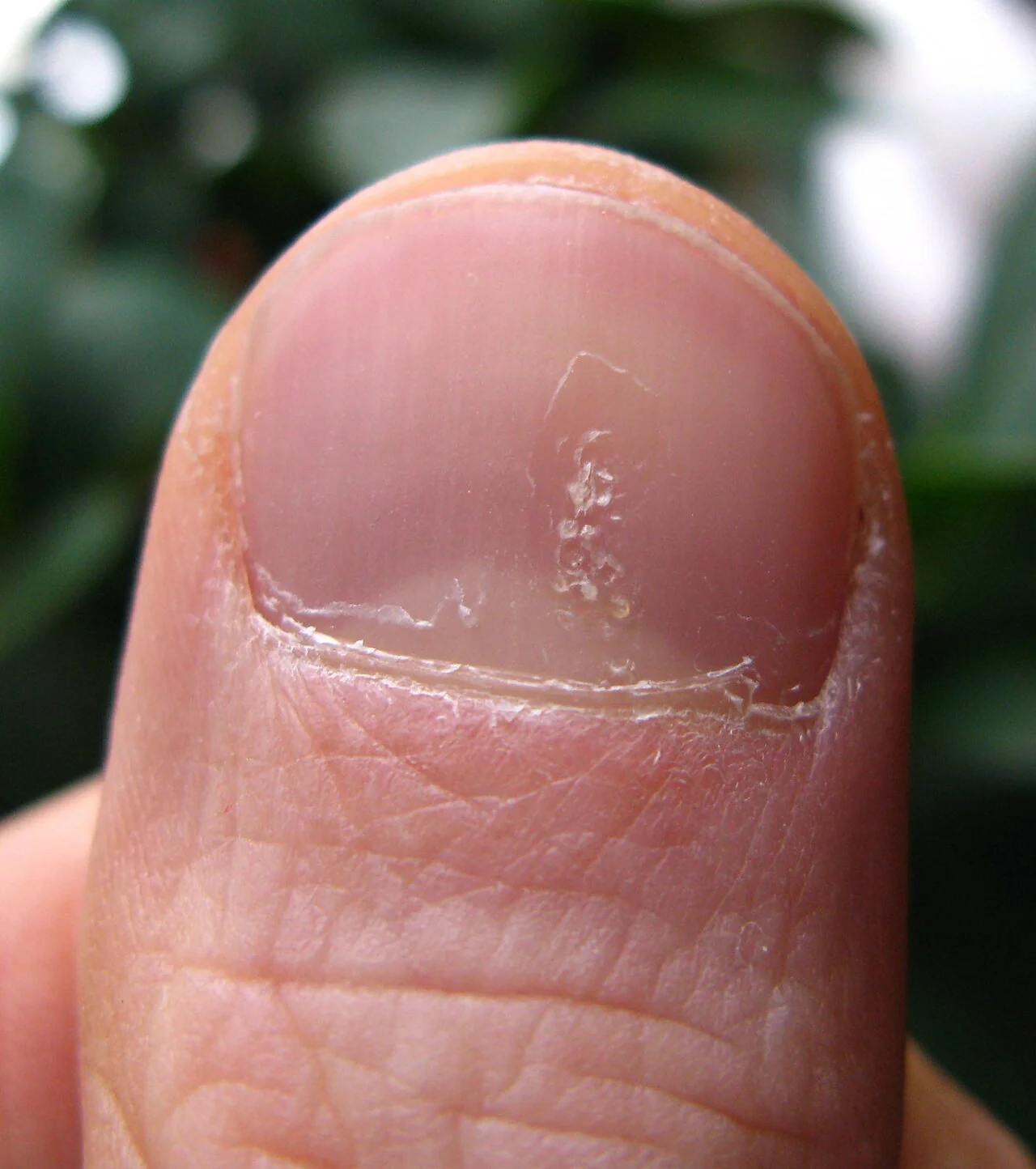
- Nail changes - pitting, onycholysis (separation of the nail from the bed), subungual hyperkeratosis, and the 'oil drop sign' (a translucent yellow-brown discolouration under the nail plate). Nail disease correlates strongly with DIP joint involvement
- Asymmetry - in contrast to the symmetry typical of rheumatoid arthritis, though the symmetrical polyarthritis pattern is an exception
- Skin psoriasis - plaques on extensor surfaces, scalp, natal cleft and umbilicus; look in these hidden sites specifically
- Eye disease - conjunctivitis and, less commonly, anterior uveitis
Examination
- Look for asymmetrical joint swelling, dactylitis, and psoriatic plaques in visible and hidden sites (scalp, natal cleft, umbilicus, behind the ears)
- Feel the DIP joints and entheseal sites (Achilles insertion, plantar fascia origin, lateral epicondyle) for tenderness and swelling
- Nails - inspect closely for pitting, onycholysis, subungual hyperkeratosis and the oil drop sign, which can be subtle
- Spine and sacroiliac joints - assess range of movement and perform provocation tests if axial disease is suspected
- Function - grip strength and hand function can be significantly affected even with modest-looking joint swelling, because of concurrent tenosynovitis and dactylitis
Differential diagnosis
- Rheumatoid arthritis - symmetrical, spares the DIPs, seropositive in most
- Osteoarthritis - DIPs affected in both, but OA is non-inflammatory, bony rather than boggy, without dactylitis, nail pitting or a raised CRP
- Gout - can also cause DIP involvement and coexist with OA; aspirate if there is doubt
- Other seronegative spondyloarthropathies - reactive arthritis and enteropathic arthritis share the enthesitis/axial pattern but have their own triggers and associations
Investigations
- Rheumatoid factor and anti-CCP - typically negative (a small minority are RF positive, which does not exclude PsA)
- CRP/ESR - may be raised but are less reliably elevated than in rheumatoid arthritis, and normal inflammatory markers do not exclude active disease
- Plain radiographs - erosive change combined with new bone formation (periostitis), producing the classic 'pencil-in-cup' deformity at the DIP joint as the phalanx is whittled to a point while the base of the adjacent bone widens; also osteolysis in arthritis mutilans
- Ultrasound or MRI - more sensitive for detecting synovitis, tenosynovitis and enthesitis, and useful where the diagnosis is unclear clinically
Screening and classification
The PEST (Psoriasis Epidemiology Screening Tool) questionnaire is used in dermatology clinics to identify patients with psoriasis who may have undiagnosed arthritis, prompting referral to rheumatology. The CASPAR criteria are used to classify established psoriatic arthritis in research and specialist practice, requiring inflammatory joint disease plus a defined score across psoriasis, nail disease, a negative RF, dactylitis and radiographic new bone formation.2
Management
Management is tailored to which pattern predominates - peripheral, axial, enthesitis/dactylitis-predominant, or skin-and-joint disease together - and involves close working between rheumatology and dermatology.3
Mild peripheral disease
NSAIDs and intra-articular corticosteroid injection for mono- or oligoarthritis, alongside physiotherapy.
Conventional synthetic DMARDs
Methotrexate is generally first-line for peripheral arthritis, particularly attractive when significant skin disease coexists since it treats both. Leflunomide, sulfasalazine and ciclosporin are alternatives, chosen partly on the basis of predominant skin versus joint involvement.
Biologic and targeted synthetic therapy
For disease inadequately controlled on conventional DMARDs, biologic therapy is escalated to, with several classes now available that also treat the skin:
- Anti-TNF (adalimumab, etanercept, infliximab, certolizumab)
- Anti-IL-17 (secukinumab, ixekizumab) - effective for both skin and joint disease, including axial involvement
- Anti-IL-12/23 (ustekinumab) and anti-IL-23 (guselkumab, risankizumab)
- JAK inhibitors (tofacitinib, upadacitinib) - oral option where biologics are unsuitable
- Apremilast - an oral PDE4 inhibitor, an option for milder disease not requiring biologic therapy
Axial psoriatic disease is managed in the same stepwise way as ankylosing spondylitis: NSAIDs first, since conventional DMARDs do not work on the axial skeleton, then a biologic (anti-TNF or anti-IL-17) if needed.
Complications
Uncontrolled disease leads to progressive joint destruction, and in the most severe pattern, arthritis mutilans, to marked functional impairment from bone resorption and digit telescoping. Like the other spondyloarthropathies, PsA carries an increased risk of cardiovascular disease related to chronic inflammation, and metabolic syndrome is common in psoriasis more broadly. Psychological impact - depression and reduced quality of life from combined skin and joint disease - is significant and should be actively asked about.
Red flags
Prognosis
Course is variable: some patients have mild, self-limiting oligoarthritis, while others develop progressive, erosive, deforming disease, particularly with delayed treatment. As with rheumatoid arthritis, early DMARD therapy aiming for low disease activity or remission substantially reduces the risk of irreversible joint damage, and the expansion of biologic options has considerably improved outcomes for both skin and joint disease over the last two decades.
References
- NICE Clinical Knowledge Summaries. Psoriatic arthritis. Available here
- Taylor W, Gladman D, Helliwell P et al. Classification criteria for psoriatic arthritis: development of new criteria from a large international study (CASPAR). Arthritis & Rheumatism. 2006. Available here
- Gossec L, Baraliakos X, Kerschbaumer A et al. EULAR recommendations for the management of psoriatic arthritis with pharmacological therapies. Annals of the Rheumatic Diseases. 2020. Available here
- NICE TA formulary. Biologics for psoriatic arthritis. Available here
- Ritchlin CT, Colbert RA, Gladman DD. Psoriatic Arthritis. New England Journal of Medicine. 2017. Available here
This article is written for revision and education. It is not clinical guidance and must not be used to make decisions about the care of a patient. Always check current NICE guidance and local protocols.