
Interstitial Lung Disease: Pattern, Cause and the Antifibrotic Era
Key points
- Interstitial lung disease: a heterogeneous group of over 200 disorders affecting the pulmonary interstitium, causing inflammation, fibrosis or both.
- Physiology: a restrictive pattern - reduced FVC and total lung capacity with a normal or raised FEV1/FVC ratio - together with a reduced transfer factor.
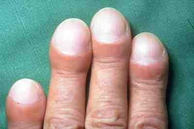
- Classic signs: progressive exertional breathlessness, a dry cough, fine bibasal end-inspiratory crackles often described as Velcro-like, and finger clubbing.
- Upper zone fibrosis: coal worker's pneumoconiosis, hypersensitivity pneumonitis, ankylosing spondylitis, radiation, tuberculosis, sarcoidosis and silicosis.
- Lower zone fibrosis: idiopathic pulmonary fibrosis, asbestosis, connective tissue disease (rheumatoid arthritis and systemic sclerosis), and drugs.
- Diagnosis: HRCT of the chest, interpreted in a specialist multidisciplinary meeting alongside the clinical picture. A confident UIP pattern on HRCT avoids the need for biopsy.
- IPF treatment: the antifibrotics pirfenidone and nintedanib slow the decline in lung function. They do not reverse fibrosis.
- The critical negative: corticosteroids and immunosuppression do not help in IPF and increase mortality. They are for inflammatory ILD, not fibrotic IPF.
Introduction
Interstitial lung disease (ILD), also called diffuse parenchymal lung disease, covers more than 200 conditions that affect the lung interstitium - the tissue between the alveolar epithelium and the capillary endothelium across which gas exchange occurs. Thickening of this space by inflammatory cells, fibrosis or both produces the same physiological consequences whatever the cause:
- Reduced lung compliance - stiff lungs that are harder to inflate, producing a restrictive ventilatory defect with reduced FVC and total lung capacity but a normal or increased FEV1/FVC ratio
- Impaired gas transfer - a reduced transfer factor (TLCO), which is often the earliest abnormality and may be reduced before spirometry changes at all
- Exercise-induced hypoxaemia - desaturation on exertion, frequently with resting saturations that look entirely normal in clinic
The clinical task has two parts. The first is recognising that the patient has ILD rather than heart failure, COPD or deconditioning - a distinction often delayed by many months. The second is identifying which ILD, because the treatments diverge completely and one of them, immunosuppression, actively harms patients with idiopathic pulmonary fibrosis.
Classification and causes
| Group | Examples |
|---|---|
| Known cause - connective tissue disease | Rheumatoid arthritis, systemic sclerosis, Sjogren syndrome, polymyositis and dermatomyositis (particularly the antisynthetase syndrome), SLE, mixed connective tissue disease |
| Known cause - drugs | Amiodarone, methotrexate, nitrofurantoin, bleomycin, busulfan, cyclophosphamide, gold, sulfasalazine, and immune checkpoint inhibitors |
| Known cause - occupational and environmental | Asbestosis, silicosis, coal worker's pneumoconiosis, berylliosis, hard metal lung disease |
| Known cause - hypersensitivity pneumonitis | Bird fancier's lung (avian proteins), farmer's lung (thermophilic actinomycetes in mouldy hay), malt worker's lung, hot tub lung, mushroom worker's lung |
| Known cause - other | Radiation pneumonitis and fibrosis, chronic aspiration, post-infectious |
| Idiopathic interstitial pneumonias | Idiopathic pulmonary fibrosis (UIP pattern), non-specific interstitial pneumonia (NSIP), cryptogenic organising pneumonia (COP), acute interstitial pneumonia, respiratory bronchiolitis-ILD and desquamative interstitial pneumonia (both smoking-related), lymphoid interstitial pneumonia |
| Granulomatous | Sarcoidosis, hypersensitivity pneumonitis, granulomatosis with polyangiitis |
| Rare and distinctive | Lymphangioleiomyomatosis (young women, cystic change, chylothorax and pneumothorax), pulmonary Langerhans cell histiocytosis (smokers, upper zone nodules and cysts), pulmonary alveolar proteinosis (crazy paving on CT), eosinophilic pneumonias |
Clinical features
Symptoms
- Progressive exertional breathlessness - insidious over months to years, and the dominant symptom. Patients typically adapt their lives around it and present late.
- Dry, non-productive cough - often intractable, and a major cause of distress in its own right
- Fatigue and reduced exercise tolerance
- Weight loss in advanced disease
- Symptoms of an underlying cause - joint pain and morning stiffness in rheumatoid arthritis, Raynaud phenomenon and skin tightening in systemic sclerosis, dysphagia and reflux, muscle weakness and a rash in myositis, erythema nodosum and eye symptoms in sarcoidosis
The history that makes the diagnosis
In ILD the history frequently contributes more than any test, and the exposures are the part most easily omitted.
- Full occupational history from leaving school onwards - every job, and specifically asbestos, silica, coal, metal dust and any dusty trade. Latency can be decades, so a job held in a patient's twenties matters.
- Complete drug history including over-the-counter and stopped drugs - amiodarone and nitrofurantoin cause fibrosis that may present after the drug has been discontinued, and nitrofurantoin taken long term for recurrent urinary infection is a classic missed cause
- Environmental exposures - birds (including a neighbour's pigeons or a feather duvet), mould and damp, hot tubs, humidifiers, air conditioning
- Smoking history - relevant to IPF, RB-ILD, DIP and Langerhans cell histiocytosis
- Radiotherapy to the chest, and the fields used
- Family history - familial pulmonary fibrosis and telomere-related disease
- A full systems review for connective tissue disease - this is not optional, since ILD may precede the joint or skin disease by years
Examination
- Fine, bibasal, end-inspiratory crackles - characteristically described as Velcro-like, and unlike the coarse crackles of bronchiectasis they do not clear on coughing
- Finger clubbing - present in around half of patients with IPF and in asbestosis, but not typically in sarcoidosis or hypersensitivity pneumonitis, which is a useful discriminator
- Reduced chest expansion
- Tachypnoea and, on a walking test, marked desaturation on exertion
- Central cyanosis in advanced disease
- Signs of pulmonary hypertension and cor pulmonale - a loud P2, a right ventricular heave, raised JVP and peripheral oedema
- Extrapulmonary signs of a cause - rheumatoid hands, sclerodactyly, telangiectasia and calcinosis, Gottron papules and mechanic's hands, lupus pernio and erythema nodosum
Investigations
Lung function
- Spirometry - a restrictive pattern: reduced FVC with a normal or raised FEV1/FVC ratio (typically above 0.7 and often above 0.8)
- Lung volumes - reduced total lung capacity and residual volume, confirming true restriction rather than poor effort
- Transfer factor (TLCO) - reduced, and often the earliest abnormality. Serial TLCO and FVC are the standard way of tracking progression and of deciding on treatment eligibility.
- Oxygen saturation at rest and on exertion, and a 6-minute walk test with desaturation measured - resting saturations are frequently normal in significant disease and give false reassurance
- Arterial blood gas in advanced disease - type 1 respiratory failure
Imaging
- Chest radiograph - may show reduced lung volumes with bibasal reticulonodular shadowing, but it is normal in up to 10% of patients with established ILD and cannot exclude the diagnosis
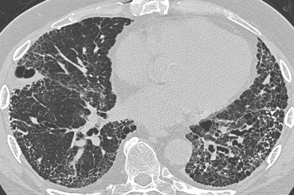
- High-resolution CT (HRCT) - the key investigation. Thin sections through the lung define the pattern, distribution and extent of disease, and frequently establish the diagnosis without biopsy.
| Pattern | Features | Typical cause |
|---|---|---|
| UIP | Subpleural, basal predominant reticulation with honeycombing, traction bronchiectasis and minimal ground glass | Idiopathic pulmonary fibrosis, and also rheumatoid and asbestos-related disease |
| NSIP | Ground-glass predominant, symmetrical, basal, with subpleural sparing and little or no honeycombing | Connective tissue disease, drugs, idiopathic |
| Organising pneumonia | Patchy peripheral or peribronchial consolidation, migratory, sometimes with the reversed halo (atoll) sign | Cryptogenic organising pneumonia, drugs, infection, connective tissue disease |
| Hypersensitivity pneumonitis | Upper and mid zone ground glass with centrilobular nodules and mosaic attenuation with air trapping on expiratory scans - the three-density sign | Antigen exposure - birds, mould, hot tubs |
| Sarcoidosis | Perilymphatic nodules along bronchovascular bundles, upper zone predominance, and bilateral hilar lymphadenopathy | Sarcoidosis |
Blood tests
- FBC, U&Es, LFTs, calcium, CRP and ESR - and note that hypercalcaemia and a raised ACE suggest sarcoidosis
- Connective tissue screen in every patient - ANA, rheumatoid factor, anti-CCP, and where indicated an extended ENA and myositis panel (including anti-Jo-1 and other antisynthetase antibodies), anti-Scl-70 and anticentromere
- ANCA - for vasculitis, particularly where there is haemoptysis or renal involvement
- Serum precipitins (specific IgG) to avian and fungal antigens where hypersensitivity pneumonitis is suspected. A positive result indicates exposure, not disease.
- Immunoglobulins and, in selected patients, HIV testing
Further investigation
- Bronchoalveolar lavage - not routinely diagnostic, but a lymphocytosis above 40% strongly supports hypersensitivity pneumonitis or sarcoidosis, and it is valuable for excluding infection and malignancy
- Transbronchial lung cryobiopsy or surgical (VATS) lung biopsy - reserved for cases where the diagnosis remains uncertain after HRCT and serology, since surgical biopsy carries real morbidity and mortality in this population
- Echocardiogram - to assess for pulmonary hypertension, which is common and prognostically important
- Right heart catheterisation where pulmonary hypertension would change management
Idiopathic pulmonary fibrosis
IPF is the commonest and most serious of the idiopathic interstitial pneumonias. It is defined by a UIP pattern in the absence of an identifiable cause, and it is a progressively fibrotic rather than inflammatory disease - which is the single fact that explains its treatment.1
- Typical patient - male, over 60, a current or former smoker, presenting with breathlessness and a dry cough over 6 to 24 months
- Associations - smoking, gastro-oesophageal reflux, chronic microaspiration, and a family history in around 5%, often with telomere-related gene variants
- Pathogenesis - repetitive alveolar epithelial injury in a susceptible individual leads to aberrant wound healing, fibroblast proliferation and excessive collagen deposition, rather than to a primarily inflammatory process
Antifibrotic therapy
- Pirfenidone - an antifibrotic with anti-inflammatory properties. Main adverse effects are photosensitivity (strict sun protection is essential), nausea, anorexia, weight loss and deranged liver function.
- Nintedanib - a tyrosine kinase inhibitor targeting fibroblast growth factor, platelet-derived growth factor and vascular endothelial growth factor receptors. Main adverse effect is diarrhoea, which is very common, along with nausea and deranged liver function; it also increases bleeding risk.
- Both slow the rate of FVC decline by roughly half. Neither reverses fibrosis nor restores lost lung function, and this needs to be explained clearly when starting them.
- NICE recommends starting them when FVC is between 50 and 80% of predicted, and discontinuing if disease progresses by more than 10% of predicted FVC in any 12-month period
- Nintedanib is also licensed for systemic sclerosis-associated ILD and for other progressive fibrosing ILDs, which has substantially widened its use
Management
Treat the cause where there is one
- Remove the antigen in hypersensitivity pneumonitis - rehoming birds, removing feather bedding, remediating mould. Complete avoidance can arrest and sometimes reverse the disease, and is far more effective than any drug.
- Stop the causative drug and consider corticosteroids in drug-induced ILD
- Remove occupational exposure, and advise on industrial injuries benefit where the disease is prescribed
- Treat the connective tissue disease - typically corticosteroids with a steroid-sparing agent such as mycophenolate, and increasingly nintedanib where the ILD is progressive despite immunosuppression. Rituximab and cyclophosphamide are used in severe or rapidly progressive disease.
- Corticosteroids for organising pneumonia, which usually responds dramatically, and for sarcoidosis where treatment is indicated
Supportive care, which applies to everyone
- Pulmonary rehabilitation - improves exercise capacity and quality of life, and is under-used in ILD
- Smoking cessation
- Ambulatory oxygen for exertional desaturation, and long-term oxygen therapy for resting hypoxaemia
- Vaccination - influenza, pneumococcal and COVID-19
- Treat gastro-oesophageal reflux, given its association with IPF and with microaspiration
- Cough management - often the most distressing symptom. Treat reflux, and consider low-dose opioids where the cough is refractory.
- Palliative care input early - for breathlessness, cough and advance care planning. Involving palliative care alongside active treatment rather than at the end is particularly valuable in IPF given the unpredictable course.
- Lung transplantation - the only intervention that improves survival in IPF. Refer early, at diagnosis in appropriate candidates, because waiting-list mortality in IPF is among the highest of any indication.
- Psychological support and patient organisations - the diagnosis carries a prognosis comparable to many cancers, and patients frequently describe feeling unsupported
Acute exacerbation of ILD
An acute exacerbation is a rapid deterioration over less than a month with new bilateral ground-glass change on HRCT, not explained by infection, heart failure or pulmonary embolism - all of which must be excluded first. It is a devastating event, with an in-hospital mortality of around 50% and a median survival after it of only a few months. Treatment is largely supportive with high-dose corticosteroids and treatment of any identified precipitant, and the evidence base is weak. It is a moment at which ceiling-of-care discussions become essential, since invasive ventilation in IPF has a very poor outcome.
Complications
- Progressive respiratory failure - the usual cause of death
- Acute exacerbations - accounting for around half of deaths in IPF
- Pulmonary hypertension and cor pulmonale - common in advanced fibrosis and a marker of poor prognosis
- Lung cancer - substantially increased in IPF and asbestosis, independent of smoking, and difficult to treat because of the poor lung reserve
- Pneumothorax and pneumomediastinum - from rupture of subpleural cysts
- Respiratory infection - each episode may precipitate an exacerbation
- Venous thromboembolism - increased risk in IPF
- Drug toxicity - photosensitivity and hepatotoxicity from pirfenidone, diarrhoea and hepatotoxicity from nintedanib, and the full range of corticosteroid and immunosuppressant complications
- Depression, anxiety and social isolation - very common, and consistently under-treated
Red flags
Prognosis
Prognosis varies enormously across the group, which is why the specific diagnosis matters so much.
- Idiopathic pulmonary fibrosis - the worst outlook, with a median survival of 3 to 5 years from diagnosis, comparable to many cancers. The course is unpredictable: some patients remain stable for years, others decline steadily, and others deteriorate abruptly with an acute exacerbation.
- NSIP - considerably better, particularly the cellular subtype, which often responds well to immunosuppression
- Cryptogenic organising pneumonia - usually responds dramatically to corticosteroids, though relapse on withdrawal is common
- Hypersensitivity pneumonitis - excellent if the antigen is identified and removed before fibrosis develops; once chronic fibrotic disease is established the prognosis approaches that of IPF
- Connective tissue disease-associated ILD - generally better than IPF, even when the HRCT pattern is UIP
- Sarcoidosis - most cases resolve spontaneously; only a minority progress to fibrosis
Across all types, the strongest adverse prognostic markers are a low or falling FVC and TLCO, desaturation on a 6-minute walk test, extensive honeycombing on HRCT, pulmonary hypertension, and older age. A fall in FVC of 10% or more, or in TLCO of 15% or more, over 6 to 12 months identifies progressive disease and is used both to define progressive pulmonary fibrosis and to trigger escalation of treatment or transplant referral.
References
- NICE CG163. Idiopathic pulmonary fibrosis in adults: diagnosis and management. 2013, updated 2017. Available here
- Raghu G, Remy-Jardin M, Richeldi L et al. Idiopathic pulmonary fibrosis (an update) and progressive pulmonary fibrosis in adults: an official ATS/ERS/JRS/ALAT clinical practice guideline. 2022. Available here
- British Thoracic Society. Interstitial lung disease guidelines and quality standards. Available here
- Idiopathic Pulmonary Fibrosis Clinical Research Network. Prednisone, azathioprine and N-acetylcysteine for pulmonary fibrosis (PANTHER-IPF). NEJM. 2012. Available here
- NICE TA379. Pirfenidone for treating idiopathic pulmonary fibrosis. 2016. Available here
- NICE TA379 and TA504. Nintedanib for treating idiopathic pulmonary fibrosis. Available here
- NICE Clinical Knowledge Summaries. Interstitial lung disease. Available here
- Yale Rosen, image contributed by Darel Heitkamp MD, CC BY-SA 2.0, via Wikimedia Commons. Available here
- IPFeditor, CC BY-SA 3.0, via Wikimedia Commons. Available here
This article is written for revision and education. It is not clinical guidance and must not be used to make decisions about the care of a patient. Always check current NICE guidance and local protocols.