
Neonatal Surgical Emergencies
Key points
- The single most important rule: bilious vomiting in a neonate is malrotation with midgut volvulus until proven otherwise - and the bowel infarcts within hours.
- Oesophageal atresia: frothy secretions, choking on the first feed, and a nasogastric tube that will not pass. Confirm with a chest radiograph showing the tube coiled in the upper pouch.
- Congenital diaphragmatic hernia: respiratory distress with a scaphoid abdomen. Intubate immediately and do not bag-mask ventilate - it inflates the herniated bowel.
- Abdominal wall defects: gastroschisis has no sac and few associated anomalies; exomphalos has a sac and frequent chromosomal and cardiac anomalies.
- Double bubble: duodenal atresia, associated with Down syndrome in around a third of cases.
- Delayed meconium: failure to pass meconium within 48 hours suggests Hirschsprung's disease, meconium ileus or an anorectal malformation.
- Initial management is common to all: nil by mouth, wide-bore nasogastric tube on free drainage, intravenous fluids, correct electrolytes, keep warm, and discuss with the regional surgical centre.
- Do not forget the anus: an anorectal malformation is found by looking, which is why inspection of the anus is part of the newborn examination.
Introduction
Neonatal surgical emergencies are individually uncommon but collectively account for a substantial share of neonatal transfers and deaths. Most present in the first 48 hours of life, and almost all announce themselves through one of three findings: bilious vomiting, failure to pass a nasogastric tube, or failure to pass meconium within 48 hours.
The generalist's job is rarely to make the precise anatomical diagnosis. It is to recognise that a baby has a surgical problem, resuscitate them properly, avoid the specific manoeuvres that make each condition worse, and get them to a paediatric surgical centre alive and stable. That sequence is what an exam question is testing.
Many of these conditions are now detected antenatally, which allows delivery to be planned at or near a surgical centre. Polyhydramnios suggests an upper gastrointestinal obstruction, an absent stomach bubble suggests oesophageal atresia, a double bubble suggests duodenal atresia, and herniated abdominal contents may be seen directly.7 A baby with an antenatally diagnosed anomaly should not be born in a unit that cannot manage it.
The common initial approach
Whatever the underlying lesion, the first hour is broadly the same and is worth being able to recite.
- Airway, breathing, circulation, with intubation where respiratory compromise is present, and thermal care - these babies cool very quickly
- Nil by mouth, with a wide-bore nasogastric or orogastric tube on free drainage and regular aspiration to decompress the stomach and prevent aspiration
- Intravenous or intraosseous access, with a 10-20 mL/kg bolus of 0.9% sodium chloride if shocked, then maintenance fluid plus replacement of measured gastric losses
- Bloods: blood gas with lactate and glucose, U&Es, FBC, coagulation, group and save, and blood cultures
- Correct hypoglycaemia and electrolyte disturbance, which are common and are frequently the thing that destabilises the baby during transfer
- Intravenous antibiotics where sepsis, perforation or bowel ischaemia is suspected4
- Intramuscular vitamin K if not already given
- Discuss immediately with the regional paediatric surgical centre and arrange neonatal retrieval
- Consider a prostaglandin E infusion if a duct-dependent cardiac lesion is a possibility, since congenital heart disease coexists with several of these anomalies8
- Talk to the parents, who have usually just been told their newborn needs an operation in another hospital
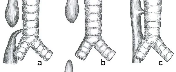
Oesophageal atresia and tracheo-oesophageal fistula
Occurs in around 1 in 3,500 births, from failure of the primitive foregut to separate cleanly into trachea and oesophagus during the fourth week of gestation.
Presentation
- Antenatal: polyhydramnios with a small or absent stomach bubble
- Frothy oral secretions and constant drooling from birth - the baby cannot swallow saliva
- Choking, coughing and cyanosis with the first feed
- Failure to pass a nasogastric tube beyond about 10 cm - use a firm 10 French tube, since a fine soft tube can coil and give a false impression of passing
- Chest and abdominal radiograph with the tube in place: it coils in the upper pouch. Gas in the abdomen indicates a distal fistula; a gasless abdomen indicates isolated atresia.
- Abdominal distension in those with a distal fistula, as ventilation forces air into the stomach
Management is nil by mouth, a Replogle tube in the upper pouch on continuous low suction to prevent aspiration of pooled saliva, nursing head-up, intravenous fluids, and transfer for surgical division of the fistula with primary oesophageal anastomosis. Long-term problems include anastomotic stricture, gastro-oesophageal reflux, recurrent fistula, and tracheomalacia producing the characteristic harsh barking cough that persists for years.
Congenital diaphragmatic hernia
Occurs in around 1 in 3,000 births. Around 85% are left-sided posterolateral (Bochdalek) defects; anterior (Morgagni) hernias are rarer and often present later or incidentally.
The critical point is that the problem is not the hernia but the lung. Abdominal viscera occupying the chest from early gestation prevent normal lung growth, producing pulmonary hypoplasia - which affects both lungs, not only the ipsilateral one - and an abnormal pulmonary vascular bed that causes severe pulmonary hypertension. Mortality is determined by these, not by the size of the defect, which is why surgical repair is deliberately delayed until the baby is physiologically stable.
- Respiratory distress and cyanosis from birth, often severe
- Scaphoid (sunken) abdomen, because the abdominal contents are in the chest
- Displaced apex beat and heart sounds, to the right in a left-sided hernia
- Absent breath sounds on the affected side, sometimes with audible bowel sounds in the chest
- Chest radiograph: bowel loops in the hemithorax, mediastinal shift, and the nasogastric tube tip seen above the diaphragm
Subsequent management involves gentle ventilation with permissive hypercapnia, treatment of pulmonary hypertension with inhaled nitric oxide, extracorporeal membrane oxygenation in selected cases at specialist centres, and delayed surgical reduction and repair of the defect. Survivors frequently have chronic lung disease, gastro-oesophageal reflux, feeding difficulty and neurodevelopmental problems.
Abdominal wall defects
| Gastroschisis | Exomphalos (omphalocele) | |
|---|---|---|
| Position | Paraumbilical, almost always to the right of a normally inserted cord | Midline, with the umbilical cord inserting into the sac itself |
| Covering | No sac - bowel is exposed, matted, oedematous and inflamed by amniotic fluid | Covered by a sac of amnion, Wharton's jelly and peritoneum, unless it has ruptured |
| Associated anomalies | Uncommon, apart from intestinal atresia in about 10% | Frequent - trisomy 13, 18 and 21, congenital heart disease, and Beckwith-Wiedemann syndrome |
| Maternal associations | Young maternal age, smoking, recreational drug use | No strong maternal association |
| Fluid and heat loss | Large, because bowel is exposed - fluid requirements are often 2-3 times maintenance | Limited while the sac is intact |
| Management | Cover the bowel immediately, primary closure or staged reduction using a preformed silo | Full workup for associated anomalies, then staged closure - or conservative epithelialisation of the sac in giant defects |
| Prognosis | Good, determined by bowel function - prolonged ileus and parenteral nutrition are common | Determined by the associated anomalies, particularly cardiac and chromosomal |
Intestinal obstruction
Duodenal atresia
Occurs in around 1 in 5,000-10,000 births and results from failure of recanalisation of the duodenal lumen. Approximately a third of affected babies have Down syndrome, and there are associations with annular pancreas, malrotation and congenital heart disease.
- Antenatal: polyhydramnios and a double bubble on ultrasound
- Vomiting within hours of birth - bilious in the 85% of cases where the atresia lies distal to the ampulla, non-bilious if proximal
- Abdominal radiograph: the double bubble of a distended stomach and proximal duodenum, with no distal gas
- Management is nasogastric decompression, fluid and electrolyte correction, echocardiography, and duodenoduodenostomy
Malrotation with midgut volvulus
During normal development the midgut herniates into the umbilical cord and returns having rotated 270 degrees anticlockwise, leaving a broad mesenteric base running from the duodenojejunal flexure in the left upper quadrant to the caecum in the right lower quadrant. In malrotation this rotation is incomplete, the mesenteric base is narrow, and abnormal peritoneal (Ladd's) bands run across the duodenum. The midgut can then twist around the superior mesenteric artery.
- Bilious vomiting is the cardinal feature, with around half presenting in the first week and most within the first month
- The baby may initially look deceptively well, with a soft abdomen and normal observations
- Later features - abdominal distension, tenderness, blood in the stool, shock and peritonitis - indicate that the bowel is already infarcting
- Upper gastrointestinal contrast study is the gold standard: the duodenojejunal flexure lies to the right of the midline and below its normal level, with a corkscrew appearance of the twisted duodenum and jejunum
- Abdominal radiograph may be entirely normal, and a normal film never excludes the diagnosis
- Ultrasound may show the whirlpool sign of vessels twisting around the superior mesenteric artery, and inversion of the normal superior mesenteric artery and vein relationship
- Management is emergency laparotomy and a Ladd's procedure: derotate the volvulus anticlockwise, divide Ladd's bands, broaden the mesentery, perform an appendicectomy, and leave the bowel in a position of non-rotation
Hirschsprung's disease
Occurs in around 1 in 5,000 births, with a male to female ratio of about 4:1. Neural crest cells fail to complete their craniocaudal migration, so ganglion cells are absent from the myenteric (Auerbach) and submucosal (Meissner) plexuses. The aganglionic segment is therefore always distal and continuous from the anus, extending proximally for a variable distance - rectosigmoid in around 80%, and total colonic in 5-10%. It is associated with Down syndrome in about 10% of cases, and with RET proto-oncogene mutations.
- Failure to pass meconium within 48 hours - the classic presentation
- Abdominal distension and bilious vomiting
- Explosive passage of stool and gas following rectal examination, as the finger temporarily dilates the contracted aganglionic segment
- Later presentation: chronic constipation from birth, faltering growth, and a history of needing suppositories or washouts from infancy6
- Suction rectal biopsy is the diagnostic gold standard - absence of ganglion cells, hypertrophied nerve trunks and increased acetylcholinesterase staining
- Contrast enema may show a transition zone with a narrow distal segment and dilated proximal bowel, but is unreliable in the neonatal period
- Anorectal manometry shows an absent rectoanal inhibitory reflex
- Management: bowel decompression with rectal washouts, then a pull-through procedure to bring ganglionated bowel down to the anus
Meconium ileus
Abnormally viscid meconium obstructs the terminal ileum. It is almost always caused by cystic fibrosis, and 10-20% of babies with cystic fibrosis present this way - often before the newborn blood spot result is available.5
- Abdominal distension, bilious vomiting and failure to pass meconium, with palpable doughy loops of bowel
- Abdominal radiograph: dilated bowel loops with a soap-bubble or ground-glass appearance in the right lower quadrant, and a relative paucity of air-fluid levels because the meconium is too viscid to layer
- A water-soluble (Gastrografin) contrast enema is both diagnostic and often therapeutic, drawing fluid into the bowel to loosen the meconium - the baby must be well hydrated before it is performed
- Surgery is required for complicated cases: volvulus, atresia, or perforation causing meconium peritonitis with intra-abdominal calcification
- Confirm cystic fibrosis with a sweat test and genetic testing, and check the newborn screening result
Anorectal malformations
These range from a covered or stenotic anus to a high malformation with a fistula to the urethra, bladder or vagina. They are found by inspecting the perineum, which is why examination of the anus is a specified part of the newborn and infant physical examination.1 Look for meconium appearing in the urine or at an abnormal perineal opening. They form part of the VACTERL association, so an echocardiogram, renal ultrasound and spinal imaging are needed. Low lesions may be repaired primarily; high lesions require a defunctioning colostomy followed by posterior sagittal anorectoplasty.
Groin and scrotal emergencies
- Incarcerated inguinal hernia - much commoner in preterm boys, presenting as an irreducible, tender groin swelling with vomiting and distension. Gentle reduction after analgesia and sedation is attempted, followed by repair on the same admission; failure to reduce means emergency surgery. The risk of incarceration is why infant inguinal hernias are repaired promptly rather than watched.
- Neonatal testicular torsion - usually extravaginal and often prenatal, presenting as a firm, hard, discoloured hemiscrotum that is characteristically non-tender. The affected testis is often already infarcted, but urgent urological assessment is still required to protect the contralateral side.
- Necrotising enterocolitis - the commonest neonatal surgical emergency of all, and covered in its own article
Putting it together
| Clue | Think of | Next step |
|---|---|---|
| Frothy secretions, nasogastric tube will not pass | Oesophageal atresia | Chest and abdominal radiograph with the tube in situ; Replogle tube on suction |
| Bilious vomiting, well-looking baby, normal film | Malrotation with volvulus | Urgent upper gastrointestinal contrast study and surgical referral |
| Double bubble, no distal gas | Duodenal atresia | Look for Down syndrome and cardiac anomalies; surgical referral |
| Multiple dilated loops with air-fluid levels | Jejunoileal atresia or distal obstruction | Contrast enema and surgical referral |
| No meconium by 48 hours, explosive stool after PR | Hirschsprung's disease | Suction rectal biopsy |
| Soap-bubble appearance in the right lower quadrant | Meconium ileus | Gastrografin enema; investigate for cystic fibrosis |
| Respiratory distress with a scaphoid abdomen | Congenital diaphragmatic hernia | Intubate, nasogastric decompression, do not bag-mask ventilate |
| Non-bilious projectile vomiting at 3-6 weeks | Pyloric stenosis - not a neonatal emergency in the same sense | Test feed and ultrasound; correct the alkalosis before surgery |
Prognosis
Outcomes in UK paediatric surgery are generally good, and have improved markedly with antenatal detection, planned delivery at or near a surgical centre, and specialist neonatal retrieval. Isolated duodenal atresia, low anorectal malformations, uncomplicated gastroschisis and rectosigmoid Hirschsprung's disease all have survival rates well above 90%.2
The conditions with the worst outlook are those where the anomaly is not the whole problem. Congenital diaphragmatic hernia carries a mortality driven by pulmonary hypoplasia and pulmonary hypertension rather than by the hernia; exomphalos carries the mortality of its associated chromosomal and cardiac anomalies; and midgut volvulus that is diagnosed late carries the mortality and lifelong morbidity of short bowel syndrome.
That last point is the one within a clinician's control. The difference between a child who has a Ladd's procedure and goes home, and a child who loses their entire midgut, is measured in hours - and usually begins with whether someone treated green vomit as an emergency or as a feeding problem.3
References
- GOV.UK. Newborn and infant physical examination (NIPE) screening programme handbook. Available here
- British Association of Paediatric Surgeons. Congenital Anomalies Surveillance System (BAPS-CASS). Available here
- Advanced Life Support Group. Advanced Paediatric Life Support: A Practical Approach to Emergencies. Available here
- NICE NG195. Neonatal infection: antibiotics for prevention and treatment. 2021. Available here
- NICE NG78. Cystic fibrosis: diagnosis and management. 2017. Available here
- NICE Clinical Knowledge Summaries. Constipation in children. Available here
- GOV.UK. Fetal anomaly screening programme handbook. Available here
- BNF for Children. Alprostadil. Available here
This article is written for revision and education. It is not clinical guidance and must not be used to make decisions about the care of a patient. Always check current NICE guidance and local protocols.