
Congenital Heart Disease
Key points
- Incidence: congenital heart disease affects around 8 per 1000 live births, making it the commonest group of congenital malformations.
- The first division: acyanotic lesions cause left-to-right shunting or obstruction; cyanotic lesions cause right-to-left shunting so that deoxygenated blood reaches the systemic circulation.
- Acyanotic: ventricular septal defect, atrial septal defect, patent ductus arteriosus, coarctation of the aorta, pulmonary and aortic stenosis.
- Cyanotic: tetralogy of Fallot, transposition of the great arteries, tricuspid atresia, total anomalous pulmonary venous drainage and truncus arteriosus.
- ASD versus VSD: ASD gives fixed splitting of the second heart sound with a pulmonary flow murmur; VSD gives a harsh pansystolic murmur at the left sternal edge, loudest when the defect is small.
- Duct-dependent lesions: present as the ductus arteriosus closes in the first days of life. Keep it open with a prostaglandin E infusion while arranging urgent transfer.
- Eisenmenger syndrome: a long-standing left-to-right shunt raises pulmonary vascular resistance until the shunt reverses, producing cyanosis. It contraindicates corrective surgery.
- Adult survivors: there are now more adults than children living with congenital heart disease in the UK, and they need lifelong specialist follow-up.
Introduction
Congenital heart disease affects approximately 8 per 1000 live births, making it the commonest group of congenital malformations. Advances in paediatric cardiac surgery mean that over 90% of affected children now survive to adulthood, and there are consequently more adults than children living with congenital heart disease in the UK. Students will therefore meet these patients in adult medicine as often as in paediatrics.
The subject looks daunting because of the number of named lesions. It becomes manageable once organised around a single question: is the patient cyanosed?
| Group | Mechanism | Examples |
|---|---|---|
| Acyanotic - shunt | Left-to-right shunt. Oxygenated blood recirculates through the lungs, causing pulmonary overcirculation and volume overload. | Ventricular septal defect, atrial septal defect, patent ductus arteriosus, atrioventricular septal defect |
| Acyanotic - obstructive | Obstruction to flow, causing pressure overload of the chamber behind it | Coarctation of the aorta, aortic stenosis, pulmonary stenosis |
| Cyanotic | Right-to-left shunt, so deoxygenated blood bypasses the lungs and enters the systemic circulation | Tetralogy of Fallot, transposition of the great arteries, tricuspid atresia, total anomalous pulmonary venous drainage, truncus arteriosus |
Aetiology and associations
Most congenital heart disease is multifactorial and no cause is identified. Recognised associations are nonetheless worth knowing, because finding one lesion should prompt a search for others.
Genetic and chromosomal
| Syndrome | Characteristic cardiac lesions |
|---|---|
| Down syndrome (trisomy 21) | Atrioventricular septal defect (the classic association), VSD, ASD |
| Turner syndrome (45,X) | Coarctation of the aorta, bicuspid aortic valve, aortic dilatation |
| DiGeorge syndrome (22q11 deletion) | Tetralogy of Fallot, truncus arteriosus, interrupted aortic arch |
| Noonan syndrome | Pulmonary stenosis, hypertrophic cardiomyopathy |
| Williams syndrome | Supravalvular aortic stenosis, peripheral pulmonary stenosis |
| Marfan syndrome | Aortic root dilatation and dissection, mitral valve prolapse |
| Edwards (18) and Patau (13) syndromes | VSD, ASD, patent ductus arteriosus |
Maternal and environmental factors
- Congenital rubella - patent ductus arteriosus and peripheral pulmonary stenosis, with cataracts and deafness
- Maternal diabetes - transposition of the great arteries, VSD, and transient hypertrophic cardiomyopathy of the newborn
- Maternal alcohol - fetal alcohol syndrome with ASD and VSD
- Maternal systemic lupus erythematosus with anti-Ro or anti-La antibodies - congenital complete heart block
- Maternal phenylketonuria
- Drugs in pregnancy - lithium (Ebstein anomaly), phenytoin, sodium valproate, warfarin, retinoids
- Maternal infection - other congenital infections
- Family history - a first-degree relative with congenital heart disease raises the risk several-fold
Acyanotic lesions
Ventricular septal defect
The commonest congenital cardiac lesion, accounting for around 30% of cases. Blood shunts from the high-pressure left ventricle to the low-pressure right ventricle.
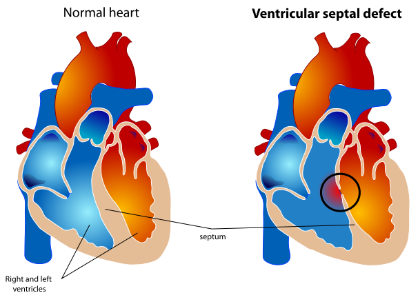
- Murmur: a harsh pansystolic murmur at the left lower sternal edge, often with a thrill
- The paradox of loudness: a small defect produces a loud murmur, because a high-velocity jet is forced through a narrow orifice. A large defect may produce a quiet murmur with a much sicker child - so murmur intensity is inversely related to severity.
- Small defects are usually asymptomatic and around half close spontaneously in the first two years
- Large defects cause heart failure in infancy - breathlessness, sweating and poor feeding, faltering growth, and recurrent chest infections - and require surgical or transcatheter closure
- Complications: heart failure, pulmonary hypertension and Eisenmenger syndrome, infective endocarditis, and aortic regurgitation from prolapse of an aortic cusp into the defect
- Acquired VSD occurs 3-5 days after myocardial infarction, presenting as a new harsh pansystolic murmur with sudden deterioration
Atrial septal defect
Often silent in childhood and frequently first diagnosed in adulthood, sometimes after a stroke.
- Types: ostium secundum (around 70%, in the region of the fossa ovalis) and ostium primum (part of the atrioventricular septal defect spectrum, associated with Down syndrome, and giving left axis deviation on the ECG)
- Murmur: an ejection systolic murmur in the pulmonary area, caused not by flow across the defect itself - which is low-pressure and silent - but by the increased volume crossing the pulmonary valve
- The classic sign: fixed splitting of the second heart sound. Normally the split widens on inspiration; in an ASD the shunt equalises the effect of respiration on the two ventricles, so the split does not vary.
- ECG: right bundle branch block with right axis deviation in secundum ASD; right bundle branch block with left axis deviation in primum ASD
- Complications: right heart dilatation and failure, atrial arrhythmia (particularly atrial fibrillation and flutter in adults), pulmonary hypertension and Eisenmenger syndrome, and paradoxical embolism causing stroke
- Treatment: transcatheter device closure for suitable secundum defects; surgical repair otherwise
- Patent foramen ovale is distinct - a residual flap-valve opening present in around 25% of adults, usually incidental, but implicated in cryptogenic stroke, where closure may be considered
Patent ductus arteriosus
The ductus arteriosus connects the pulmonary artery to the descending aorta in fetal life and normally closes within days of birth. Persistent patency causes a left-to-right shunt.
- Murmur: a continuous 'machinery' murmur below the left clavicle, present in both systole and diastole because aortic pressure exceeds pulmonary pressure throughout the cardiac cycle
- Signs: a bounding, collapsing pulse with a wide pulse pressure, from diastolic run-off into the pulmonary circulation
- Risk factors: prematurity, congenital rubella, and high altitude
- Treatment: indomethacin or ibuprofen in preterm infants (prostaglandin inhibitors promote closure), and transcatheter device closure otherwise
- Note the opposite principle: prostaglandin E1 or E2 keeps the duct open, which is life-saving in duct-dependent lesions
Coarctation of the aorta
Narrowing of the aorta, usually just distal to the left subclavian artery at the site of the ductus.
- Signs: radio-femoral delay, weak or absent femoral pulses, upper limb hypertension with lower limb hypotension, and a blood pressure difference between arms and legs
- Murmur: a systolic murmur heard over the left scapula and the back
- Associations: bicuspid aortic valve (present in up to 80%), Turner syndrome, and berry aneurysms of the circle of Willis
- Severe (preductal) coarctation presents in the neonate with shock and collapse as the duct closes, and is duct-dependent
- Mild coarctation may present in adulthood with hypertension resistant to treatment, or with the incidental finding of rib notching on chest X-ray from dilated collateral intercostal arteries
- Treatment: surgical repair or balloon angioplasty with stenting. Hypertension frequently persists after successful repair and requires lifelong follow-up.
- A useful rule: measure blood pressure in the legs as well as the arms in any young person with hypertension
Cyanotic lesions
Tetralogy of Fallot
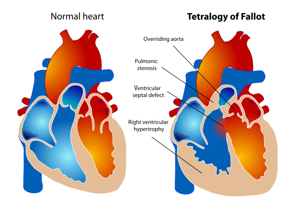
The commonest cyanotic congenital heart disease presenting beyond the neonatal period. It arises from anterior deviation of the outflow septum during development, producing four features.
- Ventricular septal defect - large and unrestrictive
- Right ventricular outflow tract obstruction (pulmonary stenosis, whether valvular, subvalvular or supravalvular) - this is the component that determines severity
- Overriding aorta - the aorta sits above the VSD, receiving blood from both ventricles
- Right ventricular hypertrophy - a consequence of the outflow obstruction
- Presentation: cyanosis, usually developing over the first weeks to months rather than at birth, with a murmur and failure to thrive
- Murmur: an ejection systolic murmur at the left sternal edge from the pulmonary stenosis, not from the VSD - a favourite exam point
- Chest X-ray: a boot-shaped heart (coeur en sabot) from right ventricular hypertrophy with a concave pulmonary artery segment, and reduced pulmonary vascular markings
- Tet spells (hypercyanotic episodes): acute worsening of right ventricular outflow obstruction causing sudden deep cyanosis, irritability and, in severe cases, syncope. Classically triggered by crying, feeding or exertion. Older children learn to squat, which increases systemic vascular resistance and reduces right-to-left shunting.
- Emergency management of a tet spell: knee-to-chest position (or squatting), oxygen, morphine to reduce agitation and hyperpnoea, intravenous fluid, and beta-blockade with propranolol. Phenylephrine may be used to raise systemic vascular resistance.
- Treatment: surgical repair, usually in the first year, closing the VSD and relieving the outflow obstruction. Adult survivors commonly develop pulmonary regurgitation and need pulmonary valve replacement, and are at risk of ventricular arrhythmia.
Transposition of the great arteries
The aorta arises from the right ventricle and the pulmonary artery from the left, creating two parallel circulations rather than one in series. Deoxygenated blood circulates around the body and oxygenated blood around the lungs.
- This is incompatible with life unless the two circulations mix, through a patent foramen ovale, VSD or patent ductus arteriosus
- Presentation: profound cyanosis within hours to days of birth, worsening dramatically as the duct closes. Cyanosis is the dominant feature and there may be no murmur at all.
- Chest X-ray: an egg-on-a-string appearance - a narrow upper mediastinum with an egg-shaped cardiac silhouette
- Immediate management: prostaglandin E infusion to maintain ductal patency, and balloon atrial septostomy to create or enlarge an atrial communication
- Definitive treatment: the arterial switch operation in the first weeks of life, with excellent long-term results
- Association: maternal diabetes
Other cyanotic lesions
- Tricuspid atresia - absence of the tricuspid valve, requiring an ASD and VSD or duct for survival, and managed by staged single-ventricle palliation culminating in the Fontan circulation
- Total anomalous pulmonary venous drainage - pulmonary veins drain into the right atrium or systemic veins rather than the left atrium; the obstructed form is a neonatal emergency with severe cyanosis and pulmonary oedema
- Truncus arteriosus - a single arterial trunk arises from the heart, with a VSD; presents with cyanosis and heart failure
- Ebstein anomaly - apical displacement of the tricuspid valve with severe tricuspid regurgitation and atrialisation of the right ventricle. Associated with maternal lithium exposure and with accessory pathways and Wolff-Parkinson-White syndrome.
- Hypoplastic left heart syndrome - duct-dependent systemic circulation requiring staged palliation
The neonate: duct-dependent circulations
The hyperoxia (nitrogen washout) test helps distinguish cardiac from respiratory causes of neonatal cyanosis: after 10 minutes of high-concentration oxygen, a PaO2 that fails to rise above approximately 20 kPa suggests a cardiac right-to-left shunt rather than lung disease. Newborn pulse oximetry screening, comparing pre-ductal (right hand) and post-ductal (foot) saturations, is now widely used to detect critical congenital heart disease before discharge.
Eisenmenger syndrome
The most important long-term complication of an uncorrected left-to-right shunt, and a favourite examination topic because it ties the whole subject together.
- A large left-to-right shunt (VSD, ASD or PDA) causes chronic pulmonary overcirculation
- Sustained high flow and pressure cause pulmonary vascular remodelling - intimal proliferation, medial hypertrophy and eventually irreversible obliterative arteriopathy
- Pulmonary vascular resistance rises until it exceeds systemic vascular resistance
- The shunt reverses to right-to-left, and the patient becomes cyanosed
- At this point the lesion is no longer correctable - closing the defect removes the pop-off valve for a suprasystemic right ventricle and causes right heart failure and death
- Clinical features: central cyanosis, clubbing, secondary polycythaemia, dyspnoea, syncope, haemoptysis, a loud P2 and signs of right heart failure. In a PDA there may be differential cyanosis and clubbing - affecting the toes but not the fingers, since the shunt enters the aorta distal to the head and arm vessels.
- Management: supportive. Avoid dehydration, high altitude and pregnancy (maternal mortality is very high). Venesection only for symptomatic hyperviscosity, and with caution because iron deficiency worsens outcomes. Pulmonary vasodilators such as bosentan and sildenafil improve symptoms and exercise capacity.
- Definitive treatment: heart-lung or lung transplantation with repair of the defect
- The lesson: significant shunts should be closed before irreversible pulmonary vascular disease develops. Eisenmenger syndrome is essentially a preventable condition, and its incidence has fallen substantially with early surgical repair.
Investigations
- Pulse oximetry - pre-ductal (right hand) and post-ductal (foot) saturations, as part of newborn screening and in any suspected case
- Four-limb blood pressure - for coarctation
- ECG - axis, chamber hypertrophy and conduction abnormalities. Left axis deviation with RBBB suggests a primum ASD; right ventricular hypertrophy suggests pulmonary stenosis or tetralogy.
- Chest X-ray - cardiac size and shape (boot-shaped in tetralogy, egg-on-a-string in transposition, snowman in supracardiac TAPVD), pulmonary vascular markings (increased in left-to-right shunts, decreased in tetralogy), and rib notching in coarctation
- Echocardiography - the definitive diagnostic investigation, defining anatomy, shunt direction and magnitude, and haemodynamics
- Hyperoxia test - to distinguish cardiac from respiratory causes of neonatal cyanosis
- Cardiac MRI and CT - for complex anatomy, right ventricular volumes, and vascular structures in adults
- Cardiac catheterisation - for haemodynamic assessment, particularly measuring pulmonary vascular resistance and its reversibility before deciding on surgery
- Genetic testing and fetal echocardiography - where a syndrome or family history is present
- Bloods - FBC (polycythaemia in cyanotic disease), iron studies, blood gas, and BNP
The adult with congenital heart disease
Adult congenital heart disease is now a subspecialty in its own right. Patients require lifelong follow-up in a specialist centre, and there are specific issues that arise repeatedly.
- Residual and recurrent lesions - pulmonary regurgitation after tetralogy repair, recoarctation, residual shunts, and prosthetic valve or conduit degeneration
- Arrhythmia - atrial flutter and fibrillation around surgical scar, and ventricular arrhythmia. This is the commonest reason for admission in adults with congenital heart disease.
- Heart failure - particularly in systemic right ventricles and in Fontan circulations
- Pulmonary hypertension
- Infective endocarditis - the risk is elevated in many lesions. UK guidance does not recommend routine antibiotic prophylaxis for dental procedures, but emphasises meticulous oral hygiene and prompt investigation of unexplained fever.1
- Pregnancy - requires pre-conception counselling and joint cardiac-obstetric care. Risk ranges from negligible in a repaired ASD to prohibitive in Eisenmenger syndrome, severe pulmonary hypertension and Marfan syndrome with a dilated aortic root.
- Contraception - oestrogen-containing methods are generally avoided in cyanotic disease and pulmonary hypertension because of thrombotic risk
- Non-cardiac surgery - needs specialist anaesthetic input and, in many cases, discussion with the congenital cardiology team
- Exercise, occupation and insurance - advice should be individualised rather than restrictive by default
- Transition from paediatric to adult services - a period of high risk for loss to follow-up, which is associated with worse outcomes
Red flags
Prognosis
Outcomes have transformed over the last five decades. Over 90% of children born with congenital heart disease now survive to adulthood, compared with around 20% in the 1940s. Small ventricular septal defects and secundum atrial septal defects, once repaired or if they close spontaneously, are compatible with a normal lifespan and normal activity.
Repaired tetralogy of Fallot has an excellent survival into middle age, though pulmonary regurgitation and right ventricular dilatation commonly require pulmonary valve replacement in the third or fourth decade, and there is a small but real risk of ventricular arrhythmia and sudden death. Transposition after arterial switch likewise has good long-term outcomes, with follow-up focused on coronary patency and neo-aortic valve function.
Single-ventricle physiology palliated with a Fontan circulation carries a more guarded prognosis. The circulation depends on passive pulmonary blood flow without a subpulmonary ventricle, and late complications include Fontan-associated liver disease, protein-losing enteropathy, arrhythmia and progressive heart failure.
Eisenmenger syndrome carries the worst outlook of the shunt lesions, with survival typically into the third or fourth decade, and it is the strongest argument for timely closure of significant left-to-right shunts. Across all lesions, the strongest predictor of good long-term outcome is continued engagement with specialist follow-up, which is why the transition from paediatric to adult services is given so much attention.
References
- NICE CG64. Prophylaxis against infective endocarditis. 2008, updated 2016. Available here
- Baumgartner H, De Backer J, Babu-Narayan SV et al. 2020 ESC Guidelines for the management of adult congenital heart disease. European Heart Journal. 2021. Available here
- NHS England. Newborn and infant physical examination (NIPE) screening programme. Available here
- British Heart Foundation. Congenital heart disease. Available here
- Regitz-Zagrosek V, Roos-Hesselink JW, Bauersachs J et al. 2018 ESC Guidelines for the management of cardiovascular diseases during pregnancy. European Heart Journal. 2018. Available here
- NICE Clinical Knowledge Summaries. Heart murmurs in children. Available here
- Resuscitation Council UK. Newborn life support guidelines. Available here
- Humpl T, Reyes JT, Holtby H et al. Beneficial effect of oral sildenafil therapy in patients with pulmonary arterial hypertension. Circulation. 2005. Available here
This article is written for revision and education. It is not clinical guidance and must not be used to make decisions about the care of a patient. Always check current NICE guidance and local protocols.