
Toxic Megacolon: Recognition and Emergency Management
Key points
- Toxic megacolon: non-obstructive dilatation of the colon (over 6 cm) accompanied by systemic toxicity, complicating severe colitis.
- Causes: most often severe ulcerative colitis; also Crohn colitis, C. difficile colitis, and other infective colitides such as Shigella and Salmonella.
- Mechanism: severe transmural inflammation impairs smooth muscle contraction via nitric oxide and inflammatory mediators, causing paralysis and dilatation.
- Precipitants: antimotility agents, opioids, anticholinergics, hypokalaemia, and colonoscopy or barium enema during severe colitis.
- Diagnosis: the Jalan criteria - radiological colonic dilatation plus systemic toxicity, with at least one additional feature such as hypotension or altered consciousness.
- Warning sign: sudden cessation of diarrhoea with worsening distension indicates deterioration, not improvement.
- Management: resuscitation, IV corticosteroids for IBD, antibiotics, bowel rest and serial abdominal X-rays with joint medical-surgical care.
- Surgery: subtotal colectomy with end ileostomy for perforation, deterioration, or failure to improve within 24-72 hours.
Introduction
Toxic megacolon is a life-threatening complication of severe colitis, defined as non-obstructive dilatation of the colon accompanied by systemic toxicity.1 The dilatation is typically most marked in the transverse colon, and a diameter exceeding 6 cm (or over 9 cm for the caecum) is the usual radiological threshold.
It is a surgical emergency in waiting. The dilated, inflamed, thin-walled colon is at high risk of perforation, which carries a mortality of up to 40%, compared with under 5% for uncomplicated toxic megacolon treated promptly. Early recognition, joint medical and surgical management, and a low threshold for colectomy are what determine outcome.
Causes
| Category | Examples |
|---|---|
| Inflammatory bowel disease | Ulcerative colitis (the commonest cause), Crohn colitis |
| Infective colitis | Clostridioides difficile, Shigella, Salmonella, Campylobacter, Entamoeba histolytica, cytomegalovirus (especially in the immunocompromised) |
| Ischaemic colitis | Following mesenteric ischaemia or a low-flow state |
| Other | Radiation colitis, obstructing colorectal carcinoma, volvulus, Behçet disease |
Precipitating factors
In a patient with severe colitis, several avoidable factors can tip them into toxic megacolon, and recognising these is a high-yield clinical point:2
- Antimotility agents such as loperamide, and opioids - these paralyse the colon and retain toxin
- Anticholinergic drugs and antidepressants with anticholinergic effects
- Hypokalaemia and other electrolyte disturbance, which impair smooth muscle function
- Colonoscopy or barium enema performed during an episode of severe colitis - avoid these, or use unprepared flexible sigmoidoscopy with minimal insufflation if endoscopic assessment is essential
- Abrupt withdrawal of corticosteroids or other maintenance therapy in inflammatory bowel disease
Pathophysiology
In severe colitis, inflammation extends beyond the mucosa into the muscularis propria, damaging the smooth muscle and the myenteric plexus. Inflammatory mediators, particularly nitric oxide released by inflammatory cells, together with interleukins and leukotrienes, inhibit smooth muscle contractility.1
The result is colonic paralysis and progressive dilatation. As the colon distends, wall tension rises in accordance with the law of Laplace, compromising mucosal blood flow and causing ischaemia. This further weakens the wall, permits bacterial translocation across the damaged mucosa - driving the systemic toxaemia and sepsis - and ultimately leads to perforation.
Clinical features
Toxic megacolon develops in a patient who is already unwell with colitis, usually within the first week of a severe flare, though it can be the presenting feature.
Abdominal features
- Abdominal distension, often marked and progressive
- Abdominal pain and tenderness, which may become generalised
- Reduced or absent bowel sounds, reflecting the ileus
- Paradoxical reduction in stool frequency: diarrhoea that had been severe may lessen or stop as the colon becomes paralysed. This is a deteriorating sign, not an improving one, and is a classic trap
- Signs of peritonism, which may be masked by corticosteroids - a critically important caveat
Systemic toxicity
- Fever above 38°C
- Tachycardia above 120 beats per minute
- Hypotension and signs of shock
- Dehydration and reduced urine output
- Altered mental state: confusion, lethargy or agitation
- Anaemia, leucocytosis and electrolyte disturbance
Diagnosis
The diagnosis is clinical and radiological, most commonly formalised using the Jalan criteria.3
| Component | Requirement |
|---|---|
| Radiological | Radiographic evidence of colonic dilatation, typically transverse colon greater than 6 cm |
| Systemic toxicity (at least 3 of) | Temperature above 38°C; heart rate above 120 beats per minute; neutrophil count above 10.5 × 10⁹/L; anaemia |
| Plus at least 1 of | Dehydration; altered consciousness; electrolyte disturbance; hypotension |
Investigations
- Abdominal X-ray: the key initial investigation, showing colonic dilatation (commonly the transverse colon), loss of haustration, mucosal islands from ulceration, and thumbprinting from mucosal oedema. Serial films, often daily or more frequently, are used to monitor progression
- Erect chest X-ray: to look for free air under the diaphragm indicating perforation
- CT abdomen and pelvis: more sensitive for perforation, abscess and the extent of colitis, and helps distinguish other causes of dilatation
- Bloods: full blood count (anaemia, leucocytosis), urea and electrolytes (hypokalaemia), CRP, albumin (low in severe disease), lactate, and group and save
- Stool studies: culture and C. difficile toxin, as infection may be the cause or a coexisting precipitant
- Blood cultures if febrile or septic
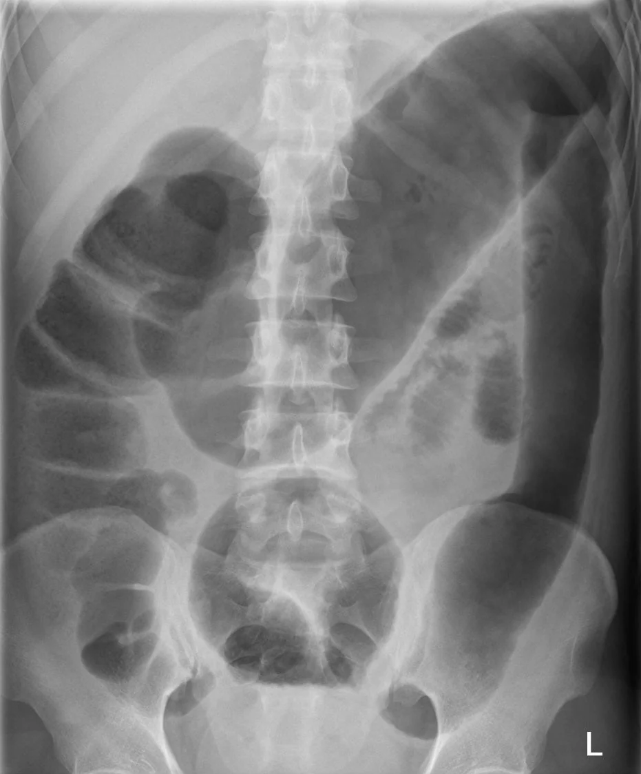
Colonoscopy and barium enema are contraindicated, as insufflation and the procedure itself substantially increase perforation risk. If endoscopic assessment is essential, a cautious unprepared flexible sigmoidoscopy with minimal air insufflation may be performed by an experienced operator.
Differential diagnosis
- Mechanical large bowel obstruction: from colorectal cancer, stricture or hernia - imaging shows a transition point, and there is no systemic toxicity of the same degree
- Sigmoid or caecal volvulus: characteristic coffee bean sign on abdominal X-ray
- Acute colonic pseudo-obstruction (Ogilvie syndrome): colonic dilatation without inflammation or systemic toxicity, typically in unwell, immobile or postoperative patients
- Severe colitis without megacolon: systemic toxicity but no significant dilatation
- Ileus from any cause, including postoperative and electrolyte-related
Management
Toxic megacolon requires admission to a high-dependency or intensive care setting, with joint medical and surgical management from the outset - the surgical team should be involved on day one, not at the point of deterioration.2
Resuscitation and supportive care
- ABCDE assessment with aggressive IV fluid resuscitation and correction of electrolytes, particularly potassium and magnesium
- Nil by mouth with bowel rest; insert a nasogastric tube for decompression if there is vomiting or ileus
- Stop all precipitating drugs: antimotility agents, opioids, anticholinergics and NSAIDs
- Broad-spectrum IV antibiotics, given the risk of bacterial translocation, sepsis and perforation
- Venous thromboembolism prophylaxis - the thrombotic risk in acute colitis is high despite the bleeding risk
- Nutritional support, with parenteral nutrition if bowel rest is prolonged
- Serial abdominal examination and X-rays, with regular observations, bloods and lactate to track the trajectory
Treating the underlying cause
- Inflammatory bowel disease: IV corticosteroids (hydrocortisone 100 mg four times daily). If there is no response by day 3, escalate to rescue therapy with infliximab or ciclosporin, with the surgical team fully involved in that decision
- C. difficile colitis: oral vancomycin plus IV metronidazole for life-threatening disease; IV metronidazole is essential here because ileus prevents oral drug reaching the colon
- Other infective causes: targeted antimicrobial therapy guided by stool and blood cultures
Some centres use positional changes, such as regular repositioning to the prone or knee-elbow position, to encourage passive decompression of colonic gas, though evidence for this is limited.
Surgery
Subtotal colectomy with end ileostomy is the operation of choice, preserving the rectum so that restorative surgery such as an ileo-anal pouch can be considered later once the patient has recovered. Indications for urgent surgery are:
- Perforation - an absolute indication
- Massive haemorrhage
- Clinical deterioration or progressive colonic dilatation despite maximal medical therapy
- Failure to improve within 24-72 hours of intensive medical treatment
- Signs of worsening sepsis or multi-organ failure
The most common and most damaging error in management is delaying surgery in a patient who is not improving. Operating on a deteriorating but not yet perforated patient carries far lower mortality than operating after perforation has occurred.
Complications
- Colonic perforation and faecal peritonitis - the feared outcome, with mortality up to 40%
- Septic shock and multi-organ failure
- Massive lower gastrointestinal haemorrhage
- Acute kidney injury from hypovolaemia and sepsis
- Venous thromboembolism
- Abdominal compartment syndrome
- Complications of emergency surgery: anastomotic and stoma problems, wound infection, and the physical and psychological adjustment to a stoma
- Malnutrition, and the long-term consequences of colectomy
Red flags
Prognosis
Prognosis depends overwhelmingly on whether perforation occurs and how promptly surgery is undertaken. Mortality for toxic megacolon recognised and treated before perforation is under 5%, but rises to around 40% once perforation has taken place.1 This steep difference is the single most important reason for early surgical involvement and a low threshold for colectomy.
Around half of patients with toxic megacolon complicating inflammatory bowel disease ultimately require colectomy, which is curative for the colonic disease in ulcerative colitis. Those managed successfully with medical therapy alone remain at risk of recurrence during subsequent flares and generally require escalation of their maintenance therapy. Outcomes are worse in older patients, those with significant comorbidity, and those in whom the diagnosis or the decision to operate is delayed.
References
- Desai J et al. Toxic Megacolon: Background, Pathophysiology, Management Challenges and Solutions. Clin Exp Gastroenterol. 2020. Available here
- Lamb CA et al. British Society of Gastroenterology consensus guidelines on the management of inflammatory bowel disease in adults. Gut. 2019. Available here
- Jalan KN et al. An experience of ulcerative colitis: toxic dilation in 55 cases. Gastroenterology. 1969. Available here
- NICE NG130. Ulcerative colitis: management. 2019. Available here
- Hellerhoff, CC BY-SA 4.0, via Wikimedia Commons. Available here
- NICE NG199. Clostridioides difficile infection: antimicrobial prescribing. 2021. Available here
- Truelove SC, Witts LJ. Cortisone in ulcerative colitis: final report on a therapeutic trial. BMJ. 1955. Available here
This article is written for revision and education. It is not clinical guidance and must not be used to make decisions about the care of a patient. Always check current NICE guidance and local protocols.