
Down Syndrome: Trisomy 21
Key points
- Down syndrome: the clinical syndrome resulting from three copies of chromosome 21 (trisomy 21), the commonest chromosomal cause of learning disability.
- Mechanism: about 95% arise from meiotic non-disjunction, strongly age-related; the remainder from unbalanced Robertsonian translocation or mosaicism.
- Recurrence risk: roughly 1%, or the mother's age-related risk if higher, after a non-disjunction trisomy; substantially higher if a parent carries a balanced translocation.
- Cardiac: congenital heart disease in ~40-50%, most often an atrioventricular septal defect - echocardiography is done in every neonate regardless of clinical examination.
- Screening: combined test at 11-14 weeks (nuchal translucency, hCG, PAPP-A) or NIPT; diagnosis requires CVS or amniocentesis.
- Surveillance: a structured, lifelong programme covering hearing, vision, thyroid, cardiac and haematological review, set out in UK Down syndrome health guidelines.
- Leukaemia risk: 10-20 fold increased risk of acute leukaemia, and a unique transient abnormal myelopoiesis in the neonatal period.
- Life expectancy: now commonly into the 60s in the UK, transformed chiefly by cardiac surgery and structured health surveillance.
Introduction
Down syndrome is the clinical syndrome caused by an extra copy of chromosome 21. It is the commonest chromosomal cause of learning disability and the commonest autosomal trisomy compatible with survival to term, with a UK live birth incidence of around 1 in 800-1,000, though this varies with maternal age and the uptake of antenatal screening.1
It is a favourite exam topic precisely because it spans the whole curriculum: cytogenetics and recurrence risk, antenatal screening, dysmorphology, congenital heart disease, and the long-term, multisystem surveillance that has transformed life expectancy over the last 50 years. This article covers the mechanism, recognition and management; the screening pathway that identifies affected pregnancies is covered in more detail in Antenatal Genetic Screening.
Aetiology and mechanism
All three mechanisms produce the same extra dose of chromosome 21 material and a similar phenotype, but they carry very different recurrence risks, which is why establishing the mechanism by karyotype is essential before counselling a family.
| Mechanism | Proportion | Karyotype | Recurrence risk |
|---|---|---|---|
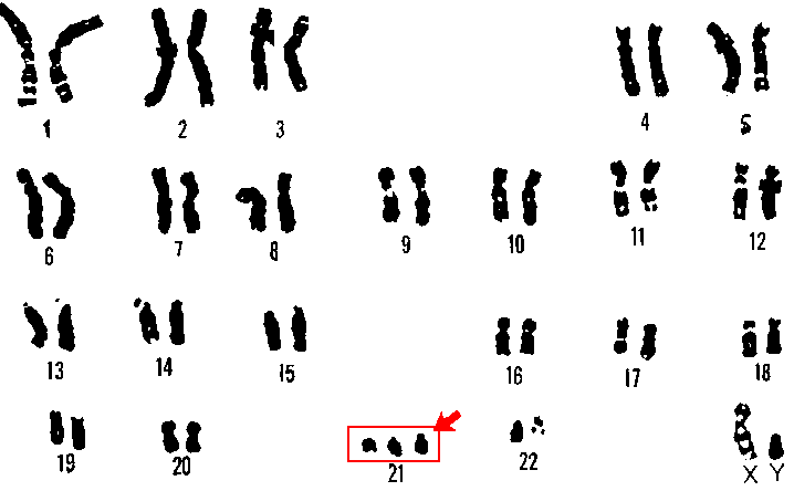
| Meiotic non-disjunction | ~95% | Free trisomy 21 (47,XX or XY,+21) | ~1%, or the mother's age-related risk if higher |
| Robertsonian translocation | ~4% | Chromosome 21 fused to another acrocentric chromosome (usually 14) | Up to ~10-15% if mother carries the balanced translocation, ~1% if father does; ~100% if a parent carries a rare 21;21 translocation |
| Mosaicism | ~1% | A mixture of trisomy 21 and normal cell lines | Low; not usually inherited, and phenotype can be milder depending on the proportion of trisomic cells |
Non-disjunction is an error during meiosis in which a pair of chromosome 21 fails to separate, so one gamete receives two copies and the resulting zygote has three. Around 90% of cases arise from an error in maternal meiosis I, and the risk rises sharply with maternal age, from roughly 1 in 1,500 at age 20 to around 1 in 100 by age 40 and higher again beyond that.2
Clinical features at birth
The dysmorphic features are individually non-specific but highly suggestive in combination, and the diagnosis is often suspected clinically at birth, prompting urgent karyotype confirmation.
- Craniofacial: brachycephaly with a flat occiput, upslanting palpebral fissures, epicanthic folds, Brushfield spots on the iris, a flat nasal bridge, a small mouth with a protruding tongue, and small, low-set ears
- Hands and feet: a single palmar (simian) crease, a short, incurved fifth finger (clinodactyly), and a wide sandal gap between the first and second toes
- Tone: significant hypotonia, contributing to poor feeding and a characteristic 'floppy' handling in the neonatal period
- Growth: birth weight and length usually within the normal range, but with growth trajectories that fall below standard centile charts thereafter, hence the use of Down syndrome-specific growth charts
- Other: a short neck with excess nuchal skin, and single umbilical artery in a minority
Differential diagnosis
Several other conditions produce a dysmorphic neonate with hypotonia, and the karyotype is what ultimately distinguishes them, though the pattern of features often gives a strong clinical steer before the result returns.
- Edwards syndrome (trisomy 18) - low birth weight, clenched overlapping fingers, rocker-bottom feet, micrognathia, and a much higher rate of major structural malformation; median survival is measured in days to weeks
- Patau syndrome (trisomy 13) - midline defects including cleft lip/palate, holoprosencephaly, polydactyly and scalp defects; similarly poor prognosis
- Congenital hypothyroidism - can present with hypotonia, a large tongue and a flat facial profile, but lacks the characteristic hand and eye findings, and is excluded by the newborn screen
- Zellweger syndrome and other peroxisomal disorders - severe hypotonia and dysmorphism, distinguished by very abnormal metabolic screening
- Prader-Willi syndrome - neonatal hypotonia and poor feeding overlap, but the facial gestalt and subsequent hyperphagic phase differ, and it is distinguished by chromosome 15 microarray or methylation testing rather than karyotype
Associated congenital anomalies and complications
The extra dose of chromosome 21 affects development across almost every system, which is why every neonate with Down syndrome receives a structured panel of assessments rather than only those with an obvious clinical problem.
Cardiac
Congenital heart disease affects 40-50% and is the leading cause of early mortality, so every baby has an echocardiogram, regardless of clinical examination findings, since a haemodynamically significant defect can be silent in the first days of life.3
- Atrioventricular septal defect (AVSD) - the single commonest lesion and highly characteristic of Down syndrome; a complete AVSD usually needs surgical repair in infancy
- Ventricular septal defect
- Atrial septal defect
- Tetralogy of Fallot and patent ductus arteriosus, less commonly
Gastrointestinal
- Duodenal atresia - presents with bilious vomiting and the classic 'double bubble' sign on abdominal X-ray
- Hirschsprung disease - delayed passage of meconium and abdominal distension
- Coeliac disease - substantially increased prevalence, screened for periodically through childhood
- Anorectal malformations, less commonly
Endocrine and haematological
- Hypothyroidism - both congenital and acquired autoimmune thyroid disease occur at increased rates, so thyroid function is checked at birth and annually thereafter
- Transient abnormal myelopoiesis (TAM) - a unique neonatal clonal blood disorder in around 10% of babies with Down syndrome, usually resolving spontaneously but carrying a risk of later leukaemia
- Acute leukaemia - a 10-20 fold increased lifetime risk compared with the general population, with acute megakaryoblastic leukaemia (AML-M7) disproportionately represented, alongside increased acute lymphoblastic leukaemia
- Polycythaemia in the neonatal period
Sensory, musculoskeletal and other
- Hearing loss - conductive (from recurrent otitis media with effusion, common due to small ear canals and eustachian tube dysfunction) and sensorineural; annual audiology assessment is routine
- Visual problems - refractive errors, strabismus, cataracts and nystagmus are all more frequent; annual ophthalmology review
- Atlantoaxial instability - ligamentous laxity increases the risk of cervical spine subluxation; symptomatic instability (neck pain, gait change, new incontinence) needs urgent imaging, though routine screening X-rays of asymptomatic children are no longer recommended
- Obstructive sleep apnoea - from a combination of relative macroglossia, midface hypoplasia and hypotonia
- Immune dysfunction - increased susceptibility to infection, contributing to the historically high rate of childhood respiratory illness
Cognitive and developmental features
Learning disability is universal but ranges from mild to severe, and is not predictable from the karyotype or from physical features. Early developmental milestones, particularly motor and speech-language, are typically delayed, and expressive language is usually more affected than comprehension.
Adults with Down syndrome are at markedly increased risk of early-onset Alzheimer's disease, related to the extra copy of the amyloid precursor protein (APP) gene, which also sits on chromosome 21. Neuropathological changes can appear from the 30s, with clinical dementia becoming increasingly common from the 40s and 50s onwards, and this is now a major focus of adult surveillance.4
Early developmental input makes a measurable difference. Structured early intervention - speech and language therapy, physiotherapy, and portage or similarly structured early-years educational support - is associated with better long-term functional communication, mobility and independence, and should start in infancy rather than waiting for a formal diagnosis of delay. Hearing and vision impairment are common, treatable, and easily missed causes of apparent additional cognitive or behavioural difficulty, which is one of the main reasons annual audiology and ophthalmology review is built into the surveillance schedule rather than left to parental concern to trigger.
Examination approach
A structured top-to-toe examination in a newborn with suspected Down syndrome should specifically document tone, the cardiac examination (while remembering its limited sensitivity for AVSD, hence the mandatory echocardiogram), abdominal examination for distension suggesting bowel obstruction, and a check for red reflexes and hip stability, alongside the dysmorphic features described above. In an older child or adult, examination should be framed around the surveillance domains - growth, hearing, vision, thyroid and cardiac status, and any new neurological or behavioural change that might signal atlantoaxial instability or, in later adulthood, emerging cognitive decline.
Health surveillance
Because problems can be asymptomatic and evolve throughout life, care is organised around a structured, lifelong surveillance schedule rather than opportunistic review, set out in UK guidelines and often coordinated through a Down syndrome health passport shared between primary and secondary care.5
| Domain | Schedule (indicative) |
|---|---|
| Echocardiogram | At birth, regardless of examination findings |
| Thyroid function | At birth, then annually |
| Hearing assessment | At birth (newborn screen) and at least annually in childhood |
| Vision/ophthalmology | By 12 months, then regularly through childhood |
| Growth | Plotted on Down syndrome-specific growth charts |
| Full blood count | At birth (screen for transient abnormal myelopoiesis) and if clinically indicated thereafter |
| Coeliac screening | From around age 2, or earlier if symptomatic |
| Sleep assessment | If symptoms suggest obstructive sleep apnoea |
| Dementia screening in adults | From around age 40, using tools adapted for baseline learning disability |
Diagnosis
Antenatally, combined or NIPT screening identifies a high-chance pregnancy, and diagnosis is confirmed by chorionic villus sampling or amniocentesis with rapid aneuploidy testing (QF-PCR) followed by full karyotype, which also identifies the mechanism. Postnatally, a clinically suspected diagnosis is confirmed the same way: rapid QF-PCR gives a result within a day or two, confirming the aneuploidy and allowing early counselling and planning, with a full karyotype following over the next one to two weeks to distinguish free trisomy from translocation or mosaicism and to inform recurrence risk counselling.6 The full antenatal pathway, including the calculation of a combined-test chance and the difference between screening and diagnostic tests, is covered in Antenatal Genetic Screening.
Breaking the diagnosis, whether antenatally or in the neonatal period, deserves particular care. Parents consistently report that how the news is delivered - promptly, by a senior clinician, in a private setting, in balanced and unhurried language that does not lead with a list of problems - shapes their experience of the whole subsequent journey. Both parents should be present where possible, and written information and a point of contact for parent support organisations should be offered at the same meeting rather than deferred to a later appointment.
Genetic counselling and recurrence risk
Recurrence risk counselling depends entirely on the mechanism identified on karyotype, which is why karyotyping - not just a rapid aneuploidy test - is performed in every case.
- Free trisomy 21 (non-disjunction): recurrence risk in a future pregnancy is approximately 1% above the mother's baseline age-related risk, or simply her age-related risk if that is already higher than 1%
- Robertsonian translocation: risk depends on which parent carries it and which chromosomes are involved - substantially higher if the mother is the carrier (around 10-15%) than the father (around 1-2%), and effectively universal if a parent carries the rare translocation involving both copies of chromosome 21 fusing together
- Mosaicism: generally a low recurrence risk, as it usually reflects a post-zygotic event rather than a parental predisposition
Prenatal diagnosis is offered in future pregnancies, and, once a family's specific mechanism is known, at-risk relatives (particularly siblings of a translocation carrier parent) can be offered their own karyotype.
Education and transition to adulthood
Most children with Down syndrome in the UK are supported through mainstream education with an Education, Health and Care Plan, though the right setting varies with the degree of learning disability and associated needs, and is reviewed regularly rather than fixed at diagnosis. Speech and language therapy and physiotherapy input from infancy materially improve functional communication and mobility outcomes.
Transition planning into adult services should begin in early adolescence, covering ongoing health surveillance, further education or supported employment, housing, and capacity and welfare arrangements as the young person reaches adulthood. Fertility is preserved in women with Down syndrome, and contraceptive and reproductive counselling should be offered as part of routine transition care; almost all men with Down syndrome are infertile.
Prognosis
Life expectancy has increased dramatically, from a median of around 25 years in the 1980s to commonly into the 60s in the UK today, driven chiefly by cardiac surgery for congenital heart disease and by structured, proactive health surveillance rather than any single treatment.7
The major determinants of outcome are the presence and severity of congenital heart disease, and, in later adulthood, the onset of Alzheimer's disease. With appropriate support most adults with Down syndrome can achieve a good quality of life, participate in education and employment to varying degrees, and live semi-independently or within supported settings.
References
- Public Health England / NHS. Congenital Anomaly and Rare Disease Registration Service (NCARDRS) annual data. Available here
- Morris JK, Mutton DE, Alberman E. Revised estimates of the maternal age specific live birth prevalence of Down's syndrome. Journal of Medical Screening. 2002. Available here
- Bergstrom S, Carr H, Petersson G et al. Trends in congenital heart defects in infants with Down syndrome. Pediatrics. 2016. Available here
- Wiseman FK, Al-Janabi T, Hardy J et al. A genetic cause of Alzheimer disease: mechanistic insights from Down syndrome. Nature Reviews Neuroscience. 2015. Available here
- Down's Syndrome Medical Interest Group (DSMIG-UK). Basic medical surveillance essentials for people with Down syndrome. Available here
- NICE. Antenatal care NG201. 2021. Available here
- Glasson EJ, Jacques A, Wong K et al. Improved survival in Down syndrome over the last 60 years. Journal of Pediatrics. 2016. Available here
This article is written for revision and education. It is not clinical guidance and must not be used to make decisions about the care of a patient. Always check current NICE guidance and local protocols.