
Aortic Dissection
Key points
- Aortic dissection: a tear in the intima allows blood to track into the media, creating a false lumen that propagates along the vessel.
- Stanford A: involves the ascending aorta, whatever the site of the tear. Around two thirds of cases, and a surgical emergency.
- Stanford B: confined to the aorta distal to the left subclavian artery. Managed medically unless complicated.
- Dominant risk factor: hypertension, present in around three quarters. Connective tissue disease, bicuspid aortic valve and cocaine account for most younger patients.
- Classic pain: sudden, maximal at onset, tearing or ripping, in the anterior chest for type A and interscapular for type B, and often migratory.
- Key signs: a blood pressure difference of more than 20 mmHg between arms, absent or unequal pulses, and a new early diastolic murmur of aortic regurgitation.
- Investigation: CT angiography of the whole aorta is the definitive test. Chest radiograph may show a widened mediastinum but is normal in a substantial minority.
- Medical treatment: beta-blockade first to reduce heart rate and the rate of rise of aortic pressure, then a vasodilator. Never a vasodilator alone.
Introduction and classification
Aortic dissection occurs when a tear in the intima allows pulsatile blood to enter the media, splitting it along its length and creating a false lumen parallel to the true one. The false lumen may rupture outwards, compress the true lumen, or occlude the origins of branch vessels - and it is this last mechanism, malperfusion, that produces the extraordinary variety of presentations.
It is uncommon, at roughly 3 to 4 cases per 100,000 per year, but it is a diagnosis every clinician must be able to consider, because untreated type A dissection kills at a rate of 1 to 2% per hour for the first 48 hours. It is also, notoriously, one of the most frequently missed diagnoses in emergency medicine, because it can convincingly imitate myocardial infarction, stroke, pulmonary embolism, renal colic and an acute abdomen.
| System | Class | Definition | Management |
|---|---|---|---|
| Stanford | Type A | Involves the ascending aorta, regardless of where the intimal tear is. About 60 to 70% of cases. | Emergency cardiothoracic surgery |
| Stanford | Type B | Does not involve the ascending aorta - begins distal to the origin of the left subclavian artery | Medical management unless complicated, then TEVAR |
| DeBakey | Type I | Originates in the ascending aorta and extends beyond the arch | Surgical (equivalent to Stanford A) |
| DeBakey | Type II | Confined to the ascending aorta | Surgical (equivalent to Stanford A) |
| DeBakey | Type III | Originates in the descending aorta, IIIa confined to the thorax and IIIb extending below the diaphragm | Equivalent to Stanford B |
Stanford is the classification to know, because it maps directly onto the treatment decision. Dissections are also described as acute within 14 days of onset, subacute from 15 to 90 days, and chronic beyond that, since the risk of complications and the surgical approach change with time.
Risk factors
Two mechanisms produce dissection: increased wall stress, and a weakened media. Most risk factors act through one or the other.
Increased aortic wall stress
- Hypertension - present in around 75% of patients and the single most important risk factor
- Cocaine and amphetamine use - the classic cause in a young patient with chest pain and a normal aorta, through acute catecholamine surge
- Intense isometric exertion, such as heavy weightlifting
- Aortic coarctation and phaeochromocytoma
- Pregnancy, particularly the third trimester and the peripartum period, and especially in women with a connective tissue disorder or bicuspid valve
- Trauma - deceleration injury classically tears the aorta at the ligamentum arteriosum, just distal to the left subclavian artery
Weakened aortic media
- Marfan syndrome - the commonest connective tissue cause, and the reason to look at a young tall patient's hands, palate and lens position
- Vascular (type IV) Ehlers-Danlos syndrome and Loeys-Dietz syndrome
- Bicuspid aortic valve, which is associated with an intrinsic aortopathy independent of the valve lesion
- Turner syndrome
- Vasculitis - giant cell arteritis and Takayasu arteritis
- Existing aortic aneurysm and a family history of dissection or thoracic aneurysm
- Iatrogenic - cardiac catheterisation, intra-aortic balloon pump, and previous cardiac or aortic surgery, which together account for a meaningful minority of cases
- Tertiary syphilis, now rare in the UK but historically important
Clinical features
Pain
- Sudden onset and maximal at the moment it starts - this instantaneous quality is the single most useful discriminator from myocardial infarction, where pain builds over minutes
- Tearing, ripping or stabbing in character, though many patients simply describe it as the worst pain of their life
- Anterior chest pain suggests type A; interscapular or back pain suggests type B
- Migratory pain that moves as the dissection propagates - from chest to back to abdomen - is highly suggestive and worth asking about directly
- Painless dissection occurs in around 5 to 10%, more often in patients with Marfan syndrome, diabetes or previous aortic surgery, and typically presents with syncope or a neurological deficit
Signs and the malperfusion syndromes
The false lumen compresses or shears off branch arteries as it propagates, so almost any organ can be affected. Recognising this pattern - severe chest pain plus an apparently unrelated ischaemic event - is what makes the diagnosis.
| Vessel affected | Consequence | Clinical sign |
|---|---|---|
| Coronary arteries, usually the right | Myocardial infarction | ST elevation, classically inferior. Thrombolysis or antiplatelets given here are catastrophic. |
| Aortic valve and root | Acute aortic regurgitation | New early diastolic murmur, wide pulse pressure, acute pulmonary oedema |
| Pericardium | Haemopericardium and cardiac tamponade | Hypotension, raised JVP, muffled heart sounds, pulsus paradoxus |
| Brachiocephalic and carotid arteries | Stroke | Hemiparesis, dysphasia, or a reduced conscious level |
| Subclavian arteries | Upper limb malperfusion | Blood pressure differential of more than 20 mmHg between arms, absent or unequal radial pulses |
| Spinal arteries | Spinal cord infarction | Paraplegia with a sensory level, often painless |
| Coeliac and mesenteric arteries | Mesenteric ischaemia | Severe abdominal pain out of proportion to the findings, rising lactate |
| Renal arteries | Renal infarction | Flank pain, haematuria, acute kidney injury, refractory hypertension |
| Iliac arteries | Acute limb ischaemia | Cold, pale, pulseless painful leg |
| Free rupture | Exsanguination | Into pericardium, left pleural cavity or mediastinum - usually fatal |
Investigations
Bedside
- Blood pressure in both arms, and comparison of all peripheral pulses. A difference of more than 20 mmHg is significant, though it is present in only about a third of cases.
- 12-lead ECG - normal in around a third, non-specific ST or T changes in another third, and frank ST elevation in a minority. A normal ECG in severe chest pain should raise rather than lower your suspicion of dissection.
- Cardiac monitoring and continuous observations
Imaging
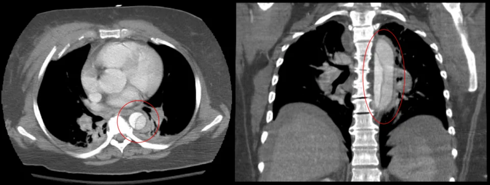
- CT angiography of the whole aorta, from the thoracic inlet to the femoral arteries, is the definitive investigation. Sensitivity and specificity both exceed 95%. It shows the intimal flap, defines the Stanford class, maps branch vessel involvement, and identifies pericardial and pleural blood.1
- Chest radiograph - may show a widened mediastinum above 8 cm, loss of the aortic knuckle, a left pleural effusion, tracheal deviation, or separation of intimal calcification from the aortic outline by more than 5 mm (the calcium sign). It is normal in 10 to 20% of dissections, so it cannot exclude the diagnosis.
- Transoesophageal echocardiography - excellent for the ascending aorta and the aortic valve, and the imaging of choice in a patient too unstable to move to the CT scanner. It also identifies tamponade and quantifies aortic regurgitation.
- Transthoracic echocardiography - quick and available at the bedside, useful for tamponade, aortic regurgitation and a dilated root, but it does not exclude dissection
- MR angiography - highly accurate but slow, and generally reserved for stable or chronic cases and follow-up
Blood tests
- FBC, U&Es, LFTs, clotting, group and crossmatch at least six units, and a venous or arterial gas with lactate
- Troponin - may be raised from coronary involvement or demand ischaemia, and a raised troponin does not exclude dissection
- D-dimer - almost always elevated, with a high sensitivity. A negative D-dimer combined with a low Aortic Dissection Detection Risk Score makes dissection unlikely, but a D-dimer must never be used alone to rule it out in a patient with a convincing history.2
- Creatinine - baseline before contrast and to detect renal malperfusion
Management
Immediate measures for all patients
- A to E assessment, high-flow oxygen if hypoxic, continuous cardiac monitoring, and two large-bore cannulae
- Urgent senior and specialist involvement - cardiothoracic surgery for type A, vascular surgery or a specialist aortic centre for type B. This call should be made on suspicion, not on confirmation.
- Adequate analgesia with intravenous morphine. Pain drives catecholamine release, so treating it is part of controlling the blood pressure, not merely humane.
- Arterial line for beat-to-beat blood pressure monitoring, sited in the arm with the higher pressure
- Crossmatch and activate the major haemorrhage protocol if there is any haemodynamic instability
- Do not give antiplatelets, anticoagulants or thrombolysis
Blood pressure and heart rate control
The therapeutic target is not simply a lower blood pressure but a lower rate of rise of aortic pressure - dP/dt - because it is the force of each pulse against the intimal flap that propagates the dissection. This is why the sequence of drugs matters.
- Intravenous beta-blocker first - labetalol or esmolol by infusion, titrated to a heart rate below 60 beats per minute and a systolic blood pressure of 100 to 120 mmHg, or the lowest pressure that maintains cerebration and urine output
- Add a vasodilator second if the pressure remains above target - sodium nitroprusside or a glyceryl trinitrate infusion
- Never give a vasodilator before beta-blockade. Vasodilatation alone causes reflex tachycardia and a greater rate of rise of aortic pressure, which accelerates propagation of the dissection.
- Where beta-blockers are contraindicated, for example in asthma, a rate-limiting calcium channel blocker such as intravenous diltiazem or verapamil is used instead
Type A dissection
This is a surgical emergency. Untreated mortality is around 1 to 2% per hour in the first 48 hours, and roughly half of patients are dead within a week. Emergency open repair replaces the ascending aorta with an interposition graft, with the aortic valve repaired or replaced and the root and arch addressed as required, usually under deep hypothermic circulatory arrest. Operative mortality is high, in the region of 15 to 25%, but far lower than the alternative.
Type B dissection
Uncomplicated type B dissection is managed medically, with strict blood pressure and heart rate control in a monitored setting, serial imaging and lifelong follow-up. Medical management gives better outcomes than surgery in this group, and the aorta remodels in many patients.
- Complicated type B - defined by rupture or impending rupture, malperfusion of a visceral, renal, spinal or limb artery, refractory pain, refractory hypertension, or rapid aortic expansion
- Complicated type B requires intervention, usually thoracic endovascular aortic repair (TEVAR), in which a covered stent graft is deployed to seal the entry tear and re-expand the true lumen
- Open surgery is reserved for cases where TEVAR is not anatomically feasible, and carries substantial risk including paraplegia
- There is growing evidence for pre-emptive TEVAR in selected uncomplicated type B dissections with high-risk features, to promote favourable remodelling and prevent late aneurysmal degeneration
Long-term management
- Lifelong antihypertensive therapy, usually including a beta-blocker, with a target of below 130/80 mmHg
- Surveillance imaging with CT or MR at intervals, since the false lumen can dilate aneurysmally over years
- Smoking cessation and cardiovascular risk reduction
- Avoidance of heavy isometric exercise and competitive contact sport
- Genetic assessment and screening of first-degree relatives where a heritable aortopathy is suspected, which is a commonly forgotten step
- Cardiology review before any future pregnancy in a woman with a connective tissue disorder or previous dissection
Complications
- Rupture into the pericardium, mediastinum or left pleural cavity - the commonest cause of death
- Cardiac tamponade, particularly with type A. Pericardiocentesis is generally avoided because relieving the tamponade can restore the pressure that drives further bleeding; the treatment is surgery.
- Acute severe aortic regurgitation with pulmonary oedema and cardiogenic shock
- Myocardial infarction from coronary ostial involvement
- Stroke and spinal cord infarction with paraplegia
- Mesenteric, renal and limb ischaemia from branch vessel malperfusion
- Late aneurysmal dilatation of the false lumen, which is why surveillance is lifelong
- Post-operative complications - bleeding, stroke, renal failure, prolonged ventilation, and paraplegia after descending aortic repair or TEVAR
Red flags
Prognosis
Type A dissection carries a mortality of approximately 1 to 2% per hour for the first 48 hours if untreated, with around half of patients dead by seven days and the great majority within a month. Emergency surgery reduces in-hospital mortality to roughly 15 to 25%, and survivors who reach discharge generally do well, with five-year survival in the region of 70%.
Uncomplicated type B dissection managed medically has an in-hospital mortality of around 10%, considerably better than the 20 to 30% seen when it becomes complicated and requires intervention. The longer-term issue for these patients is aneurysmal degeneration of the false lumen, which occurs in perhaps a quarter to a third over five years and is the reason surveillance imaging must not lapse.
The determinant of survival that is actually within a junior clinician's control is the time to diagnosis. Registry data consistently show a substantial proportion of dissections are not suspected on first assessment, and the delay is measured in hours. Asking two questions - was the pain maximal the moment it started, and is the blood pressure the same in both arms - costs nothing and is what turns a missed diagnosis into a survivable one.
References
- Isselbacher EM, Preventza O, Hamilton Black J et al. 2022 ACC/AHA guideline for the diagnosis and management of aortic disease. Circulation. 2022. Available here
- Nazerian P, Mueller C, Soeiro AM et al. Diagnostic accuracy of the aortic dissection detection risk score plus D-dimer for acute aortic syndromes (ADvISED). Circulation. 2018. Available here
- Erbel R, Aboyans V, Boileau C et al. ESC guidelines on the diagnosis and treatment of aortic diseases. European Heart Journal. 2014. Available here
This article is written for revision and education. It is not clinical guidance and must not be used to make decisions about the care of a patient. Always check current NICE guidance and local protocols.