
Ehlers-Danlos Syndrome: Classification and Management
Key points
- Ehlers-Danlos syndrome: a group of heritable connective tissue disorders characterised by joint hypermobility, skin hyperextensibility and tissue fragility.
- Classification: the 2017 international classification recognises 13 subtypes; hypermobile, classical and vascular are the three that matter for finals.
- Hypermobile EDS (hEDS): by far the commonest, diagnosed on clinical criteria alone because no causative gene has been identified.
- Classical EDS (cEDS): COL5A1 or COL5A2 variants; marked skin hyperextensibility with widened atrophic 'cigarette paper' scarring.
- Vascular EDS (vEDS): COL3A1 variants; arterial, bowel and uterine rupture - rare, but the subtype that causes sudden death in early adulthood.
- Beighton score: a 9-point bedside assessment of generalised joint hypermobility; the threshold is age- and sex-dependent.
- Management: no disease-modifying treatment; physiotherapy focused on joint stabilisation, pain management, and screening for associated dysautonomia and gastrointestinal symptoms.
- Vascular EDS specifics: celiprolol reduces arterial events; avoid arteriography and elective surgery where possible, and manage pregnancy as high risk.
Introduction
Ehlers-Danlos syndrome (EDS) is not one condition but a group of heritable disorders of connective tissue, unified by a triad of joint hypermobility, skin hyperextensibility and tissue fragility. The underlying defect lies in collagen or in the enzymes and proteins that process and cross-link it, and the particular collagen affected determines which tissues fail and how dangerously.1
The clinically important point, and the one exams test, is that the subtypes differ enormously in prognosis. Hypermobile EDS is common, causes considerable pain and disability, but does not shorten life. Vascular EDS is rare, may look deceptively mild on examination, and carries a substantial risk of sudden arterial or bowel rupture in early adulthood. Treating them as a single entity is the central error to avoid.
The 2017 international classification recognises 13 subtypes, twelve of which have an identified molecular basis. This article concentrates on the three encountered in UK practice and examinations, with the rarer subtypes summarised only insofar as they illustrate the underlying principle.
Classification
The 2017 nosology replaced the older Villefranche numbering (types I-X), so a modern answer should use the descriptive names rather than roman numerals.2
| Subtype | Gene / inheritance | Defining features | Prognosis |
|---|---|---|---|
| Hypermobile (hEDS) | No gene identified; autosomal dominant pattern in families | Generalised joint hypermobility, chronic widespread pain, recurrent subluxation; skin soft and mildly stretchy but scarring is normal | Normal life expectancy; morbidity from pain, fatigue and dysautonomia |
| Classical (cEDS) | COL5A1 or COL5A2 (type V collagen); autosomal dominant | Marked skin hyperextensibility, widened atrophic 'cigarette paper' scars, easy bruising, joint hypermobility | Normal life expectancy; wound healing and joint problems dominate |
| Vascular (vEDS) | COL3A1 (type III collagen); autosomal dominant | Thin translucent skin with visible veins, characteristic facies, acrogeria, arterial/bowel/uterine rupture; hypermobility often limited to small joints | Median survival around 50 years; arterial rupture is the usual cause of death |
Other subtypes include kyphoscoliotic EDS (PLOD1, autosomal recessive, with congenital hypotonia, progressive scoliosis and scleral fragility), arthrochalasia and dermatosparaxis types, and several ultra-rare forms. Their existence matters mainly as a reminder that a patient with connective tissue features not fitting the common three warrants specialist genetic assessment rather than a default label of hEDS.
Clinical features
Joints
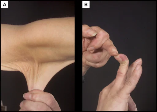
- Generalised joint hypermobility, assessed formally with the Beighton score
- Recurrent subluxation and dislocation, often of the shoulder, patella, and temporomandibular joint, sometimes occurring with minimal or no trauma
- Chronic joint pain, which is frequently widespread, disproportionate to imaging findings, and the dominant symptom in hypermobile EDS
- Early osteoarthritis from repeated joint microtrauma and instability
- Soft tissue injury - recurrent sprains, tendinopathy and slow recovery from injury
Skin
- Hyperextensibility - skin that stretches easily and then springs back, tested by lifting the skin over a neutral site such as the volar forearm
- Atrophic scarring - wide, thin, papery scars, classically described as 'cigarette paper', most marked in classical EDS
- Easy bruising, often with no recalled trauma, which not infrequently raises unwarranted safeguarding concerns before the diagnosis is known
- Fragility and poor wound healing - wounds that dehisce, and sutures that cut through tissue
- Thin, translucent skin with clearly visible subcutaneous veins over the chest and abdomen - a specific pointer to vascular EDS
Systemic and associated features
A substantial part of the symptom burden in hypermobile EDS comes from associations outside the joints and skin, which are easily missed if the consultation focuses only on musculoskeletal complaints.
- Postural orthostatic tachycardia syndrome (PoTS) and other forms of dysautonomia - dizziness, palpitations and presyncope on standing, and a major contributor to fatigue
- Chronic fatigue, frequently as disabling as the pain itself
- Gastrointestinal dysmotility - reflux, bloating, constipation and symptoms overlapping with irritable bowel syndrome
- Pelvic floor dysfunction, urinary incontinence and, in women, dyspareunia
- Anxiety and depression, both as an association and as a consequence of chronic pain and prolonged diagnostic delay
- Mast cell activation symptoms - flushing, urticaria and food or drug intolerance, reported in a proportion of patients
The Beighton score
The Beighton score is a nine-point bedside assessment of generalised joint hypermobility. One point is scored for each of the following, with bilateral manoeuvres scoring one point on each side.
- Passive dorsiflexion of the fifth finger beyond 90 degrees - one point each hand
- Passive apposition of the thumb to the flexor aspect of the forearm - one point each hand
- Hyperextension of the elbow beyond 10 degrees - one point each arm
- Hyperextension of the knee beyond 10 degrees - one point each leg
- Forward flexion of the trunk with knees straight, placing the palms flat on the floor - one point
| Group | Threshold |
|---|---|
| Prepubertal children and adolescents | 6 or more out of 9 |
| Adults up to age 50 | 5 or more out of 9 |
| Adults over 50 | 4 or more out of 9 |
Vascular Ehlers-Danlos syndrome
Vascular EDS accounts for a small minority of cases but a large majority of EDS-related deaths, and deserves separate treatment because both its recognition and its management differ fundamentally from the other subtypes. It results from pathogenic variants in COL3A1, encoding type III collagen, the predominant collagen of arterial walls, bowel and uterus.3
Recognition
- Thin, translucent skin with a clearly visible venous pattern over the chest, abdomen and limbs
- Characteristic facial appearance - a thin, pinched nose, thin lips, hollow cheeks and prominent eyes
- Acrogeria - aged-looking, wrinkled skin over the hands and feet, disproportionate to the patient's age
- Extensive bruising at unusual sites, often the presenting concern
- Small joint hypermobility only - typically the fingers and toes, so the Beighton score may be unimpressive
- A family history of sudden death, arterial rupture or bowel perforation in a young relative - one of the most valuable single clues
Management principles
- Celiprolol, a beta-blocker with partial beta-2 agonist activity, is the one drug with trial evidence of reduced arterial rupture and dissection, and is started at diagnosis in adults4
- Avoid invasive arteriography where possible - catheter-based arterial studies risk vessel injury in fragile tissue; use non-invasive CT or MR angiography for surveillance instead
- Avoid elective surgery where a conservative alternative exists, and ensure any surgical team is explicitly informed of the diagnosis in advance, as tissues hold sutures poorly
- Avoid colonoscopy and other invasive endoscopic procedures unless there is a compelling indication
- Blood pressure control, avoidance of heavy lifting and contact sport, and a medical alert card or bracelet so that emergency teams know the diagnosis immediately
- Pre-pregnancy counselling covering both the 50% transmission risk and the substantial maternal risk of pregnancy itself
Investigations
Investigation is driven by which subtype is suspected, and a key point for exams is that hypermobile EDS has no confirmatory test - it is a clinical diagnosis made against the 2017 criteria, and a normal genetic panel does not exclude it.
- Clinical diagnostic criteria - the 2017 hEDS criteria require generalised joint hypermobility, plus a specified combination of systemic features, family history and musculoskeletal complications, plus exclusion of other diagnoses
- Genetic testing - targeted panels or COL3A1 sequencing where classical, vascular or a rarer subtype is suspected; this is the priority investigation whenever vascular EDS is a possibility
- Echocardiography - to assess the aortic root and screen for mitral valve prolapse, particularly in classical and vascular subtypes
- CT or MR angiography - for arterial surveillance in vascular EDS, in preference to catheter arteriography
- Active stand test or tilt-table testing - where PoTS is suspected clinically
- Investigations to exclude mimics - inflammatory markers and autoantibodies if an inflammatory arthropathy is possible, and consideration of Marfan syndrome and Loeys-Dietz syndrome where the habitus or aortic findings suggest them
Differential diagnosis
- Marfan syndrome - overlapping hypermobility and aortic risk, but distinguished by tall marfanoid habitus, ectopia lentis and aortic root dilatation rather than diffuse medium-vessel fragility
- Loeys-Dietz syndrome - aggressive arterial aneurysm and dissection with hypertelorism and bifid uvula; an important differential for vascular EDS specifically
- Osteogenesis imperfecta - blue sclerae and fragility fractures dominate, with joint laxity a secondary feature
- Hypermobility spectrum disorder - symptomatic hypermobility not meeting full hEDS criteria; the distinction matters less for management than for accurate labelling
- Fibromyalgia and chronic fatigue syndrome - overlap heavily with hEDS in presentation, and can coexist with it rather than being mutually exclusive
- Non-accidental injury - the easy bruising and fragile skin of EDS have led to wrongful safeguarding referrals, and conversely EDS should never be assumed as an explanation for injuries without proper assessment
Management
There is no disease-modifying treatment for any EDS subtype. Management is multidisciplinary, symptom-directed, and - in hypermobile EDS particularly - built around helping patients function well with a chronic condition rather than around eliminating symptoms.
Musculoskeletal
- Physiotherapy is the cornerstone - low-resistance, progressive strengthening to stabilise joints through improved muscular control, with careful avoidance of end-range stretching, which worsens instability
- Occupational therapy - joint protection strategies, pacing, and aids or splints where they improve function
- Avoid high-impact and contact sports; swimming and controlled resistance work are generally better tolerated
- Orthopaedic surgery is approached cautiously - repairs frequently fail in lax tissue, and conservative management is preferred where possible
Pain and fatigue
Chronic pain in EDS is often mixed nociceptive and neuropathic, with a significant central sensitisation component, so escalating simple analgesia rarely succeeds alone. Management follows general chronic pain principles: a multidisciplinary pain service, careful use of neuropathic agents, structured pacing, and psychological therapies aimed at function rather than at cure. Opioids should be avoided or minimised, given long-term harm and poor efficacy in this population, and they may worsen the associated gastrointestinal dysmotility.
Associated conditions
- PoTS - increased fluid and salt intake, compression garments, graded recumbent exercise, and drug treatment such as ivabradine, beta-blockers or fludrocortisone under specialist guidance
- Gastrointestinal symptoms - dietary modification, laxatives and prokinetics, managed along irritable bowel syndrome lines
- Mental health - active screening for and treatment of anxiety and depression, acknowledging the impact of long diagnostic delay, which frequently runs to many years
- Wound care - deep sutures, left in longer than usual, with tension-free closure and clear communication of the diagnosis to any operating team
Genetic counselling
Most clinically relevant subtypes - hypermobile, classical and vascular - follow autosomal dominant inheritance, so each child of an affected parent has a 50% chance of inheriting the condition, with male-to-male transmission possible. Expressivity varies considerably even within a family, so a mildly affected parent cannot be reassured that a child will be similarly mild.
For vascular EDS, once a COL3A1 variant is identified, cascade testing of first-degree relatives is a priority rather than an option, because identifying an affected relative allows celiprolol, surveillance and emergency planning to begin before a first catastrophic event. This is one of the clearest examples in genetics where predictive testing changes management immediately, and it is why the usual caution about testing for untreatable adult-onset conditions does not apply here. Kyphoscoliotic and dermatosparaxis subtypes are autosomal recessive, with the 1-in-4 recurrence risk and relevance of consanguinity that implies.
Prognosis
Prognosis diverges sharply by subtype. Hypermobile and classical EDS carry a normal life expectancy, with the burden falling on chronic pain, fatigue, recurrent injury and the resulting effects on education, employment and mental health - a burden that is real and frequently underestimated by clinicians precisely because it is not life-threatening.
Vascular EDS historically carried a median survival of around 48-50 years, with arterial rupture the usual cause of death and a first major vascular event often occurring before the age of 40. Contemporary outcomes appear better with earlier diagnosis, celiprolol, blood pressure control and specialist follow-up, though the condition remains serious and the evidence base is limited by its rarity.3,4
References
- Malfait F, Francomano C, Byers P et al. The 2017 international classification of the Ehlers-Danlos syndromes. American Journal of Medical Genetics Part C. 2017. Available here
- Ehlers-Danlos Support UK. Types of EDS and diagnostic criteria. Available here
- Byers PH. Vascular Ehlers-Danlos Syndrome. GeneReviews, NCBI Bookshelf. Available here
- Ong KT, Perdu J, De Backer J et al. Effect of celiprolol on prevention of cardiovascular events in vascular Ehlers-Danlos syndrome (BBEST): a prospective randomised, open, blinded-endpoints trial. The Lancet. 2010. Available here
- Royal College of General Practitioners / NICE Clinical Knowledge Summaries. Joint hypermobility. Available here
This article is written for revision and education. It is not clinical guidance and must not be used to make decisions about the care of a patient. Always check current NICE guidance and local protocols.