
Marfan Syndrome: Diagnosis and Management
Key points
- Marfan syndrome: an autosomal dominant disorder of the extracellular matrix protein fibrillin-1, causing a multisystem connective tissue phenotype.
- Gene: FBN1 on chromosome 15; around 25% of cases arise from a new mutation with no family history.
- Cardinal risk: progressive aortic root dilatation predisposing to aortic dissection and rupture - the leading cause of death, and the reason for lifelong echocardiographic surveillance.
- Skeletal features: tall stature, long limbs and digits (arachnodactyly), pectus deformity, scoliosis and joint hypermobility.
- Ocular features: lens dislocation (ectopia lentis), typically upward, and severe myopia.
- Diagnosis: the revised Ghent nosology, which weights aortic root dilatation and ectopia lentis most heavily and requires molecular or systemic scoring for the rest.
- Medical management: beta-blockers or losartan (or both) slow the rate of aortic root dilatation; genotype and phenotype guide the threshold for prophylactic aortic root surgery.
- Activity advice: avoid contact sports and heavy isometric exertion because of aortic and lens dislocation risk; moderate aerobic activity is encouraged.
Introduction
Marfan syndrome is an autosomal dominant disorder of connective tissue caused by pathogenic variants in FBN1, the gene encoding fibrillin-1, a major structural protein of the extracellular matrix microfibrils that give elastic tissue its integrity. It affects roughly 1 in 5,000-10,000 people and, because fibrillin-1 is expressed throughout the body, produces a distinctive combination of skeletal, ocular and cardiovascular features.1
It is a condition where getting the diagnosis right, and then following through on lifelong surveillance, genuinely changes survival: aortic dissection was historically the leading cause of premature death, and modern management - medical therapy, surveillance imaging and appropriately timed prophylactic surgery - has extended life expectancy from around 45 years in the mid-20th century to close to normal today.2
Aetiology and pathophysiology
Fibrillin-1 microfibrils provide structural scaffolding in connective tissue and also regulate the bioavailability of transforming growth factor beta (TGF-beta) by sequestering it in the extracellular matrix. Loss of normal fibrillin-1 function both weakens tissue mechanically and releases excess active TGF-beta signalling, which is now understood to drive much of the vascular and skeletal pathology, rather than mechanical weakness alone. This dual mechanism is why angiotensin receptor blockers, which reduce TGF-beta signalling, have a therapeutic role alongside mechanically-focused beta-blockade.
Inheritance is autosomal dominant with high penetrance but markedly variable expressivity - even within the same family carrying the identical variant, severity can range from mild to life-threatening in childhood. Around 25% of cases arise from a de novo mutation, so the absence of a family history does not exclude the diagnosis, and paternal age is a recognised risk factor for new mutations, as with other conditions arising from single-gene changes in sperm.
Clinical features
Features cluster in three systems - skeletal, ocular and cardiovascular - and the diagnostic approach (the Ghent nosology, below) is built around systematically scoring findings across all three, since no single feature is sensitive or specific enough alone.
Skeletal
- Tall stature with a disproportionately long arm span exceeding height, and a reduced upper-to-lower body segment ratio
- Arachnodactyly - long, slender fingers and toes
- Pectus carinatum or excavatum - chest wall deformity from abnormal costal cartilage growth
- Scoliosis, sometimes progressive and requiring bracing or surgery
- Joint hypermobility, though usually less pronounced than in Ehlers-Danlos syndrome
- Pes planus (flat feet) and hindfoot deformity
- Reduced elbow extension and a high-arched (narrow, 'Gothic') palate with dental crowding
- Facial features - a long, narrow face (dolichocephaly), enophthalmos, downslanting palpebral fissures and retrognathia, though these are non-specific
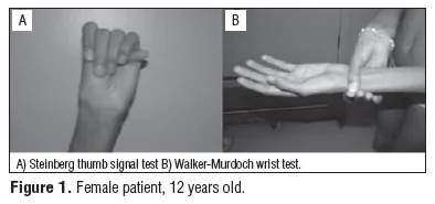
- Walker-Murdoch (wrist) sign - the thumb and fifth finger overlap when wrapped around the opposite wrist
- Steinberg (thumb) sign - the thumb, folded across the palm inside a clenched fist, protrudes beyond the ulnar border of the hand
Ocular
- Ectopia lentis - dislocation or subluxation of the lens, classically upward and temporal, present in around 60% and one of the two most heavily weighted features in the diagnostic criteria
- Severe myopia, from an abnormally long globe
- Increased risk of retinal detachment, early cataract and glaucoma
Cardiovascular
- Aortic root dilatation, typically at the sinuses of Valsalva, present in the majority and progressive over time - the feature that drives prognosis and management
- Aortic dissection or rupture - the major life-threatening complication, risk rising sharply once the aortic root exceeds a critical diameter
- Mitral valve prolapse, sometimes with regurgitation
- Aneurysm elsewhere in the arterial tree, though far less commonly than the aortic root
Diagnosis: the revised Ghent nosology
The 2010 revised Ghent criteria simplified diagnosis by giving the greatest weight to the two most specific findings - aortic root dilatation/dissection and ectopia lentis - and using a systemic score for the many other, less specific features.3
| Family history | Findings | Diagnosis |
|---|---|---|
| No known FBN1 variant or family history | Aortic root dilatation/dissection AND ectopia lentis | Marfan syndrome |
| No known FBN1 variant or family history | Aortic root dilatation/dissection AND a systemic score of 7 or more | Marfan syndrome |
| No known FBN1 variant or family history | Ectopia lentis AND an identified FBN1 variant known to cause aortic disease | Marfan syndrome |
| A relative meeting the above criteria | Ectopia lentis alone, OR a systemic score of 7 or more, OR aortic root dilatation | Marfan syndrome |
The systemic score sums points across a checklist of skeletal, skin, pulmonary and other features (wrist and thumb signs, pectus deformity, scoliosis, pneumothorax, dural ectasia, myopia, mitral valve prolapse, and more), with different features weighted differently. It is a scoring tool used by specialists rather than something to memorise item by item, but knowing that it exists - and that no single feature outside aortic disease and ectopia lentis is diagnostic alone - is the exam-relevant point.
Examination approach
An OSCE Marfan station rewards a systematic sweep across the three key systems rather than jumping straight to the wrist and thumb signs.
- General inspection - tall, slim build, long limbs, and observe gait and posture for scoliosis
- Hands - arachnodactyly, and perform the Walker-Murdoch and Steinberg signs
- Arm span versus height - measure both; arm span exceeding height is characteristic
- Chest - inspect for pectus carinatum or excavatum
- Spine - inspect for scoliosis, ask the patient to bend forward (Adam's forward bend test)
- Face - long, narrow face, high-arched palate, downslanting palpebral fissures
- Eyes - ask about visual acuity and known lens dislocation; fundoscopy and slit-lamp assessment are for the ophthalmologist, but visual acuity can be screened at the bedside
- Cardiovascular - auscultate for the mid-systolic click and late systolic murmur of mitral valve prolapse, and for the early diastolic murmur of aortic regurgitation if the root is significantly dilated
- Joints - assess hypermobility (Beighton score) and elbow extension
Differential diagnosis
- Loeys-Dietz syndrome - another autosomal dominant TGF-beta pathway disorder, with more aggressive and diffuse arterial aneurysm disease, hypertelorism, bifid uvula/cleft palate, and dissection risk at smaller aortic diameters, making early recognition particularly important
- Ehlers-Danlos syndrome (vascular type) - arterial rupture and dissection can occur, but joint hypermobility and skin fragility are more prominent, and the mechanism (COL3A1) and typical vessel involvement (medium arteries, not chiefly the aortic root) differ
- Homocystinuria - marfanoid habitus with downward lens dislocation, learning disability and a thrombotic tendency; autosomal recessive, screened by plasma homocysteine
- MASS phenotype (myopia, mitral valve prolapse, mild aortic dilatation, striae, skeletal features) - overlapping features but does not meet full Ghent criteria and does not progress to aggressive aortic disease
- Familial thoracic aortic aneurysm syndromes without the syndromic features - isolated aortic disease without skeletal or ocular involvement
Investigations
- Transthoracic echocardiography - first-line and repeated imaging of the aortic root, the cornerstone of both diagnosis and surveillance
- Slit-lamp ophthalmological examination - to detect ectopia lentis, which can be asymptomatic
- MRI or CT aortography - for full assessment of the aorta beyond the root, particularly before major surgery or if transthoracic views are inadequate
- FBN1 genetic testing - confirms the diagnosis and enables cascade testing of relatives
- Spinal imaging - for scoliosis assessment, and MRI of the lumbosacral spine if dural ectasia (a systemic score feature) is suspected
- Plasma homocysteine - if homocystinuria is a realistic differential, particularly with downward lens dislocation or learning disability
Management
Medical therapy
Beta-blockers (traditionally atenolol) have long been first-line, reducing the rate of aortic root dilatation by lowering the rate of ventricular pressure rise (dP/dt) and, over time, aortic wall shear stress. Losartan, an angiotensin II receptor blocker, works through the TGF-beta pathway rather than a haemodynamic mechanism, and trial evidence supports its use either as an alternative when beta-blockers are not tolerated or in combination.4 The choice and combination is individualised, but the principle for an exam answer is that both classes have evidence, working by different mechanisms, and either or both are appropriate depending on tolerance and the treating centre's protocol.
Surveillance
Echocardiography is repeated at least annually (more frequently if the aortic root is enlarging or approaching the surgical threshold, or in pregnancy), tracking the aortic root diameter over time against normal ranges for age and body surface area. Ophthalmological review continues periodically through life, and spinal surveillance continues through the period of scoliosis risk in adolescence.
Surgery
Prophylactic aortic root replacement is considered once the aortic root reaches a defined threshold, generally around 45-50 mm in an adult, adjusted downward for a rapid rate of growth, a strong family history of dissection at a smaller diameter, significant aortic regurgitation, or planned pregnancy, and adjusted for body size in children.5 Operating before dissection occurs, on a controlled elective basis, carries a far lower mortality than emergency repair of an established dissection, which is the entire rationale for lifelong surveillance in the first place.
Lifestyle and activity advice
- Avoid contact and collision sports, and heavy isometric exertion (e.g. heavy weightlifting) - both raise aortic wall stress and carry a risk of lens dislocation from direct trauma or Valsalva-related pressure changes
- Moderate aerobic activity is encouraged rather than restricted altogether, since deconditioning carries its own harms and most low-to-moderate intensity aerobic exercise is considered safe
- Pregnancy carries increased aortic dissection risk from haemodynamic changes and should be planned with specialist joint cardio-obstetric input; a dilated aortic root above a defined threshold is a relative or absolute contraindication to pregnancy depending on the exact measurement and rate of change
- Genetic counselling for reproductive planning, given the 50% transmission risk to each child of an affected parent
Genetic counselling and family screening
Because inheritance is autosomal dominant, each child of an affected parent has a 50% chance of inheriting the variant, irrespective of sex, and male-to-male transmission occurs. Variable expressivity means a mildly affected parent can have a severely affected child, so counselling must avoid implying that a child's course will mirror the parent's.
- First-degree relatives of an affected person should be offered screening - clinical assessment, echocardiography and ophthalmological review - even if they appear well, since aortic dilatation is asymptomatic until it dissects
- Once an FBN1 variant is identified in the proband, cascade genetic testing is faster and more definitive than repeated clinical assessment, and can discharge relatives who test negative from lifelong surveillance
- A negative clinical screen in childhood does not exclude the diagnosis, because features evolve with growth - relatives who have not had genetic testing need repeated assessment through adolescence rather than a single reassuring echocardiogram
- Reproductive options for an affected person include prenatal diagnosis by chorionic villus sampling or amniocentesis, and pre-implantation genetic testing with IVF, both of which require the familial variant to have been identified first
- Around a quarter of cases are de novo, so unaffected parents of an affected child usually have a low recurrence risk - but gonadal mosaicism means this is not zero, and prenatal testing may still be offered in subsequent pregnancies
Complications
Aortic dissection and rupture remain the dominant life-threatening complication. Other complications include progressive aortic or mitral regurgitation and heart failure, retinal detachment, spontaneous pneumothorax (from apical bullae, a recognised systemic score feature), and the cumulative musculoskeletal burden of scoliosis, joint pain and reduced mobility over a lifetime.
Prognosis
Untreated, historical life expectancy was significantly reduced, chiefly from aortic dissection in early-to-mid adulthood. With modern medical therapy, structured surveillance and appropriately timed prophylactic surgery, life expectancy now approaches that of the general population, making this one of the clearer examples in genetics of a single, well-organised surveillance pathway materially changing survival rather than simply managing symptoms.2
References
- Dietz H. Marfan Syndrome. GeneReviews, NCBI Bookshelf. Available here
- Silverman DI, Burton KJ, Gray J et al. Life expectancy in the Marfan syndrome. American Journal of Cardiology. 1995. Available here
- Loeys BL, Dietz HC, Braverman AC et al. The revised Ghent nosology for the Marfan syndrome. Journal of Medical Genetics. 2010. Available here
- Lacro RV, Dietz HC, Sleeper LA et al. Atenolol versus losartan in children and young adults with Marfan's syndrome. New England Journal of Medicine. 2014. Available here
- European Society of Cardiology. Guidelines for the diagnosis and treatment of aortic diseases. 2014, updated 2024. Available here
This article is written for revision and education. It is not clinical guidance and must not be used to make decisions about the care of a patient. Always check current NICE guidance and local protocols.