
IgA Vasculitis (Henoch-Schonlein Purpura)
Key points
- IgA vasculitis: an IgA-mediated small vessel vasculitis, formerly called Henoch-Schonlein purpura. It is the commonest vasculitis of childhood, with a peak age of 3 to 10 years.
- The classic tetrad: palpable purpura (required for diagnosis), arthralgia or arthritis, abdominal pain, and renal involvement.
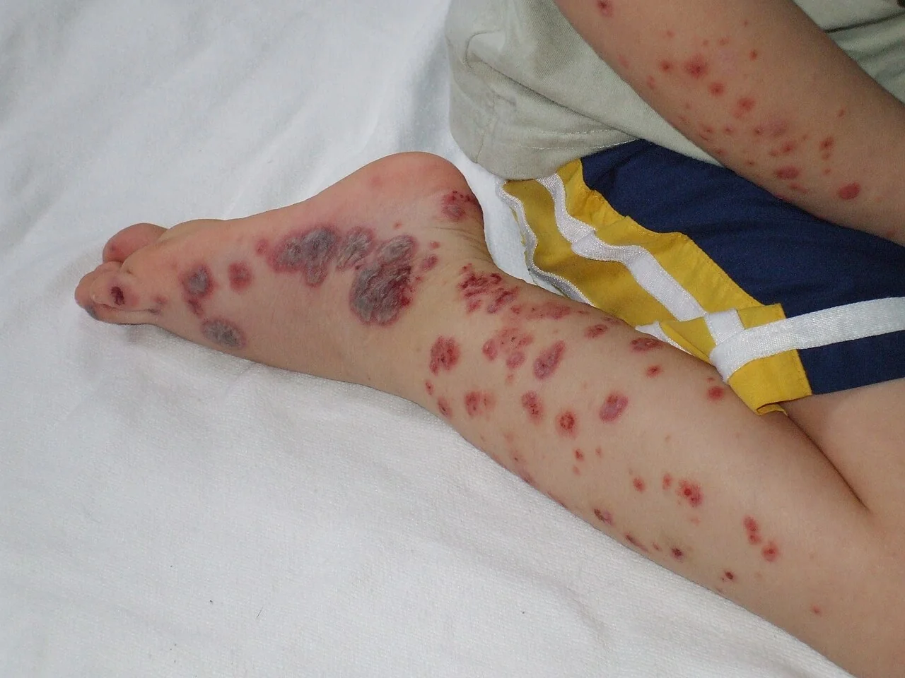
- The rash is the diagnostic feature: palpable, non-blanching purpura distributed symmetrically over the extensor surfaces of the legs and the buttocks - a gravity- and pressure-dependent distribution.
- Trigger: typically follows an upper respiratory tract infection by 1 to 3 weeks. It shares its pathogenesis with IgA nephropathy, of which it is essentially the systemic form.
- Diagnosis is clinical: made by the EULAR/PRINTO/PRES criteria - purpura plus at least one of abdominal pain, arthritis, renal involvement or biopsy showing IgA deposition. Investigations mainly exclude mimics.
- Must exclude meningococcal sepsis: a non-blanching rash in an unwell, febrile child is meningococcal disease until proven otherwise - the most important immediate differential.
- Management is supportive: rest, hydration and simple analgesia. Corticosteroids are reserved for severe abdominal pain, significant nephritis or other severe organ involvement - they do not prevent nephritis.
- Follow-up is the exam point: urine dipstick and blood pressure must be monitored for at least 6 to 12 months, because nephritis can appear weeks after the rash has resolved.
Introduction
IgA vasculitis is an immune complex-mediated small vessel vasculitis in which IgA1-containing complexes deposit in the walls of small vessels - principally capillaries, venules and arterioles - in the skin, joints, gut and glomeruli, provoking a neutrophilic inflammatory response.1
It was known for many years as Henoch-Schonlein purpura, and the Chapel Hill consensus renamed it to reflect its mechanism. It is the commonest vasculitis of childhood, with an incidence of around 10 to 20 per 100,000 children per year, a peak age of 3 to 10 years, a slight male predominance, and a striking seasonality - most cases occur in autumn and winter, following the pattern of respiratory infections.
Adults can be affected, and when they are the disease is generally more severe, with a higher rate of significant renal involvement and a greater likelihood of progressing to chronic kidney disease. An adult presenting with this picture also warrants more thought about alternative diagnoses and, in older patients, about an underlying malignancy.
Aetiology
The trigger is usually an infection, and around half to two-thirds of cases follow an upper respiratory tract infection by 1 to 3 weeks.
- Infections - most commonly a viral URTI; also group A Streptococcus, Mycoplasma, Helicobacter pylori, parvovirus B19, varicella and hepatitis
- Vaccinations - a rare and temporally associated trigger
- Drugs - antibiotics (penicillins, cephalosporins), NSAIDs, ACE inhibitors
- Insect bites and cold exposure, occasionally reported
- Malignancy - a recognised association in adults, particularly with solid tumours (lung, prostate, gastrointestinal) and haematological malignancy. New IgA vasculitis in an older adult should prompt consideration of an occult cancer.
- Often no trigger is identified
The pathogenesis involves abnormally galactose-deficient IgA1, which is recognised as foreign, forms circulating immune complexes with IgG, and deposits in small vessel walls where it activates the alternative complement pathway. The resulting leucocytoclastic vasculitis produces the characteristic purpura, and mesangial deposition produces the nephritis.
Clinical features
The classic presentation is a tetrad, though not all four components are present in every patient and they may appear at different times. Purpura is required for the diagnosis and is the presenting feature in the majority.
Skin
- Palpable purpura - the cardinal sign. The lesions are raised (hence 'palpable') and non-blanching, starting as urticarial or erythematous macules and evolving into purpura and ecchymoses over hours to days
- Distribution is characteristically symmetrical over the extensor surfaces of the legs and the buttocks, and over pressure-bearing and dependent areas - it may extend to the arms and trunk but classically spares the face and mucous membranes
- The distribution reflects gravitational and hydrostatic pressure, which is why in a non-ambulant infant it may appear predominantly on the back and buttocks
- Subcutaneous oedema of the scalp, hands, feet, periorbital region and scrotum, particularly in younger children
- Crops occur in waves over days to weeks, with new lesions appearing as older ones fade to brown, so lesions of differing age coexist
- Bullous or necrotic lesions occur in a minority and indicate more severe disease
Joints
- Arthralgia or arthritis in around two-thirds, often an early feature that may precede the rash
- Typically oligoarticular and involving the large joints of the lower limbs - knees and ankles
- Painful, swollen and warm, but transient and non-deforming - it resolves completely without joint damage, which is a reassuring point for families
Gastrointestinal
- Colicky abdominal pain in up to two-thirds, caused by vasculitis and submucosal oedema and haemorrhage of the bowel wall
- Nausea and vomiting, and gastrointestinal bleeding - melaena or frank blood per rectum
- Intussusception is the important complication - classically ileo-ileal rather than the usual ileocolic, which means it may be missed on ultrasound of the right iliac fossa if the sonographer is not warned. Suspect it with severe or worsening pain, a palpable mass or bloody stools.
- Less commonly bowel infarction, perforation, pancreatitis or protein-losing enteropathy
- Abdominal pain may precede the rash, which is a well-recognised trap - such children are sometimes investigated or even operated on for a surgical abdomen before the diagnosis becomes apparent
Renal
- Renal involvement occurs in around 20 to 50% of children and is the principal determinant of long-term prognosis
- Usually presents as isolated microscopic haematuria, with or without proteinuria
- May progress to nephritic syndrome, nephrotic syndrome, hypertension or, rarely, rapidly progressive glomerulonephritis
- Typically develops within the first 4 to 6 weeks of the illness, but can appear up to 6 months later - which is the entire reason for prolonged urine surveillance
- Histology is identical to IgA nephropathy - mesangial IgA deposition with mesangial proliferation, and crescents in severe cases
Other features
- Scrotal pain and swelling in boys - which must be distinguished from testicular torsion, and may require Doppler ultrasound to do so
- Low-grade fever and malaise - but a high fever should prompt reconsideration of the diagnosis
- Rarely pulmonary haemorrhage, seizures, headache or other CNS involvement, and carditis
Differential diagnosis
| Condition | Distinguishing features |
|---|---|
| Meningococcal septicaemia | Unwell, febrile, rapidly spreading rash, shock - as above |
| Immune thrombocytopenic purpura (ITP) | Low platelet count and a flat, non-palpable purpura with mucosal bleeding and epistaxis; the child is otherwise well. The platelet count is the key discriminator - it is normal or raised in IgA vasculitis. |
| Non-accidental injury | Bruising of varying ages in unusual sites, an inconsistent history, or a distribution that does not fit; must always be considered in a child with unexplained bruising |
| Haemolytic uraemic syndrome | Diarrhoea (often bloody), anaemia with thrombocytopenia and schistocytes, and AKI |
| Other systemic vasculitides | ANCA-associated vasculitis (positive ANCA, upper airway and lung involvement), polyarteritis nodosa, cryoglobulinaemic vasculitis (usually adults) |
| Systemic lupus erythematosus | Positive ANA and anti-dsDNA, low complement, malar rash, multisystem involvement |
| Acute haemorrhagic oedema of infancy | A benign leucocytoclastic vasculitis in infants under 2, with large targetoid purpuric plaques and facial involvement, but no visceral involvement |
| Drug-induced (leucocytoclastic) vasculitis | Temporal relationship to a new drug; may look identical, so a careful drug history matters |
| Coagulopathy or leukaemia | Abnormal clotting or full blood count; consider if there is pallor, lymphadenopathy, hepatosplenomegaly or bone pain |
Investigations
IgA vasculitis is a clinical diagnosis. Investigations serve mainly to exclude mimics and to assess renal involvement, rather than to confirm the diagnosis.
Essential investigations
- Urine dipstick - in every patient at every review; the single most important test, looking for haematuria and proteinuria
- Blood pressure - also at every review; hypertension indicates renal involvement
- Urine protein:creatinine ratio if the dipstick shows protein, to quantify it
- U&Es and creatinine - to assess renal function
- Full blood count - the platelet count is normal or raised, which excludes ITP and other thrombocytopenic causes; also checks for anaemia and the abnormalities of leukaemia
- Clotting screen - normal, excluding coagulopathy
- CRP and ESR - often mildly raised
- Blood cultures if there is any suspicion of sepsis
Selected further investigations
- Serum IgA - raised in around half, but this is neither sensitive nor specific and does not make the diagnosis
- Throat swab and ASOT - if a streptococcal trigger is suspected
- Complement C3 and C4 - normal in IgA vasculitis; a low complement should prompt consideration of lupus or post-infectious glomerulonephritis
- ANA, ANCA - to exclude other vasculitides and connective tissue disease where the presentation is atypical or the patient is an adult
- Abdominal ultrasound - if abdominal pain is severe, to look for intussusception (remembering that it is often ileo-ileal, so the sonographer must be told what is suspected) and to exclude other pathology
- Doppler ultrasound of the testes - if there is scrotal pain, to exclude torsion
- Skin biopsy - rarely needed, but shows leucocytoclastic vasculitis with IgA deposition on immunofluorescence; useful in atypical or adult cases
- Renal biopsy - indicated for significant or persistent proteinuria, nephrotic syndrome, hypertension with renal impairment, or a rising creatinine, since the findings (particularly the presence of crescents) guide immunosuppression
- Investigation for occult malignancy in older adults with new IgA vasculitis
Management
Most children need only supportive care and can be managed at home, since the disease is self-limiting in the great majority. The clinical work lies in recognising the minority who need more, and in maintaining the follow-up that detects late nephritis.
Supportive management
- Rest, adequate hydration and reassurance - explaining the expected course, including that the rash comes in crops and that recurrence is common, prevents a great deal of anxiety
- Simple analgesia - paracetamol for joint and abdominal pain
- NSAIDs may be used for arthralgia in patients with normal renal function and no gastrointestinal bleeding, but should be avoided where there is renal involvement or GI haemorrhage
- Elevation of the legs may reduce dependent purpura and oedema
- Most patients can be managed in primary care, with arrangements for regular review
Corticosteroids
Managing nephritis
- Isolated microscopic haematuria with normal blood pressure and function - monitor only; no treatment is needed and it usually resolves
- Persistent proteinuria - ACE inhibitor or ARB to reduce proteinuria and protect renal function, with nephrology input
- Nephrotic syndrome, significant persistent proteinuria, or impaired renal function - refer to paediatric nephrology; renal biopsy and immunosuppression (corticosteroids, and in severe or crescentic disease cyclophosphamide, mycophenolate, azathioprine or rituximab) may be needed
- Rapidly progressive glomerulonephritis with crescents - treated aggressively as for any crescentic nephritis, with high-dose corticosteroids and cyclophosphamide, sometimes with plasma exchange
- Blood pressure control throughout
Follow-up
Complications
- IgA vasculitis nephritis - the most important complication, ranging from isolated haematuria to rapidly progressive glomerulonephritis, and the determinant of long-term outcome
- Chronic kidney disease and end-stage renal disease - in a small minority of children, and a larger proportion of adults
- Intussusception - classically ileo-ileal; may cause obstruction and require reduction or surgery
- Gastrointestinal haemorrhage, bowel ischaemia, infarction and perforation
- Pancreatitis and protein-losing enteropathy (rare)
- Orchitis and scrotal involvement - and the diagnostic problem of excluding torsion
- Nephrotic syndrome with its thrombotic and infective complications
- Hypertension - which may persist after the acute illness
- Recurrence - occurs in around a third, usually milder and within the first few months
- Pulmonary haemorrhage and CNS involvement (seizures, encephalopathy) - rare but serious
- Complications of corticosteroid treatment where prolonged courses are needed
Red flags
Prognosis
The prognosis in children is excellent. IgA vasculitis is a self-limiting illness in the great majority: the rash and joint symptoms typically resolve within 4 to 6 weeks, and most children recover completely without any long-term sequelae. Recurrence occurs in around a third, usually within the first few months and usually milder and shorter than the initial episode.
Renal involvement determines the long-term outlook, and this is where the clinical attention belongs. Although 20 to 50% of children develop some renal abnormality, the great majority have isolated microscopic haematuria that resolves spontaneously. Fewer than 1 to 2% of all children with IgA vasculitis progress to end-stage renal disease, but the risk is concentrated in those with heavier proteinuria at presentation.
The features predicting a poorer renal outcome are consistent: nephrotic-range proteinuria, nephritic syndrome at presentation, impaired renal function, hypertension, and crescents or significant chronic damage on biopsy. Conversely, a child with isolated haematuria, normal blood pressure and normal renal function has a very low risk of long-term problems.
Adults do considerably less well than children. They have a higher frequency of significant nephritis, are more likely to develop chronic kidney disease, and more often require immunosuppression - and in older adults the possibility of an associated malignancy adds a further prognostic dimension. This age difference is worth remembering, because the reassuring statistics quoted for childhood disease should not be transferred to an adult patient.
References
- Ozen S, Pistorio A, Iusan SM et al. EULAR/PRINTO/PRES criteria for Henoch-Schonlein purpura. Annals of the Rheumatic Diseases. 2010. Available here
- NICE Clinical Knowledge Summaries. Henoch-Schonlein purpura. Available here
- Jennette JC, Falk RJ, Bacon PA et al. 2012 Revised International Chapel Hill Consensus Conference Nomenclature of Vasculitides. Available here
- NICE NG51. Sepsis: recognition, diagnosis and early management. 2016, updated 2024. Available here
- KDIGO 2021 Clinical Practice Guideline for the Management of Glomerular Diseases. Available here
- Okwikikim (English Wikipedia), public domain, via Wikimedia Commons. Available here
This article is written for revision and education. It is not clinical guidance and must not be used to make decisions about the care of a patient. Always check current NICE guidance and local protocols.