
Renal Cancer
Key points
- Renal cell carcinoma (RCC): accounts for around 85% of primary renal malignancies, arising from the proximal convoluted tubule. Clear cell is the commonest subtype (~75-80%).
- The classic triad: haematuria, loin pain and a palpable flank mass - but this occurs in under 10% and usually indicates advanced disease. Over half are now found incidentally on imaging done for another reason.
- Risk factors: smoking, obesity, hypertension, end-stage renal disease and long-term dialysis, and inherited syndromes - notably von Hippel-Lindau.
- Paraneoplastic syndromes: RCC is famous for these: polycythaemia (EPO), hypercalcaemia (PTHrP), hypertension (renin) and Stauffer syndrome - reversible non-metastatic hepatic dysfunction.
- The varicocoele sign: a new left-sided varicocoele that does not empty on lying flat suggests tumour obstructing the left renal vein, into which the left testicular vein drains.
- Diagnosis: contrast-enhanced CT of the abdomen and pelvis is the key investigation. Biopsy is often avoided in operable disease as imaging is characteristic and there is a small seeding risk.
- Spread: invades the renal vein and inferior vena cava (a characteristic behaviour), and metastasises to lung ('cannonball' metastases), bone, liver and brain.
- Treatment: surgery is the only curative option - partial or radical nephrectomy. RCC is chemotherapy- and radiotherapy-resistant; advanced disease is treated with immunotherapy and tyrosine kinase inhibitors.
Introduction
Renal cancer accounts for around 13,000 new UK diagnoses each year. Renal cell carcinoma (RCC) makes up about 85% of cases and arises from the renal tubular epithelium, specifically the proximal convoluted tubule. It is roughly twice as common in men and peaks between 60 and 70 years.1
RCC has been called the 'internist's tumour' because of its remarkable capacity to present with systemic and paraneoplastic manifestations rather than urological ones - fever, weight loss, anaemia, polycythaemia, hypercalcaemia or deranged liver function in the absence of metastases. It is worth holding this in mind, because a patient may be investigated at length for a systemic illness before the kidney is considered.
The other defining feature of modern practice is incidental detection. More than half of RCCs are now discovered by chance on CT or ultrasound performed for unrelated reasons, at an earlier stage than symptomatic presentations - which has substantially improved overall outcomes.
| Tumour | Proportion / notes |
|---|---|
| Clear cell RCC | 75-80% of RCC. Associated with VHL gene loss on chromosome 3p. Lipid- and glycogen-rich cytoplasm gives the clear appearance |
| Papillary RCC | ~10-15%. Often multifocal; generally better prognosis than clear cell |
| Chromophobe RCC | ~5%. Best prognosis of the common subtypes; associated with Birt-Hogg-Dubé syndrome |
| Collecting duct / medullary carcinoma | Rare and aggressive. Renal medullary carcinoma is associated with sickle cell trait |
| Transitional cell carcinoma of the renal pelvis | ~10% of renal tumours; behaves like bladder cancer (smoking, aromatic amines) and presents with haematuria |
| Wilms tumour (nephroblastoma) | The commonest renal tumour of childhood, typically under 5 years, presenting as an abdominal mass |
| Oncocytoma / angiomyolipoma | Benign. Angiomyolipoma is associated with tuberous sclerosis and can bleed catastrophically (Wunderlich syndrome) |
Risk factors
Acquired
- Smoking - roughly doubles the risk and is the most important modifiable factor
- Obesity - a strong and consistent association, particularly in women
- Hypertension - independent of antihypertensive treatment
- End-stage renal disease and long-term dialysis - acquired cystic kidney disease develops in patients on prolonged dialysis and carries a substantially increased risk of RCC
- Male sex and increasing age
- Occupational exposures - cadmium, asbestos, trichloroethylene, petroleum products
- Renal transplantation and long-term immunosuppression
Inherited syndromes
- Von Hippel-Lindau (VHL) syndrome - autosomal dominant, from loss of the VHL tumour suppressor gene on chromosome 3p. Causes bilateral and multifocal clear cell RCC presenting at a young age, together with cerebellar and retinal haemangioblastomas and phaeochromocytoma. The commonest inherited cause and the one to know
- Hereditary papillary renal carcinoma - MET proto-oncogene mutations
- Birt-Hogg-Dubé syndrome - chromophobe RCC and oncocytomas, with fibrofolliculomas and pulmonary cysts
- Tuberous sclerosis - angiomyolipomas and an increased RCC risk
- Hereditary leiomyomatosis and RCC (HLRCC) - fumarate hydratase mutations; aggressive tumours
- Sickle cell trait - renal medullary carcinoma in young Black patients
Suspect an inherited syndrome where RCC presents under 50 years, is bilateral or multifocal, or where there is a family history or other syndromic features - such patients warrant genetic referral.
Clinical features
Local features
- Haematuria - visible or non-visible; the commonest single symptom
- Loin or flank pain - a dull persistent ache; colicky pain can occur if clot passes down the ureter
- Palpable flank mass - a firm, smooth mass that moves with respiration and is ballotable
- The classic triad of all three occurs in under 10% and indicates advanced disease. Its rarity is itself an examination point - do not wait for it
The left varicocoele sign
Paraneoplastic and systemic features
Present in around 20-30% of patients, and often the reason for presentation:
- Polycythaemia - from inappropriate erythropoietin (EPO) secretion. A raised haemoglobin in a patient with a renal mass is classic; note that anaemia is actually more common overall (from chronic disease and haematuria)
- Hypercalcaemia - from PTH-related peptide (PTHrP) secretion or from bone metastases
- Hypertension - from renin secretion or arteriovenous shunting within the tumour
- Stauffer syndrome - reversible, non-metastatic hepatic dysfunction with raised ALP and transaminases, hepatosplenomegaly and abnormal clotting, in the absence of liver metastases. It resolves after nephrectomy, and is thought to be IL-6 mediated
- Pyrexia of unknown origin, night sweats, weight loss, anorexia and fatigue
- Raised ESR and CRP, thrombocytosis, and amyloidosis (AA type)
- Cushing syndrome from ACTH secretion (rare), and polymyalgia-like syndromes
Metastatic disease
RCC characteristically invades the renal vein and inferior vena cava, sometimes extending as a tumour thrombus into the right atrium. Distant metastases go to lung (classically producing 'cannonball' metastases on chest X-ray), bone (typically lytic and painful), liver and brain. RCC is also notorious for late recurrence, sometimes many years after apparently curative surgery.
Investigations
- Contrast-enhanced CT of the abdomen and pelvis - the key investigation, both for diagnosis and staging. It characterises the lesion, assesses renal vein and IVC involvement, nodal disease and the contralateral kidney. Renal masses are graded by the Bosniak classification for cystic lesions
- Ultrasound - often the first test performed, distinguishing a simple cyst (needing no further action) from a solid or complex mass (needing CT)
- CT chest - for lung metastases as part of staging
- MRI - useful for assessing IVC tumour thrombus extent and where contrast CT is contraindicated
- Urine dipstick and cytology - haematuria; cytology if a transitional cell tumour of the renal pelvis is suspected
- FBC - looking for anaemia or polycythaemia
- U&Es and creatinine - baseline function, critical when planning nephrectomy
- Bone profile including calcium - hypercalcaemia
- LFTs - deranged in Stauffer syndrome or with liver metastases
- LDH, ESR/CRP - prognostic markers used in metastatic risk scores
- Bone scan or CT head - only if clinically indicated by symptoms
- Renal biopsy - often avoided in a patient with a resectable mass and characteristic imaging, because of small risks of bleeding and tract seeding and the risk of sampling error. It is indicated before ablative therapy or systemic treatment, in small indeterminate masses under surveillance, and where lymphoma or metastasis from another primary is suspected
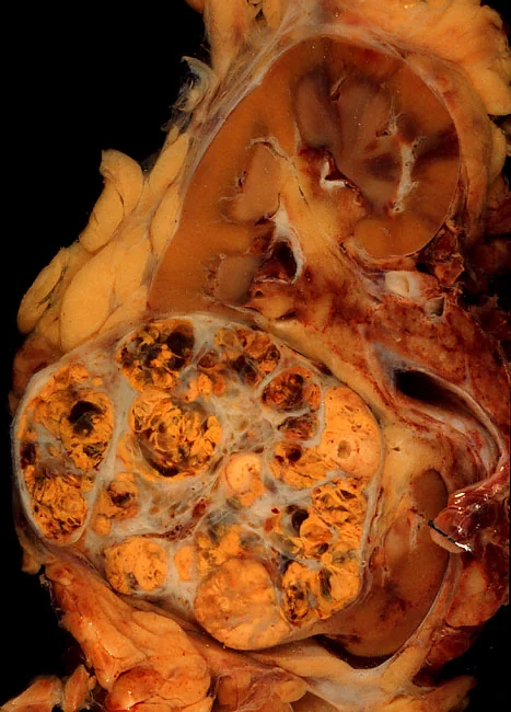
Staging
| Stage | Description |
|---|---|
| T1 | Confined to the kidney, 7 cm or less (T1a ≤4 cm, T1b >4-7 cm) |
| T2 | Confined to the kidney, more than 7 cm |
| T3 | Extends into major veins (renal vein, IVC) or perinephric tissues, but not beyond Gerota's fascia and not into the ipsilateral adrenal |
| T4 | Invades beyond Gerota's fascia, including contiguous extension into the ipsilateral adrenal gland |
| N1 | Metastasis in regional lymph nodes |
| M1 | Distant metastasis |
Gerota's fascia is the key anatomical boundary - tumour confined within it (T1-T3) remains potentially curable by surgery, whereas extension beyond it (T4) implies far more advanced disease. Note also that venous invasion alone does not preclude cure: a tumour thrombus extending into the IVC can still be resected with good outcomes, which is unusual among solid tumours.
Management
Localised disease
- Partial nephrectomy (nephron-sparing surgery) - preferred wherever technically feasible, particularly for T1 tumours, in patients with a solitary kidney, bilateral tumours, CKD or inherited syndromes. It preserves renal function without compromising cancer outcomes
- Radical nephrectomy - removal of the kidney with perinephric fat within Gerota's fascia, for larger or more central tumours. Performed laparoscopically or robotically where possible. The adrenal gland is preserved unless directly involved
- Percutaneous ablation (radiofrequency or cryoablation) - for small tumours in patients unfit for surgery; requires prior biopsy
- Active surveillance - reasonable for small renal masses (under 3-4 cm) in elderly or comorbid patients, since many grow very slowly and competing causes of death predominate
- Surgical resection of IVC tumour thrombus - technically demanding but potentially curative
Advanced and metastatic disease
- Immune checkpoint inhibitors - nivolumab, pembrolizumab, ipilimumab; combination immunotherapy is now standard first-line treatment for many patients with metastatic disease and has substantially improved survival
- Tyrosine kinase inhibitors targeting VEGF signalling - sunitinib, pazopanib, axitinib, cabozantinib; used alone or combined with immunotherapy. Side effects include hypertension, hand-foot syndrome, diarrhoea, fatigue and hypothyroidism
- mTOR inhibitors - everolimus, temsirolimus; later-line options
- Cytoreductive nephrectomy - considered in selected patients with metastatic disease and good performance status
- Metastasectomy - resection of solitary or oligometastatic deposits can give prolonged survival, reflecting RCC's unusual biology
- Palliative radiotherapy - for bone pain, spinal cord compression and brain metastases
- Bone-protective agents - bisphosphonates or denosumab for skeletal metastases
- Best supportive and palliative care throughout
Complications
- Metastatic disease - lung ('cannonball' lesions), bone, liver and brain; RCC is notable for late recurrence years after nephrectomy
- Renal vein and IVC tumour thrombus, occasionally extending to the right atrium and causing pulmonary embolism
- Paraneoplastic syndromes - polycythaemia, hypercalcaemia, hypertension, Stauffer syndrome, amyloidosis
- Anaemia from chronic disease and blood loss
- Severe haematuria and clot retention
- Chronic kidney disease after nephrectomy, particularly with pre-existing impairment - the principal argument for nephron-sparing surgery
- Spontaneous retroperitoneal haemorrhage - especially with angiomyolipomas (Wunderlich syndrome)
- Pathological fracture and spinal cord compression from bone metastases
- Treatment toxicity - hypertension, hypothyroidism, hand-foot syndrome and diarrhoea from TKIs; immune-related adverse events (colitis, hepatitis, pneumonitis, thyroiditis, hypophysitis) from checkpoint inhibitors
- Venous thromboembolism
Red flags
Prognosis
Prognosis depends principally on stage at diagnosis, and has improved substantially in recent decades - partly through better systemic therapy, but largely because incidental detection finds tumours earlier. Localised disease (T1) treated surgically has a 5-year survival above 90%, and small tumours removed by partial nephrectomy do particularly well. Overall 5-year survival across all stages in the UK is around 70%.
Metastatic disease carries a much poorer outlook, historically with 5-year survival under 15%. This has changed meaningfully with combination immunotherapy and targeted agents, which have extended median survival considerably, though the disease remains incurable in most. Prognostic scores (such as the IMDC criteria, using performance status, time from diagnosis to treatment, haemoglobin, calcium, neutrophils and platelets) stratify patients into favourable, intermediate and poor risk groups and guide systemic treatment choice.
Two features of RCC's natural history deserve emphasis. First, late recurrence is characteristic - relapse can occur a decade or more after apparently curative nephrectomy, so follow-up is prolonged. Second, RCC shows unusual biological behaviour at both extremes: spontaneous regression of metastases is rare but genuinely documented, and resection of solitary metastases can produce long-term survival, which is why metastasectomy is considered in RCC when it would not be in most other solid tumours. Histological subtype also matters, with chromophobe and papillary tumours generally carrying a better prognosis than clear cell, and collecting duct and medullary carcinomas a considerably worse one.
References
- NICE NG12. Suspected cancer: recognition and referral. 2015, updated 2023. Available here
- Cancer Research UK. Kidney cancer statistics. Available here
- European Association of Urology. Guidelines on Renal Cell Carcinoma. Available here
- Ljungberg B, Albiges L, Abu-Ghanem Y et al. EAU Guidelines on Renal Cell Carcinoma: 2022 Update. European Urology. Available here
- Motzer RJ, Tannir NM, McDermott DF et al. Nivolumab plus ipilimumab in advanced renal cell carcinoma (CheckMate 214). NEJM. 2018. Available here
- NICE Clinical Knowledge Summaries. Haematuria. Available here
- Ed Uthman, MD, public domain, via Wikimedia Commons. Available here
This article is written for revision and education. It is not clinical guidance and must not be used to make decisions about the care of a patient. Always check current NICE guidance and local protocols.