
Spinal Cord Compression: Diagnosis and Management
Key points
- Spinal cord compression: pressure on the spinal cord from tumour, infection, disc or trauma, threatening permanent neurological deficit - a time-critical emergency.
- Metastatic spinal cord compression (MSCC): the commonest cause in adults; commonly from breast, lung, prostate, renal, thyroid and myeloma.
- Cardinal warning symptom: new or progressive back pain, often worse on lying flat, coughing or straining - typically precedes neurological signs by weeks.
- Neurological pattern: upper motor neurone signs below the level of the lesion, a sensory level, and bladder or bowel disturbance.
- Investigation of choice: urgent MRI of the whole spine within 24 hours - the whole spine, because multiple levels are often involved.
- Immediate treatment: dexamethasone (with gastric protection) as soon as MSCC is suspected, unless lymphoma is likely and tissue is needed first.
- Definitive treatment: surgical decompression or radiotherapy, decided by MDT according to tumour type, stability, prognosis and fitness.
- Prognostic rule: the neurological status at the time of treatment is the single best predictor of outcome - a patient who is still walking is likely to keep walking.
Introduction
Spinal cord compression occurs when the cord (or, below L1-L2, the cauda equina) is compressed by tumour, infection, haematoma, disc material or displaced bone. It is an emergency because neurological damage becomes irreversible once established - the window in which function can be preserved is measured in hours to days, not weeks.1
Metastatic spinal cord compression (MSCC) is the commonest cause in adults and complicates around 5-10% of cancers, sometimes as the first presentation of an undiagnosed malignancy. The defining clinical challenge is that back pain precedes neurological signs by weeks, so the opportunity to intervene comes and goes before weakness appears - which is why recognising the pain pattern, rather than waiting for weakness, is the whole skill.
The spinal cord ends at approximately L1-L2 in adults. Compression above this level produces cord (upper motor neurone) signs; below it, the cauda equina is affected, producing lower motor neurone signs and the distinct clinical picture of cauda equina syndrome.
Metastatic spinal cord compression is not a rare event: it affects several thousand patients each year in the UK, and in a meaningful minority it is the first presentation of an undiagnosed cancer rather than a complication of known disease. That last point is easily forgotten, and it is the reason new progressive back pain with any neurological feature warrants imaging even in a patient with no cancer history.
Aetiology
- Metastatic disease - the commonest cause; extradural deposits in a vertebral body collapse or extend backwards into the canal. Classic primaries are breast, lung, prostate, kidney and thyroid, plus myeloma and lymphoma
- Primary spinal tumours - much less common; intradural or intramedullary lesions such as meningioma, schwannoma or ependymoma
- Infection - epidural abscess (consider in intravenous drug use, diabetes, immunosuppression, bacteraemia or recent spinal instrumentation) and tuberculosis of the spine (Pott's disease)
- Degenerative disease - severe cervical or thoracic spondylosis causing cervical spondylotic myelopathy, a chronic, progressive form of compression
- Central disc prolapse - a large central disc herniation, most consequential in the cervical spine or at the cauda equina
- Trauma - vertebral fracture or dislocation with retropulsion of bone
- Haematoma - epidural haematoma, particularly in anticoagulated patients or after spinal procedures
Clinical features
Pain - the earliest and most important symptom
- New, progressive back pain, often severe and unrelenting
- Worse on lying flat, at night, or on coughing, sneezing or straining (all of which raise epidural venous pressure)
- Band-like radicular pain around the chest or abdomen at the level of the lesion
- Localised spinal tenderness on percussion
- Pain typically precedes neurological signs by weeks - this is the window for intervention
Neurological features
- Limb weakness below the level of the lesion, often initially described as heaviness, clumsiness, or difficulty on stairs
- Upper motor neurone signs below the level: increased tone, hyperreflexia, extensor plantar responses, clonus (note that in the acute phase 'spinal shock' may produce flaccidity and areflexia, which can be misleading)
- A sensory level - a level on the trunk below which sensation is altered, which is close to specific for cord pathology
- Bladder and bowel dysfunction - urinary hesitancy, retention with overflow incontinence, or constipation; usually a late sign in cord compression, and a sign that damage may already be advanced
- Gait disturbance and unsteadiness
Clinical examination
- Inspect and palpate the spine for deformity, and percuss for focal tenderness marking the level
- Tone, power, reflexes and plantar responses in all four limbs - looking for upper motor neurone signs below the lesion and comparing upper with lower limbs to help localise
- Sensory testing to identify a sensory level, testing methodically from the feet upwards on the trunk
- Perianal sensation and anal tone (with consent and a chaperone) - essential where cauda equina syndrome is possible
- Bladder assessment - palpate for a distended bladder and check a post-void residual volume
- Gait, if it is safe to assess
- General examination for a primary malignancy - breasts, chest, abdomen, prostate, lymph nodes, skin
- Fever and signs of infection if epidural abscess is a consideration
Differential diagnosis
- Cauda equina syndrome - below the level of the cord, with lower motor neurone signs, saddle anaesthesia and early sphincter involvement
- Transverse myelitis - inflammatory cord lesion, often with a more subacute onset, associated with MS or NMOSD, and with a normal or non-compressive MRI appearance
- Spinal cord infarction - abrupt onset (anterior spinal artery syndrome spares dorsal column function: proprioception and vibration preserved)
- Guillain-Barre syndrome - ascending weakness with areflexia, but no sensory level and no back pain of this character
- Mechanical back pain or simple radiculopathy - by far commoner, but lacks red flags, a sensory level or upper motor neurone signs3
- Motor neurone disease - progressive weakness with combined UMN/LMN signs, but no sensory level and no sphincter disturbance until very late
- Vitamin B12 deficiency (subacute combined degeneration) - a slowly progressive myelopathy with dorsal column and corticospinal involvement
Investigations
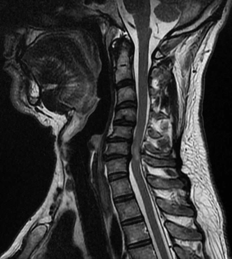
- MRI whole spine with contrast - defines the level, extent and cause of compression, and identifies additional asymptomatic levels
- CT - used if MRI is contraindicated, and for assessing bony anatomy and spinal stability before surgery
- Bloods: FBC, U&Es, LFTs, bone profile (hypercalcaemia is common in malignancy), CRP, clotting; myeloma screen (serum protein electrophoresis, free light chains) where relevant
- Blood cultures - if epidural abscess is suspected
- Investigations to find a primary if malignancy is not already known - CT chest/abdomen/pelvis, PSA, mammography, and biopsy of the most accessible lesion
- Biopsy - important where the diagnosis is uncertain, and specifically before steroids if lymphoma is a serious possibility, since steroids can obscure the histology
Management
Immediate
- Dexamethasone (typically 16 mg daily, with proton pump inhibitor cover and blood glucose monitoring) as soon as MSCC is suspected - it reduces peritumoural oedema and can preserve or improve neurological function while definitive treatment is arranged
- Flat bed rest with log-rolling until spinal stability has been assessed, if the spine may be unstable
- Adequate analgesia, often requiring opioids
- Urgent discussion with the acute oncology service, spinal surgery and clinical oncology - MSCC coordinator pathways exist in most UK trusts specifically to expedite this
- VTE prophylaxis, catheterisation if in retention, and pressure area care
- Antibiotics immediately if epidural abscess is suspected, after blood cultures
Definitive treatment
| Option | Best suited to | Notes |
|---|---|---|
| Surgical decompression and stabilisation4 | Spinal instability, bony compression or retropulsion, single-level disease, unknown primary needing tissue, or progression during radiotherapy | Requires reasonable performance status and expected survival of at least a few months; often followed by post-operative radiotherapy |
| Radiotherapy | Radiosensitive tumours (myeloma, lymphoma, small cell lung, prostate, breast), multi-level disease, or patients unfit for surgery | The most commonly used definitive treatment; can be given urgently |
| Systemic therapy | Highly chemosensitive or hormone-sensitive tumours | Used alongside local treatment rather than instead of it in the acute setting |
| Best supportive care | Very poor performance status or short prognosis | Focus on analgesia, comfort and dignity, with palliative care input |
Ongoing care
- Rehabilitation - physiotherapy and occupational therapy, with spinal injury rehabilitation input where deficit is established
- Bladder and bowel management - catheterisation or intermittent self-catheterisation, and a structured bowel regimen
- Bisphosphonates or denosumab - for skeletal-related events in relevant malignancies
- Palliative care involvement early, alongside active treatment
- Psychological support - the sudden loss of mobility and independence is profound, and often occurs alongside a new or advancing cancer diagnosis
Complications
- Permanent paraplegia or tetraplegia
- Permanent bladder and bowel dysfunction, and sexual dysfunction
- Pressure ulcers from immobility and sensory loss
- Venous thromboembolism
- Respiratory compromise with high cervical lesions
- Autonomic dysreflexia with lesions above T6
- Spinal instability and progressive deformity
- Complications of steroids - hyperglycaemia, gastric ulceration, proximal myopathy, neuropsychiatric effects, infection risk
Red flags
Prognosis
The single most powerful predictor of neurological outcome is neurological function at the time treatment is started. Patients who are still ambulant when treated are very likely to remain ambulant; those who have already lost the ability to walk have a much lower chance of regaining it, and patients who are paraplegic with complete sensory loss rarely recover useful function. This is precisely why the pathway is built around urgency rather than around waiting for a diagnosis to declare itself.2
Overall survival in MSCC is driven by the underlying malignancy rather than the compression itself, and median survival after an episode of MSCC is often only a matter of months - though this varies enormously between tumour types, being considerably better in myeloma, lymphoma, breast and prostate cancer than in lung cancer. Compression from a benign or infective cause, treated promptly, can have an excellent outcome.
Two aspects of ongoing care are easily overlooked once the acute emergency has passed. Spinal stability must be formally assessed, because a patient with an unstable metastatic deposit may be at risk of further cord injury from ordinary movement, and will need bracing or surgical stabilisation as well as radiotherapy. Rehabilitation should begin early, even in patients with a limited prognosis, since improvements in transfers, seating and continence management have a substantial effect on dignity and on the ability to be cared for at home.
Finally, an episode of MSCC is a marker of advanced malignancy, and it is an appropriate point to revisit the overall treatment plan - discussing prognosis honestly, involving palliative care alongside oncology, and establishing the patient's preferences about future care while they are able to express them.
A recurring theme in national audits is that delay usually occurs at the point of first contact rather than within the specialist pathway - back pain in a patient with cancer being managed as musculoskeletal, or a referral deferred until the following working day. Most trusts therefore operate a 24-hour MSCC coordinator service specifically so that any clinician, in any setting, can escalate directly without needing to arrange the imaging themselves. Knowing that this route exists, and using it early, is one of the most useful things a junior clinician can do in this condition.
References
- NICE NG234. Spinal metastases and metastatic spinal cord compression. 2023. Available here
- Levack P, Graham J, Collie D et al. Don't wait for a sensory level - listen to the symptoms: a prospective audit of the delays in diagnosis of malignant cord compression. Clinical Oncology. 2002. Available here
- NICE NG59. Low back pain and sciatica in over 16s: assessment and management. 2016, updated 2020. Available here
- Patchell RA, Tibbs PA, Regine WF et al. Direct decompressive surgical resection in the treatment of spinal cord compression caused by metastatic cancer. The Lancet. 2005. Available here
This article is written for revision and education. It is not clinical guidance and must not be used to make decisions about the care of a patient. Always check current NICE guidance and local protocols.