
Hypernatraemia
Key points
- Hypernatraemia: serum sodium above 145 mmol/L. Far less common than hyponatraemia, but carries higher mortality because of the frail populations it affects.
- It is a water deficit: hypernatraemia is almost always a deficit of water relative to sodium, not an excess of salt. Total body sodium is usually normal or low.
- It requires two failures: an intact thirst mechanism with access to water will correct almost any water loss. So hypernatraemia implies water loss PLUS an inability to drink - the very young, the elderly, the confused, the sedated and the dependent.
- Commonest causes: inadequate intake in dependent patients, GI losses, osmotic diuresis (hyperglycaemia, HHS), diabetes insipidus, and excessive insensible loss from fever and burns.
- The urine tells you the cause: concentrated urine (osmolality >700) means the kidney is responding appropriately to extrarenal loss. Inappropriately dilute urine (<300) with hypernatraemia means diabetes insipidus.
- Symptoms: thirst (if the mechanism is intact), lethargy, irritability, weakness, confusion, and in severe cases seizures and coma from cerebral cell shrinkage.
- The correction limit: do not lower sodium faster than 10-12 mmol/L in 24 hours (8-10 in chronic cases). Over-rapid correction causes cerebral oedema and seizures.
- Route matters: oral or enteral water is preferred where possible. If IV is needed, use 5% dextrose or 0.45% saline, with 0.9% saline first if the patient is shocked.
Introduction
Hypernatraemia is a serum sodium above 145 mmol/L. It is considerably less common than hyponatraemia but is clinically important because it is associated with mortality of 40-60% in hospitalised patients - a figure that reflects the frailty and severity of illness of the people who develop it far more than the direct toxicity of the sodium itself.1
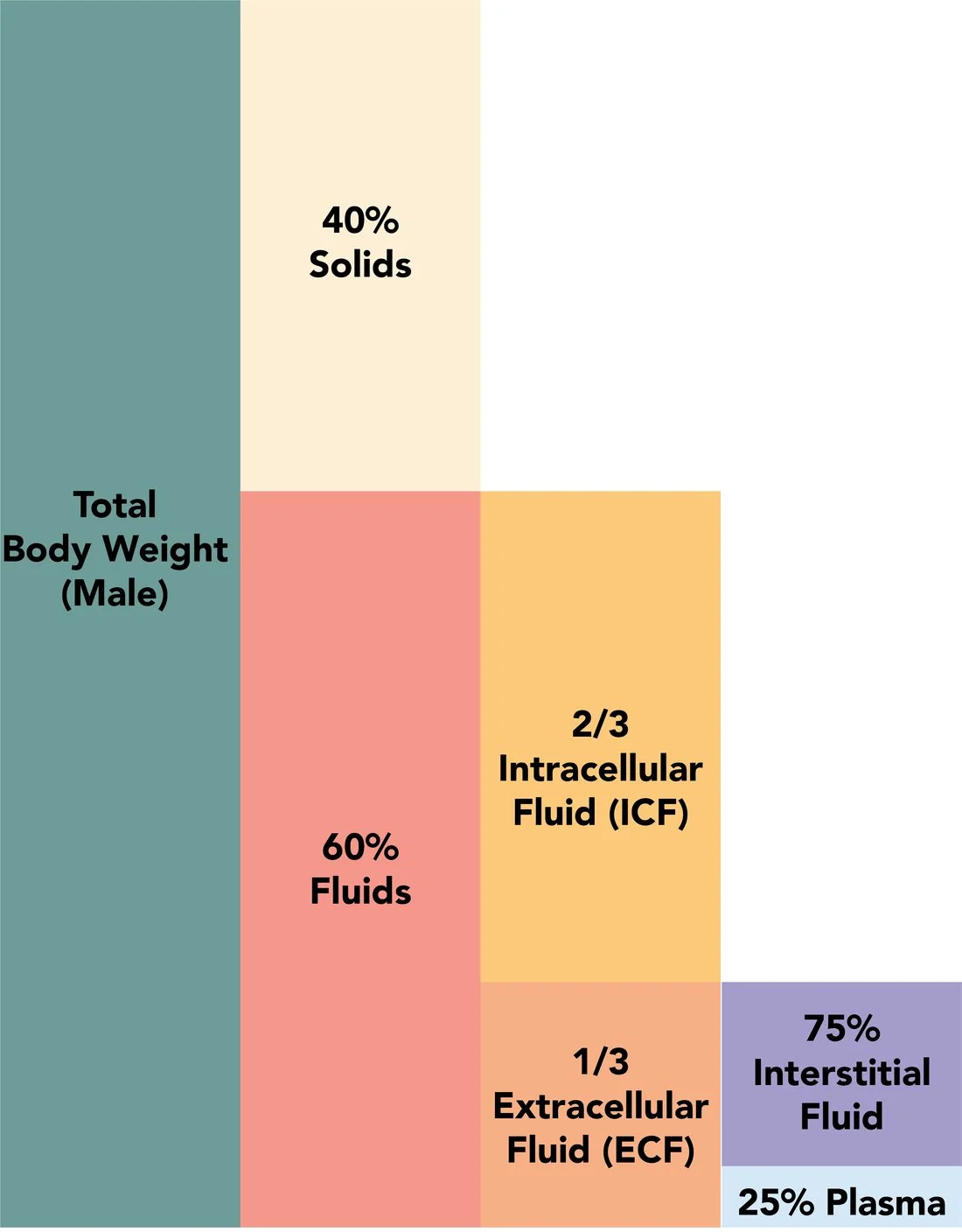
Because sodium is the principal extracellular osmole, a rise in serum sodium raises plasma osmolality and draws water out of cells by osmosis. In the brain this causes cell shrinkage, which produces the neurological symptoms and, in severe or rapid cases, can tear bridging veins to cause intracranial haemorrhage. As with hyponatraemia, the brain adapts over 48 hours by generating intracellular osmolytes - and that adaptation is what makes rapid correction dangerous.
Causes
Causes are best grouped by volume status, exactly as for hyponatraemia, because this determines the fluid used for replacement.
| Volume status | Mechanism | Causes |
|---|---|---|
| Hypovolaemic (water loss exceeding sodium loss) - the commonest pattern | Loss of hypotonic fluid | Renal: osmotic diuresis (hyperglycaemia, HHS, mannitol, high-protein feeds), loop diuretics, post-obstructive diuresis, recovery phase of AKI. Extrarenal: diarrhoea (especially in infants), vomiting, sweating and fever, burns, and losses from fistulae or stomas |
| Euvolaemic (pure water loss) | Loss of water without sodium | Diabetes insipidus - cranial (pituitary surgery, trauma, tumours, infiltration) or nephrogenic (lithium, hypercalcaemia, hypokalaemia, CKD, demeclocycline). Also increased insensible losses through skin and lungs (fever, tachypnoea, hot environments, mechanical ventilation), and inadequate intake in dependent patients |
| Hypervolaemic (sodium gain) - uncommon | Excess sodium administration or retention | Hypertonic saline, sodium bicarbonate in resuscitation, hypertonic feeds or incorrectly made-up infant formula, salt poisoning (accidental or non-accidental), primary hyperaldosteronism and Cushing syndrome (usually only mild hypernatraemia), and near-drowning in sea water |
Clinical features
Symptoms are predominantly neurological, and reflect cellular dehydration of the brain. As with hyponatraemia, the rate of change matters more than the absolute value.
- Thirst - the earliest and most prominent symptom, but only if the thirst mechanism is intact and the patient can communicate. Its absence in a dependent or obtunded patient is precisely why the diagnosis is missed
- Lethargy, weakness and irritability
- Confusion and delirium
- Nausea and vomiting
- Muscle twitching, hyperreflexia and tremor; in severe cases rigidity
- Seizures and coma in severe hypernatraemia (typically above 158-160 mmol/L, or with rapid onset)
- Intracranial haemorrhage - subarachnoid or subdural, from tearing of bridging veins as the brain shrinks; a particular risk in infants
- Polyuria and polydipsia - pointing towards diabetes insipidus or osmotic diuresis
- Signs of volume depletion - dry mucous membranes, reduced skin turgor, tachycardia, postural hypotension, oliguria, sunken eyes and, in infants, a depressed fontanelle
- In infants: a high-pitched cry, irritability alternating with lethargy, poor feeding and fever - hypernatraemic dehydration in a breastfed neonate with inadequate feeding is a recognised and serious presentation
A practical point on examination: hypernatraemic dehydration preserves circulating volume relatively well, because water is drawn out of cells into the extracellular space. Signs of shock therefore appear later than in isotonic dehydration - which means a child or adult may be considerably more depleted than they look.
Investigations
- U&Es, creatinine and eGFR - confirm hypernatraemia and assess renal function
- Serum osmolality - raised, confirming true hypertonicity
- Urine osmolality - the single most discriminating test (see below)
- Urinary sodium - helps establish whether losses are renal or extrarenal
- Glucose - hyperglycaemia causing osmotic diuresis is a very common contributor; exclude HHS, which combines profound hyperglycaemia, hyperosmolality and hypernatraemia
- Fluid balance chart and daily weights - essential, and often more informative than any blood test; weight loss quantifies the water deficit
- Calcium and potassium - hypercalcaemia and hypokalaemia both cause nephrogenic DI
- Lithium level - if the patient takes lithium
- Paired serum and urine osmolality with a water deprivation test - to diagnose and classify DI, followed by desmopressin to separate cranial from nephrogenic
- MRI pituitary - if cranial DI is suspected; the normal posterior pituitary bright spot is characteristically absent
- Review the drug chart and feed prescription - hypertonic feeds, sodium-containing drugs and diuretics are frequently implicated
Management
The principles are: restore circulating volume first if the patient is shocked, then replace the water deficit slowly, replace ongoing losses, and treat the underlying cause - including the reason the patient could not drink.2
Step 1 - resuscitate if shocked
If there is haemodynamic compromise, give 0.9% sodium chloride to restore circulating volume, even though it is relatively hypotonic compared with the patient's plasma and will not lower the sodium quickly. Perfusion takes priority over the sodium concentration. Once the patient is haemodynamically stable, switch to correcting the water deficit.
Step 2 - estimate the water deficit
Step 3 - replace the water at a safe rate
| Situation | Fluid | Notes |
|---|---|---|
| Patient can drink or has enteral access | Oral or nasogastric water | Preferred route wherever possible - safest, most physiological, and self-limiting. Often the whole treatment in a dependent patient |
| Haemodynamically compromised | 0.9% sodium chloride | Restore perfusion first. Switch once stable |
| Euvolaemic / pure water deficit | 5% dextrose | Provides free water once the glucose is metabolised. Monitor blood glucose - infusing large volumes can cause hyperglycaemia and a further osmotic diuresis |
| Hypovolaemic with ongoing losses | 0.45% sodium chloride | Provides both water and some sodium; a common practical choice |
| Sodium overload (hypervolaemic) | Stop the sodium source; loop diuretic plus water replacement | Dialysis if there is renal failure or severe overload |
| Cranial diabetes insipidus | Desmopressin plus water replacement | Monitor closely - sodium can then fall rapidly |
| Nephrogenic diabetes insipidus | Remove the cause (stop lithium, correct calcium and potassium); thiazide diuretic and low-salt diet, sometimes with NSAIDs | Paradoxically, thiazides reduce urine output in nephrogenic DI |
Step 4 - treat the cause and prevent recurrence
- Identify and address why the patient could not drink - this is the step most often omitted. Ensure fluids are within reach, that assistance with drinking is provided and documented, and consider a fluid balance chart and daily weights
- Treat hyperglycaemia, particularly HHS, which requires careful simultaneous management of glucose, sodium and osmolality
- Review medications - lithium, diuretics, hypertonic feeds and sodium-containing preparations
- Dietitian review of enteral feed composition and free water flushes; inadequate water flushes with concentrated feeds is a classic hospital cause
- Speech and language therapy assessment where swallowing is impaired
- In infants, review feeding technique and adequacy, and check formula is being made up correctly; consider safeguarding if salt poisoning is a possibility
- Refer to endocrinology for diabetes insipidus
Complications
- Seizures, coma and death - from severe cellular dehydration of the brain
- Intracranial and subarachnoid haemorrhage - from tearing of bridging veins as the brain shrinks away from the skull; particularly in infants and with rapid onset
- Cerebral venous sinus thrombosis - promoted by haemoconcentration and dehydration
- Cerebral oedema and seizures from over-rapid correction - the principal iatrogenic hazard, and the mirror image of osmotic demyelination in hyponatraemia
- Central pontine myelinolysis - rarely reported with rapid osmotic shifts in hypernatraemia too, although far more characteristic of hyponatraemia correction
- Acute kidney injury - from volume depletion
- Rhabdomyolysis - a recognised complication of severe hypernatraemia
- Hyperglycaemia - from large-volume 5% dextrose infusion, potentially worsening the osmotic diuresis
- Venous thromboembolism - from haemoconcentration and immobility
- Pressure ulcers, falls and functional decline in the frail dependent patients who typically develop it
- Long-term neurological deficit - particularly in infants who have had severe hypernatraemic dehydration or intracranial haemorrhage
Red flags
Prognosis
Mortality in hospitalised patients with hypernatraemia is high - commonly quoted at 40-60% in adults, and higher still in those who develop it after admission rather than presenting with it. This figure should be interpreted carefully: hypernatraemia is only rarely the direct cause of death. It is far more often a marker of severe underlying illness, frailty and dependency - the patients who cannot drink are the patients who are already sickest.
Hospital-acquired hypernatraemia carries particular significance. Unlike community-acquired cases, which usually reflect an acute illness with fluid loss, hypernatraemia developing during an admission generally means a dependent patient did not receive the water they needed. It is therefore treated in many trusts as a marker of care quality, and it is largely preventable through attention to fluid balance charts, assisted drinking, adequate free water with enteral feeds, and daily weights.
Outcome depends principally on the underlying cause and on the rate of correction. Hypernatraemia from a reversible cause in an otherwise well patient corrects completely with water and carries no lasting consequence. The two situations that cause avoidable harm are failure to recognise it - allowing sodium to climb to levels causing seizures and intracranial haemorrhage - and over-enthusiastic correction, which causes cerebral oedema. Infants and children are the most vulnerable at both ends, having the highest risk of intracranial haemorrhage from severe hypernatraemia and of cerebral oedema from rapid correction, which is why paediatric correction is deliberately slower and why severe paediatric hypernatraemic dehydration can leave permanent neurological deficit.
References
- Adrogué HJ, Madias NE. Hypernatremia. New England Journal of Medicine. 2000. Available here
- NICE CG174. Intravenous fluid therapy in adults in hospital. 2013, updated 2017. Available here
- NICE NG29. Intravenous fluid therapy in children and young people in hospital. 2015, updated 2020. Available here
- Society for Endocrinology. Emergency Endocrine Guidance: diabetes insipidus (arginine vasopressin deficiency) in hospital. Available here
- Joint British Diabetes Societies. The Management of the Hyperosmolar Hyperglycaemic State (HHS) in Adults. Available here
- NHS England / NPSA. Risks of hypernatraemia and inadequate hydration in hospital inpatients. Available here
- Alan Sved and David Walsh, CC BY-SA 4.0, via Wikimedia Commons. Available here
This article is written for revision and education. It is not clinical guidance and must not be used to make decisions about the care of a patient. Always check current NICE guidance and local protocols.