
Cerebral Venous Sinus Thrombosis: Diagnosis and Management
Key points
- Cerebral venous sinus thrombosis (CVST): thrombosis of one or more of the dural venous sinuses, impairing venous drainage and CSF resorption.
- Typical patient: a younger woman, often with a prothrombotic state - pregnancy, the puerperium, the combined oral contraceptive pill, or an inherited thrombophilia.
- Presentation: headache is almost universal (often progressive, worse lying flat), frequently with seizures, focal deficit, or a reduced conscious level - the pattern is much more variable than arterial stroke.
- First investigation: CT venography or MR venography, since a plain CT or MRI can look deceptively normal.
- Key sign: the 'empty delta sign' on contrast CT - a filling defect in the superior sagittal sinus.
- Management: anticoagulation (usually with a DOAC or LMWH/warfarin) is started even when there is an associated haemorrhagic infarct, since the underlying problem is thrombosis.
- Refractory cases: endovascular thrombectomy or thrombolysis is considered for deterioration despite anticoagulation.
- Prognosis: generally good with treatment - most patients recover well, though a minority are left with a deficit, seizures, or chronic raised intracranial pressure.
Introduction
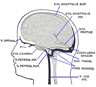
Cerebral venous sinus thrombosis (CVST) is thrombosis of one or more of the dural venous sinuses - most commonly the superior sagittal sinus or the transverse sinuses - which drain venous blood from the brain and are responsible for CSF resorption via the arachnoid granulations.1
Occlusion causes two overlapping problems: impaired venous drainage, which raises venous pressure and can cause venous infarction (often haemorrhagic, since the tissue remains perfused by unobstructed arteries while unable to drain), and impaired CSF resorption, which raises intracranial pressure independently of any infarct.
CVST is rare compared with arterial stroke but disproportionately affects younger people, and particularly women, which makes it an important cause of stroke-like presentations in a population where arterial stroke is otherwise uncommon. Its presentation is notoriously variable, and it is a classic 'do not miss' diagnosis in headache and stroke-mimic teaching.
CVST accounts for well under 1% of all strokes, with an incidence of roughly 1-2 per 100,000 per year, but the mean age at presentation is around 35 - decades younger than for arterial stroke - and around three-quarters of adult cases occur in women, reflecting the contribution of pregnancy, the puerperium and hormonal contraception. Any stroke-like presentation in a young woman should therefore prompt active consideration of a venous rather than arterial mechanism.
Aetiology
CVST is fundamentally a disease of thrombosis, and almost always has an identifiable prothrombotic trigger, unlike most arterial strokes.
- Pregnancy and the puerperium - a major risk period, particularly the weeks after delivery
- Combined oral contraceptive pill and hormone replacement therapy
- Inherited thrombophilia - factor V Leiden, protein C/S deficiency, antithrombin deficiency, prothrombin gene mutation
- Antiphospholipid syndrome
- Malignancy - both direct effect and paraneoplastic hypercoagulability
- Dehydration
- Infection - local spread from otitis media, mastoiditis or sinusitis (especially to the transverse/sigmoid sinuses or cavernous sinus), or as part of severe systemic sepsis
- Head injury or neurosurgical procedures, including direct trauma to a sinus
- Inflammatory conditions - inflammatory bowel disease, Behçet disease, nephrotic syndrome
- Haematological disorders - polycythaemia, essential thrombocythaemia
- A cause is not found in a minority despite thorough investigation
Risk factors
- Female sex, especially of reproductive age
- Pregnancy and the puerperium
- Combined oral contraceptive use
- Known thrombophilia or personal/family history of venous thromboembolism
- Recent head or neck infection, particularly otitis media, mastoiditis or sinusitis
- Dehydration and recent significant illness
- Active malignancy
Clinical features
Presentation is genuinely variable, reflecting the sinus involved and whether venous infarction has occurred, but a few patterns recur.
Understanding why the presentation is so variable comes back to venous anatomy. Unlike the arterial supply, the cerebral venous system has extensive collateral drainage, so occlusion of a single sinus may be entirely compensated, produce only headache from raised pressure, or cause frank infarction - depending on how good the collaterals are in that individual. This is why CVST has no single stereotyped syndrome in the way that an MCA infarct does.
It also explains the characteristically haemorrhagic nature of venous infarcts. In arterial stroke, tissue dies because it is not perfused. In venous infarction the arterial supply continues to deliver blood into tissue that cannot drain, so hydrostatic pressure rises until vessels rupture. The infarct is therefore congested and frequently bleeds, and it does not respect arterial territory boundaries - a combination that should immediately raise the possibility of a venous cause on imaging.
- Headache - present in the great majority of patients, often subacute and progressive over days, classically worse lying flat and in the morning, and may be the only symptom for some time
- Signs of raised intracranial pressure - papilloedema, visual obscurations, nausea and vomiting; can closely mimic idiopathic intracranial hypertension
- Seizures - considerably more common than in arterial stroke, and can be focal or generalised
- Focal neurological deficit - which may not fit a single arterial territory, or may fluctuate, or affect both sides (as with superior sagittal sinus thrombosis causing bilateral leg weakness)
- Reduced consciousness - in more extensive thrombosis or with significant venous infarction
- Cavernous sinus thrombosis specifically - a distinct and dramatic presentation with painful proptosis, chemosis, ophthalmoplegia (III, IV, VI palsies) and sensory loss in the ophthalmic/maxillary trigeminal distribution, usually from spreading facial or sinus infection
Clinical examination
- Fundoscopy - papilloedema is common and should always be checked
- Focal neurology - which may be bilateral, or fluctuate, unlike typical arterial stroke
- Conscious level
- Signs of the underlying cause - mastoid tenderness, signs of otitis media, or (in cavernous sinus thrombosis) proptosis, chemosis and ophthalmoplegia
- Signs of an underlying prothrombotic state - recent pregnancy, features of a connective tissue or inflammatory disease
Differential diagnosis
- Idiopathic intracranial hypertension - very similar presentation with headache and papilloedema, but CVST must be actively excluded with venography before this diagnosis is accepted
- Arterial ischaemic stroke - CVST can mimic it, but the demographic (young, female) and non-arterial pattern of deficit are clues
- Subarachnoid haemorrhage - both cause headache and can coexist, since CVST itself can occasionally cause a subarachnoid bleed
- Meningitis or encephalitis - fever and more prominent meningism, though CVST can also cause fever if related to local infection
- Migraine - especially if there is a personal history, though new significant change in pattern warrants imaging
- Primary seizure disorder
Investigations
Plain, non-contrast CT or MRI can appear normal or show only subtle changes, and a normal scan does not exclude CVST - dedicated venous imaging is required whenever the diagnosis is suspected.
The empty delta sign deserves a word of caution as well as recognition. It is specific when clearly present, but it is only visible on contrast-enhanced imaging, appears in a minority of cases, and can be mimicked by a normally high-splitting superior sagittal sinus. It should therefore be treated as a useful confirmatory clue rather than something whose absence provides reassurance.
A similar caution applies to the hyperdense sinus on plain CT. Thrombus appears dense, but so does normal sinus blood in a patient with a high haematocrit or dehydration, and the finding is easily over- or under-called. Neither sign is a substitute for dedicated venography where the clinical suspicion is real.
- CT venography or MR venography - the definitive investigations, demonstrating a filling defect in the affected sinus
- Contrast CT may show the 'empty delta sign' - a triangular filling defect in the superior sagittal sinus surrounded by enhancing dura, representing thrombus within the sinus
- Non-contrast CT may show a hyperdense sinus (thrombus) or venous infarcts, which are frequently haemorrhagic and do not respect arterial boundaries
- D-dimer - a normal D-dimer makes CVST less likely but does not reliably exclude it, particularly when the presentation is isolated headache or has been present for some time
- Thrombophilia screen - once the acute event is over (results can be altered by acute thrombosis and anticoagulation), particularly in the absence of an obvious provoking factor
- Lumbar puncture - may show a raised opening pressure; performed once haemorrhage and significant mass effect have been excluded, and can help distinguish CVST from idiopathic intracranial hypertension
- Screen for the underlying cause - inflammatory markers, blood cultures if infection is suspected, pregnancy test
Management
Anticoagulation
Anticoagulation is the cornerstone of treatment, and is started even in the presence of a haemorrhagic venous infarct, because the underlying process driving further damage is ongoing thrombosis, and the haemorrhage results from venous congestion rather than a bleeding tendency - a point that surprises many students and is frequently tested.2
- Low molecular weight heparin, followed by long-term warfarin or (increasingly) a DOAC
- Duration is typically 3-6 months for a provoked event, longer or indefinite if there is a strong unprovoked thrombophilia or recurrent CVST
- Pregnant patients are anticoagulated with LMWH throughout pregnancy and the puerperium, since warfarin and DOACs are contraindicated in pregnancy
Supportive and specific measures
- Seizure control - antiepileptic treatment for seizures at presentation; prophylactic use in the absence of a seizure is not routine
- Managing raised intracranial pressure - analgesia, and specific measures (acetazolamide, therapeutic lumbar puncture, or optic nerve sheath fenestration) if papilloedema threatens vision, similarly to idiopathic intracranial hypertension
- Treat the underlying cause - antibiotics and source control for infective causes (mastoiditis, sinusitis, cavernous sinus thrombosis), stopping the combined oral contraceptive pill
- Endovascular thrombectomy or catheter-directed thrombolysis - considered for patients who deteriorate despite adequate anticoagulation, though evidence is less robust than for arterial thrombectomy
- Decompressive craniectomy - for large venous infarcts causing life-threatening mass effect refractory to medical management
Complications
- Venous infarction, frequently haemorrhagic and bilateral
- Raised intracranial pressure and, if untreated, visual loss from chronic papilloedema
- Seizures and post-thrombotic epilepsy
- Recurrent CVST or other venous thromboembolism
- Death, in a minority, usually from extensive infarction or uncontrolled raised intracranial pressure
Red flags
CVST is most often missed because it does not resemble arterial stroke and the patient does not fit the expected demographic. The combination of a young patient, a prothrombotic risk factor and any of the features below should prompt dedicated venous imaging.
Prognosis
Overall prognosis is better than for arterial stroke of comparable severity: most patients treated with anticoagulation make a good functional recovery, and mortality is low with modern management. Outcome is worse with extensive thrombosis, deep venous system involvement, significant haemorrhagic infarction, or a markedly reduced conscious level at presentation. A minority of patients are left with persistent headache, seizures, or chronic raised intracranial pressure requiring ongoing management.3
Follow-up has two distinct strands that are easily conflated. The first is haematological: establishing whether an underlying thrombophilia or occult malignancy explains the event, which determines how long anticoagulation continues and whether family members need testing. The second is neurological and ophthalmological: monitoring for persisting raised intracranial pressure, since papilloedema can outlast recanalisation and threaten vision even in a patient whose headache has settled. Repeat venography is usually performed after several months to document recanalisation and provide a baseline for any future event.
Counselling about future risk matters, particularly in the typical patient - a young woman. The combined oral contraceptive pill is contraindicated afterwards, and any future pregnancy requires planned thromboprophylaxis with low molecular weight heparin through pregnancy and the puerperium. CVST is not, however, a contraindication to pregnancy itself, and framing it that way avoids unnecessary distress.
One further point worth knowing is vaccine-induced immune thrombotic thrombocytopenia (VITT), a rare syndrome recognised following adenoviral-vector COVID-19 vaccines, in which CVST occurred alongside thrombocytopenia and anti-PF4 antibodies. It is now of limited day-to-day relevance but remains clinically important as the exception to standard management: heparin is avoided in favour of non-heparin anticoagulants, and immunoglobulin is given. It is a useful reminder to check the platelet count in any patient presenting with unusual-site thrombosis.
References
- Ferro JM, Bousser MG, Canhão P et al. European Stroke Organization guideline for the diagnosis and treatment of cerebral venous thrombosis. European Stroke Journal. 2017. Available here
- Saposnik G, Barinagarrementeria F, Brown RD et al. Diagnosis and management of cerebral venous thrombosis - a statement for healthcare professionals. Stroke. 2011. Available here
- NICE CKS. Stroke and TIA. Available here
This article is written for revision and education. It is not clinical guidance and must not be used to make decisions about the care of a patient. Always check current NICE guidance and local protocols.