
Neurofibromatosis: Types 1 and 2
Key points
- Neurofibromatosis type 1: autosomal dominant, NF1 gene on chromosome 17 encoding neurofibromin, a tumour suppressor; affects about 1 in 3,000.
- NF1 diagnosis: clinical, requiring two or more of seven criteria - café-au-lait macules, neurofibromas, axillary/inguinal freckling, optic glioma, Lisch nodules, bony dysplasia, or an affected first-degree relative.
- Neurofibromatosis type 2: autosomal dominant, NF2 gene on chromosome 22 encoding merlin; far rarer at about 1 in 30,000.
- NF2 hallmark: bilateral vestibular schwannomas causing progressive sensorineural hearing loss, tinnitus and imbalance.
- De novo mutation: around half of NF1 cases arise new, so an absent family history does not exclude the diagnosis.
- Commonest NF1 morbidity: learning difficulties and ADHD, present in a substantial minority, not the tumours.
- Malignant transformation: malignant peripheral nerve sheath tumour arises in plexiform neurofibromas - suspect on rapid growth, new persistent pain or a new neurological deficit.
- Management: no cure; lifelong surveillance including annual blood pressure and ophthalmology, with MEK inhibitors for inoperable plexiform neurofibromas.
Introduction
Neurofibromatosis types 1 and 2 share a name for historical reasons but are entirely separate conditions, caused by different genes on different chromosomes, presenting differently and requiring different surveillance. Both are autosomal dominant tumour predisposition syndromes affecting tissues derived from the neural crest, and both result from loss of a tumour suppressor gene.1
NF1 (formerly von Recklinghausen disease) is roughly ten times commoner, presents in childhood with skin findings, and its main day-to-day burden is neurodevelopmental rather than oncological. NF2 presents in adolescence or early adulthood with hearing loss from bilateral vestibular schwannomas, and its burden is neurological and surgical. Conflating the two is the single commonest error in exam answers on this topic.
Neurofibromatosis type 1: genetics and pathophysiology
NF1 is caused by loss-of-function variants in the NF1 gene on chromosome 17, which encodes neurofibromin. Neurofibromin is a GTPase-activating protein that switches off Ras signalling; losing it leaves Ras constitutively active, driving cell proliferation. This is why NF1 behaves as a tumour predisposition syndrome, and why MEK inhibitors, which act downstream in the same Ras-MAPK pathway, have become an effective targeted therapy.
- Autosomal dominant inheritance with near-complete penetrance - virtually everyone with a pathogenic variant shows some feature by adulthood
- Highly variable expressivity, even within one family carrying an identical variant, ranging from a few café-au-lait macules to disfiguring plexiform tumours
- Around 50% of cases are de novo, one of the highest new-mutation rates of any genetic condition, reflecting the large size of the gene
- Tumours arise by a second hit to the remaining normal allele in a somatic cell, following Knudson's two-hit model of tumour suppressor loss
- Segmental (mosaic) NF1 occurs where the mutation arises post-zygotically, producing features confined to one body region
NF1: clinical features and diagnostic criteria
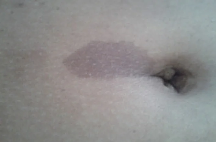
Diagnosis is clinical, requiring two or more of the following criteria. Features accumulate with age, so a young child who does not yet meet criteria may still do so later - café-au-lait macules usually appear first, neurofibromas typically not until puberty.2
| Criterion | Detail |
|---|---|
| Café-au-lait macules | Six or more, over 5 mm in greatest diameter before puberty, or over 15 mm after puberty |
| Neurofibromas | Two or more neurofibromas of any type, or one plexiform neurofibroma |
| Freckling | Axillary or inguinal freckling (Crowe's sign) - freckling in non-sun-exposed skin folds |
| Optic pathway glioma | Usually presenting in early childhood, often asymptomatic and found on screening |
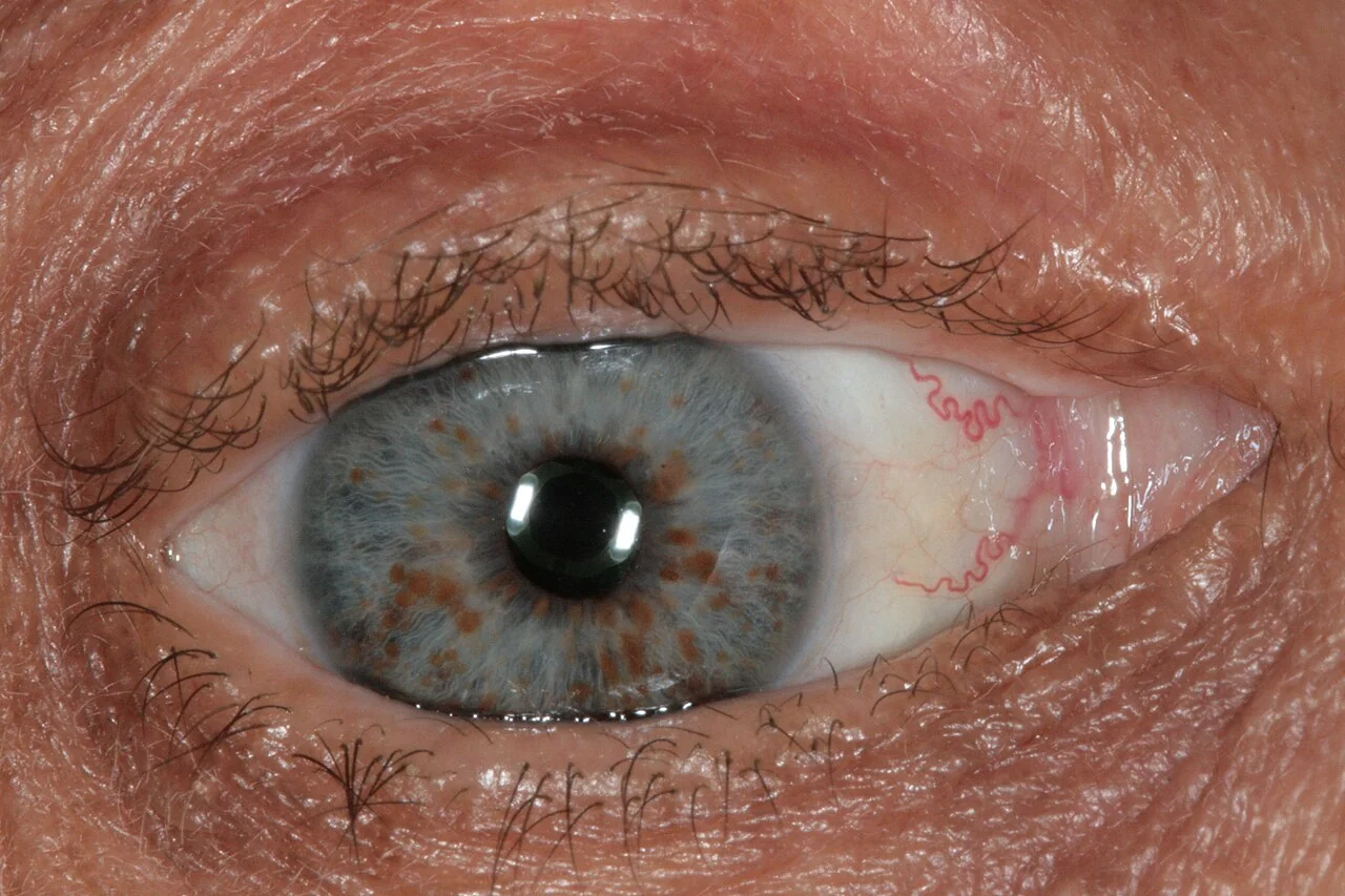
| Iris findings | Two or more Lisch nodules on slit-lamp examination, or two or more choroidal abnormalities on optical coherence tomography |
| Distinctive bony lesion | Sphenoid wing dysplasia, or anterolateral bowing of the tibia with or without pseudarthrosis |
| Family history | A first-degree relative meeting the above criteria |
Neurofibromas
- Cutaneous neurofibromas - soft, fleshy, skin-coloured or violaceous papules and nodules that appear from puberty and increase in number through adult life; benign, but a major source of psychological distress and stigma
- Subcutaneous neurofibromas - firmer, deeper nodules along peripheral nerves, which can be tender
- Plexiform neurofibromas - diffuse, infiltrating tumours involving multiple nerve fascicles, often congenital, sometimes with overlying hyperpigmentation or hypertrichosis. These are the lesions that can undergo malignant transformation, and they can cause disfigurement and compress vital structures
Neurodevelopmental features
It is worth stating plainly, because it is frequently overlooked in favour of the more visually striking findings: the commonest source of morbidity in NF1 is not tumour but learning difficulty. Specific learning difficulties affect a substantial proportion, ADHD is markedly over-represented, and autism spectrum traits are more common than in the general population. General intelligence is usually within the normal range but shifted slightly downwards. Early educational assessment and support matter more to most families than tumour surveillance does.
NF1: complications
Complications span multiple systems and drive the surveillance schedule.
- Optic pathway glioma - in around 15% of children, usually low-grade pilocytic astrocytoma, most often presenting before age 6; many are asymptomatic and never need treatment, but they can cause visual loss or precocious puberty if the chiasm or hypothalamus is involved
- Malignant peripheral nerve sheath tumour (MPNST) - a lifetime risk of roughly 8-13%, arising within plexiform neurofibromas, and the leading cause of NF1-related death
- Hypertension - a specific and examinable association, which may be essential, or secondary to renal artery stenosis or phaeochromocytoma; this is why blood pressure is checked at every review
- Scoliosis - common, and can be rapidly progressive dystrophic scoliosis requiring surgical intervention
- Tibial dysplasia and pseudarthrosis - anterolateral bowing of the tibia which may fracture and fail to unite
- Epilepsy, headache, and other central nervous system tumours including brainstem and cerebellar gliomas
- Breast cancer - women with NF1 have an increased risk under the age of 50, and enhanced surveillance is now offered in the UK
- Short stature and macrocephaly - both common and generally benign, but worth recognising so they are not over-investigated
Neurofibromatosis type 2
NF2 results from loss-of-function variants in the NF2 gene on chromosome 22, encoding merlin (also called schwannomin), another tumour suppressor. It is much rarer, at roughly 1 in 25,000-33,000, and around half of cases are de novo, with mosaicism relatively common in that group.3
Clinical features
- Bilateral vestibular schwannomas - the defining feature, arising on the vestibular portion of the eighth cranial nerve and presenting with progressive sensorineural hearing loss, tinnitus and imbalance, typically in the late teens to thirties
- Other schwannomas on any cranial or peripheral nerve, and spinal tumours causing cord or root compression
- Meningiomas, often multiple, and ependymomas of the spinal cord
- Juvenile posterior subcapsular cataract - an early and useful diagnostic clue, sometimes preceding the tumours
- Peripheral neuropathy and, in children, a mononeuropathy such as facial palsy or a squint as the first presentation
- Skin features are minimal - a few café-au-lait macules or skin plaques may occur, but nothing like the burden seen in NF1
Management
Care is delivered through specialist national NF2 services. Management balances tumour control against preservation of hearing and facial nerve function, and watchful waiting with serial MRI is often preferred to early surgery for stable tumours. Bevacizumab, an anti-VEGF antibody, can shrink vestibular schwannomas and improve hearing in selected patients. Surgery is used for tumours causing brainstem compression or progressive deficit, and auditory brainstem implants or cochlear implants can restore some useful hearing after tumour removal. Hearing rehabilitation, including early sign language teaching before hearing is lost, is a core part of care rather than an afterthought.
Comparing NF1 and NF2
| NF1 | NF2 | |
|---|---|---|
| Gene / chromosome | NF1, chromosome 17 (neurofibromin) | NF2, chromosome 22 (merlin) |
| Incidence | ~1 in 3,000 | ~1 in 30,000 |
| Typical age at presentation | Early childhood | Late teens to thirties |
| Skin | Café-au-lait macules, freckling, cutaneous neurofibromas | Minimal skin involvement |
| Eye | Lisch nodules (iris hamartomas) | Juvenile posterior subcapsular cataract |
| Hallmark tumour | Neurofibroma (including plexiform); optic glioma | Bilateral vestibular schwannoma; meningioma, ependymoma |
| Main morbidity | Learning difficulty; MPNST is the main mortality risk | Hearing loss, balance and cranial nerve deficits |
| Memory aid | Seventeen letters in 'von Recklinghausen' | Type 2, chromosome 22, bilateral (two) acoustic neuromas |
Investigations
NF1 remains a clinical diagnosis in most cases, with genetic testing reserved for atypical presentations, very young children not yet meeting criteria, and reproductive planning. NF2 is more reliant on imaging and molecular confirmation.
- Slit-lamp examination - for Lisch nodules in NF1 and cataract in NF2; both require an ophthalmologist rather than bedside inspection
- MRI brain and orbits - for optic pathway glioma in NF1; MRI internal auditory meati with contrast is the key investigation for suspected NF2
- Whole-body MRI - increasingly used to quantify internal plexiform neurofibroma burden in NF1, which correlates with MPNST risk
- Genetic testing - NF1 or NF2 sequencing and deletion/duplication analysis; note that mosaicism can produce a negative blood test in a genuinely affected person, particularly in NF2
- Blood pressure measurement - at every clinical contact in NF1, with plasma or urinary metanephrines and renal artery imaging if hypertension is found
- Spinal imaging - if scoliosis or neurological signs suggest cord or root involvement
- Audiology - baseline and serial testing in NF2, and where any hearing concern arises in NF1
Differential diagnosis
- Legius syndrome - café-au-lait macules and freckling without neurofibromas, Lisch nodules or tumour risk; caused by SPRED1 variants and an important benign mimic that spares patients unnecessary surveillance
- McCune-Albright syndrome - café-au-lait macules with irregular 'coast of Maine' borders (versus the smooth 'coast of California' borders in NF1), plus precocious puberty and fibrous dysplasia
- Isolated café-au-lait macules - one or two are common and entirely normal in the general population
- Schwannomatosis - multiple schwannomas with pain as the dominant symptom, but without bilateral vestibular schwannomas
- Sporadic unilateral vestibular schwannoma - far commoner than NF2 and not associated with a tumour predisposition syndrome
- Tuberous sclerosis - another neurocutaneous syndrome, distinguished by ash-leaf macules, shagreen patches, facial angiofibromas, epilepsy and cortical tubers
Management and surveillance in NF1
There is no cure. Care is coordinated through a multidisciplinary team, and, as with the other conditions in this topic, the value lies in structured lifelong surveillance that detects treatable complications before they cause irreversible harm.4
| Domain | Approach |
|---|---|
| Blood pressure | At every clinical review, from childhood - the single highest-yield routine measurement |
| Ophthalmology | Annual review through childhood for optic pathway glioma; visual acuity and fields |
| Growth and development | Height, weight, head circumference and pubertal staging - precocious puberty suggests chiasmatic involvement |
| Neurodevelopment | Proactive educational assessment; do not wait for school failure |
| Spine | Clinical examination for scoliosis at each review through growth |
| Skin/tumour review | Annual clinical examination, with a low threshold for imaging if a lesion changes |
| Breast screening | Enhanced surveillance offered to women with NF1 from age 40 (or earlier per local protocol) |
Treatment
- Symptomatic and supportive care for the great majority - educational support, physiotherapy, pain management and psychological support
- Surgical excision of cutaneous neurofibromas causing pain, functional problems or significant distress, accepting that they frequently recur and that the total burden cannot be surgically cleared
- MEK inhibitors (selumetinib) for symptomatic, inoperable plexiform neurofibromas in children - a genuine advance, shrinking tumours by targeting the Ras-MAPK pathway left unchecked by neurofibromin loss5
- Chemotherapy (typically carboplatin and vincristine) for progressive optic pathway glioma; radiotherapy is generally avoided in NF1 because it increases the risk of second malignancy and vasculopathy
- Orthopaedic input for scoliosis and tibial pseudarthrosis
Genetic counselling
Both conditions are autosomal dominant, so an affected person has a 50% chance of transmitting the variant to each child, with male-to-male transmission possible. Because expressivity is so variable in NF1, counselling must be explicit that the severity in a child cannot be predicted from the parent's phenotype.
- Around half of cases are de novo, so unaffected parents of an affected child have a low recurrence risk - though gonadal mosaicism means it is not zero
- Parents of a newly diagnosed child should be examined carefully, including slit-lamp examination, since a mildly affected parent is easily missed
- Prenatal diagnosis and pre-implantation genetic testing are available once the familial variant is identified, but counselling must acknowledge that testing confirms the variant, not the severity
- Mosaicism should be considered where features are segmental or where a clinically affected person tests negative on blood, particularly in NF2, since this alters the recurrence risk to offspring
Prognosis
Most people with NF1 have a near-normal life expectancy, though population studies show it reduced by several years on average, driven chiefly by MPNST and, to a lesser extent, by cardiovascular disease and other malignancy. The everyday impact is dominated by learning difficulty, the cosmetic and psychological burden of cutaneous neurofibromas, and the anxiety of living with a tumour predisposition syndrome - all of which deserve as much clinical attention as the surveillance imaging.
NF2 carries a more guarded prognosis, with reduced life expectancy related to the cumulative burden of intracranial and spinal tumours and the risks of repeated neurosurgery. Outcomes have improved substantially with specialist centralised care, serial imaging in place of early aggressive surgery, bevacizumab, and hearing rehabilitation planned before hearing is lost rather than after.
References
- Friedman JM. Neurofibromatosis 1. GeneReviews, NCBI Bookshelf. Available here
- Legius E, Messiaen L, Wolkenstein P et al. Revised diagnostic criteria for neurofibromatosis type 1 and Legius syndrome. Genetics in Medicine. 2021. Available here
- Evans DG. NF2-Related Schwannomatosis. GeneReviews, NCBI Bookshelf. Available here
- NHS England. Service specification: complex neurofibromatosis type 1 service. Available here
- Gross AM, Wolters PL, Dombi E et al. Selumetinib in children with inoperable plexiform neurofibromas. New England Journal of Medicine. 2020. Available here
This article is written for revision and education. It is not clinical guidance and must not be used to make decisions about the care of a patient. Always check current NICE guidance and local protocols.