
Soft Tissue Injury: Sprains, Strains and Tendon Rupture
Key points
- Sprain vs strain: a sprain injures a ligament, a strain injures a muscle or tendon. Both are graded I (mild, minimal fibre disruption) to III (complete rupture).
- First-line management: POLICE - Protection, Optimal Loading, Ice, Compression, Elevation - has replaced RICE; early controlled movement outperforms strict rest.
- Ankle sprain: usually an inversion injury tearing the anterior talofibular ligament; use the Ottawa Ankle Rules to decide who needs an X-ray.
- ACL rupture: a twisting or pivoting injury with an audible pop and rapid haemarthrosis within hours. Lachman's test is the most sensitive examination finding.
- Achilles rupture: sudden pain as if kicked in the calf during push-off activity, positive Simmonds-Thompson test. Associated with fluoroquinolone antibiotics and steroid injection - an exam favourite.
- Rotator cuff: supraspinatus is most commonly affected; painful arc and weak resisted abduction, degenerative in older patients, traumatic in younger ones.
- Investigation: clinical diagnosis is usually sufficient; X-ray to exclude fracture per Ottawa-type rules, ultrasound or MRI for soft tissue detail when the diagnosis is unclear.
- Do not miss: a hot swollen joint is septic arthritis or haemarthrosis until proven otherwise, and an acutely locked knee needs urgent assessment.
Introduction
Soft tissue injuries of ligament, muscle and tendon are among the commonest presentations in emergency departments, sports medicine clinics and general practice. Terminology matters and is frequently tested: a sprain is an injury to a ligament, the fibrous band connecting bone to bone that stabilises a joint, whereas a strain is an injury to a muscle or its tendon, the structure connecting muscle to bone.1
Both are described using the same three-grade severity scale, based on the degree of fibre disruption rather than the tissue involved. This grading determines the expected healing time and guides how aggressively the injury needs to be investigated and treated.
| Grade | Pathology | Clinical features |
|---|---|---|
| I (mild) | Stretching with microscopic, minimal fibre disruption | Mild pain and swelling, little or no loss of function, joint or muscle remains stable |
| II (moderate) | Partial tear with macroscopic fibre disruption | Moderate pain, swelling and bruising, some loss of function and mild-to-moderate instability |
| III (severe) | Complete rupture of the ligament, muscle or tendon | Severe pain (sometimes surprisingly little, as nerve fibres are also torn), marked swelling, gross instability or a palpable defect, and significant loss of function |
The overwhelming majority of soft tissue injuries are managed conservatively and heal without long-term sequelae. The exam and clinical priorities are the same: recognise the mechanism and pattern that suggest a specific structure is injured, apply validated clinical decision rules to avoid missing a fracture, and know which patients need onward referral rather than simple advice.
General management principles
POLICE, not RICE
For decades the standard first-aid advice was RICE - Rest, Ice, Compression, Elevation. This has been superseded by POLICE: Protection, Optimal Loading, Ice, Compression, Elevation.2 The change reflects evidence that prolonged rest and immobilisation delay recovery: connective tissue and muscle both respond to graduated mechanical loading by remodelling more effectively, and early controlled movement within the limits of pain reduces stiffness, muscle wasting and time to return to function compared with strict rest.
- Protection - short-term avoidance of further damage, for example a brace, crutches or strapping in the first day or two
- Optimal Loading - the key change from RICE. Movement and weight-bearing are reintroduced as early as symptoms allow, guided by pain, rather than enforced through complete immobilisation
- Ice - reduces pain and swelling in the acute phase, applied for around 15-20 minutes at a time with a barrier to prevent skin burns
- Compression - a support bandage limits swelling and provides proprioceptive feedback
- Elevation - above the level of the heart where practical, to reduce oedema
Analgesia
Paracetamol and, where not contraindicated, a short course of an NSAID such as ibuprofen are both used for pain relief. There is a long-standing theoretical debate about whether NSAIDs impair soft tissue healing - the early inflammatory phase is part of the normal repair process, and blunting it with a prostaglandin inhibitor could in theory slow remodelling. In practice this effect has not been shown to be clinically significant for the short courses (a few days) typically used for a simple sprain or strain, and NSAIDs remain in common use for analgesia alongside topical preparations, with paracetamol as an alternative where NSAIDs are contraindicated or the theoretical concern is felt to outweigh the benefit.3
When to refer
Most grade I and II injuries are managed with POLICE, analgesia and safety-netting alone. Referral to physiotherapy is appropriate for persistent pain, recurrent instability, or to guide a structured rehabilitation and return-to-sport programme. Referral to orthopaedics is indicated for suspected grade III (complete) rupture where surgical repair may be needed, for instability that fails to settle with rehabilitation, for a suspected fracture or joint dislocation, and for any red flag feature covered later in this article.
Ankle sprains
Ankle sprains are by far the commonest soft tissue injury encountered in clinical practice, accounting for a large proportion of sports injury and emergency department attendances. The typical mechanism is an inversion injury - the foot rolls inwards, often on uneven ground or landing awkwardly from a jump, forcing the ankle into plantarflexion and inversion.
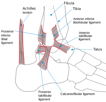
This mechanism loads the lateral ligament complex, which is injured far more often than the medial (deltoid) ligament because it is intrinsically weaker and the inversion mechanism is much more common than eversion. The lateral complex has three components, and they are injured in a predictable order as force increases:
- Anterior talofibular ligament (ATFL) - the weakest and most anterior component, and by far the most commonly injured
- Calcaneofibular ligament (CFL) - injured with greater force, alongside the ATFL
- Posterior talofibular ligament (PTFL) - the strongest, rarely injured except in severe trauma or dislocation
The Ottawa Ankle Rules
Clinical assessment alone cannot reliably distinguish a severe sprain from a fracture, and imaging every ankle injury is neither necessary nor efficient. The Ottawa Ankle Rules are a validated clinical decision tool with a sensitivity approaching 100% for clinically significant fractures, allowing safe exclusion of fracture without an X-ray in patients who do not meet the criteria.4
| Zone | Criteria for X-ray |
|---|---|
| Ankle X-ray series | Pain in the malleolar zone AND bone tenderness along the distal 6 cm of the posterior edge or tip of the lateral malleolus, OR bone tenderness along the distal 6 cm of the posterior edge or tip of the medial malleolus, OR inability to weight bear for four steps both immediately after the injury and in the emergency department |
| Foot X-ray series | Pain in the midfoot zone AND bone tenderness at the base of the fifth metatarsal, OR bone tenderness at the navicular, OR inability to weight bear for four steps both immediately after the injury and in the emergency department |
Grading and management
Ankle sprains are graded I to III using the same scheme as other ligament injuries, based on swelling, bruising, tenderness and the degree of instability on examination (anterior drawer and talar tilt testing, usually deferred until swelling has settled). Grade I and II injuries are managed with POLICE, analgesia and early weight-bearing as tolerated, with most patients returning to normal activity within two to six weeks. Grade III injuries, or those with persistent instability, may need a period of immobilisation in a below-knee cast or removable boot followed by structured physiotherapy, and recurrent instability despite rehabilitation is an indication for orthopaedic referral to consider lateral ligament reconstruction.
Knee ligament and meniscal injuries
Anterior cruciate ligament (ACL) injury
The ACL is injured by a twisting or pivoting movement, classically a sudden change of direction or deceleration with the foot planted, and is a common non-contact sports injury in football, netball and skiing. It can also occur with a direct blow to the knee. The classic history is an audible or felt 'pop' at the moment of injury, followed by rapid-onset swelling from haemarthrosis developing within hours - a useful discriminator from meniscal injury, where swelling tends to develop more slowly over 24 hours as it is a synovial effusion rather than a haemarthrosis.
Lachman's test is the single most sensitive examination test for ACL rupture and should be performed in preference to, or alongside, the anterior drawer test. The knee is flexed to around 20-30 degrees, the femur is stabilised, and the tibia is pulled anteriorly; increased anterior translation with a soft or absent endpoint compared with the other side is positive. The anterior drawer test is performed with the knee flexed to 90 degrees and is less sensitive acutely, partly because pain, swelling and hamstring guarding limit the required flexion.5
Management depends on the patient's functional demands, age, activity level and the degree of instability. Structured physiotherapy focused on quadriceps and hamstring strengthening and proprioception is appropriate for lower-demand patients or those willing to modify activity, and can allow the knee to function well despite the deficient ligament. Surgical reconstruction, usually with an autograft, is favoured in younger and more active patients, particularly those involved in pivoting sports, or where there is ongoing instability affecting activities of daily living.
Collateral ligament injuries
The medial collateral ligament (MCL) is injured by a valgus force to the knee, typically a blow to the lateral side of a planted leg, common in contact sports such as rugby and football. The lateral collateral ligament (LCL) is injured less often, by a varus force. Both are assessed by applying the corresponding stress (valgus for MCL, varus for LCL) with the knee in slight flexion and comparing laxity and endpoint with the uninjured side. Unlike the ACL, isolated collateral ligament injuries are usually managed conservatively with bracing and physiotherapy, as they have a good intrinsic healing capacity; surgery is reserved for combined multi-ligament injuries or grade III tears with gross instability.
Meniscal tears
The menisci are fibrocartilaginous shock absorbers, injured by a twisting movement on a loaded, weight-bearing knee, often with the knee partly flexed - a classic combination is standing up and twisting from a squat. Unlike the ACL, meniscal tears can also occur through degenerative wear in older patients with minimal or no trauma. Clinical features are joint line tenderness, and mechanical symptoms of locking or catching where a torn fragment intermittently obstructs full extension. McMurray's test - flexing the knee fully, then extending it while applying rotational stress and palpating the joint line - is positive if it reproduces pain or a palpable/audible click.
| Structure | Mechanism | Key history | Key test |
|---|---|---|---|
| ACL | Twisting/pivoting, sudden deceleration | Audible pop, rapid haemarthrosis within hours | Lachman's test (most sensitive) |
| MCL | Valgus stress | Medial pain, may have felt a pop | Valgus stress test |
| LCL | Varus stress | Lateral pain | Varus stress test |
| Meniscus | Twisting on a loaded knee | Joint line pain, locking or catching, delayed swelling | McMurray's test |
Achilles tendon rupture
Achilles tendon rupture typically occurs in a middle-aged 'weekend warrior' - someone in their thirties to fifties who is intermittently rather than regularly active, during sudden forceful push-off or sprinting activity, for example lunging for a shot in badminton or squash. The tendon is thought to be relatively less vascular and more prone to degenerative change with age, which combines with the sudden eccentric load to cause rupture.
Two drug associations are heavily favoured in exams. Fluoroquinolone antibiotics (such as ciprofloxacin) carry a well-documented, MHRA-flagged risk of tendon rupture, particularly the Achilles, especially in patients over 60, on corticosteroids, or with a transplant history.6 A history of corticosteroid injection near the tendon is also a recognised risk factor, thought to weaken the tendon through local catabolic effects.
The classic history is sudden, sharp pain in the back of the calf or heel, 'as if kicked', sometimes with an audible pop, occurring during the precipitating activity. Walking is still possible in many cases, because other plantarflexors (tibialis posterior, the peroneals, flexor hallucis longus) partially compensate, which can mislead an unwary examiner into under-diagnosing rupture.
Management is either conservative, with a period of immobilisation in an equinus (plantarflexed) cast or removable boot that is gradually brought to neutral over several weeks, or surgical repair. The choice depends on the patient's age, activity level, rupture characteristics (for example a large gap) and shared decision-making, since both approaches, followed by a structured rehabilitation programme, achieve comparable functional outcomes, though surgery carries a lower re-rupture rate at the cost of surgical complications such as wound infection.7
Rotator cuff injury
The rotator cuff comprises four muscles - supraspinatus, infraspinatus, teres minor and subscapularis - that dynamically stabilise the glenohumeral joint. Supraspinatus is the most commonly affected, both because of its role in initiating abduction and because it passes through the relatively narrow subacromial space, predisposing it to impingement.
In older patients the mechanism is usually degenerative, arising from chronic tendon wear and subacromial impingement rather than a single injury, and may present insidiously. In younger patients, cuff tears are more often traumatic, following a fall onto an outstretched arm or a forceful eccentric load, and are more likely to need surgical assessment.
The classic finding is a painful arc of abduction, typically between around 60 and 120 degrees, with pain easing again above this range, alongside weakness on resisted abduction and external rotation corresponding to the affected tendon(s). A full-thickness tear may produce a positive drop-arm sign, where the patient cannot control lowering the arm from full abduction.
Ultrasound is a first-line, accessible and dynamic investigation for suspected rotator cuff tear, with MRI reserved for surgical planning or where ultrasound is equivocal.8 Management progresses from analgesia and physiotherapy-led rehabilitation, through subacromial corticosteroid injection for symptom control, to arthroscopic or open surgical repair for full-thickness tears that fail conservative management or in younger, higher-demand patients.
Muscle strains
Muscle strains follow the same grading system as ligament sprains and are common in sport, particularly at muscle-tendon junctions subjected to rapid eccentric loading. The hamstring is the classic example, torn during explosive sprinting in football, athletics and rugby, presenting with sudden posterior thigh pain, localised tenderness and bruising, and a palpable defect in grade III tears. Other frequently affected sites include the quadriceps, calf (gastrocnemius, sometimes called 'tennis leg' when the medial head tears) and adductors. Management follows the same POLICE-based principles as other soft tissue injuries, with graded return to sport guided by pain and function; recurrence is common if rehabilitation and a gradual return to full training load are rushed.
Investigations
Most soft tissue injuries are diagnosed clinically, from the mechanism, history and examination findings described above, without the need for imaging.
- Plain radiographs - to exclude fracture, guided by validated decision rules such as the Ottawa Ankle Rules rather than performed indiscriminately
- Ultrasound - dynamic, accessible assessment of tendon and superficial ligament integrity, particularly useful for the Achilles and rotator cuff
- MRI - the most detailed assessment of ligament, meniscal and tendon injury, used when the diagnosis remains unclear clinically, when surgery is being considered, or to characterise the extent of injury (for example the size of a rotator cuff tear or an ACL tear with associated bone bruising)
- Joint aspiration - where a hot, swollen joint raises concern for septic arthritis or to distinguish a haemarthrosis (suggesting significant internal derangement such as ACL rupture) from a simple effusion
Complications
Most grade I and II injuries recover fully, but complications become more likely with higher-grade injuries, delayed diagnosis, or inadequate rehabilitation.
- Chronic instability - recurrent ankle sprains are common after an inadequately rehabilitated lateral ligament injury, and an ACL-deficient knee is prone to recurrent giving-way and further meniscal damage
- Early osteoarthritis - untreated instability, particularly of the ACL-deficient knee, accelerates degenerative joint change over subsequent years
- Re-rupture - a risk after both conservative and surgical management of Achilles rupture, and after ACL reconstruction, particularly with premature return to sport
- Missed compartment syndrome or fracture - assuming a significant limb injury is 'just a sprain' without adequate examination or imaging where indicated is a recognised cause of delayed diagnosis and avoidable morbidity
- Stiffness and muscle wasting - more likely with prolonged immobilisation, which is precisely why optimal loading has replaced strict rest
- Chronic tendinopathy - can follow an inadequately rehabilitated strain, particularly at the Achilles and patellar tendons
Red flags and differentials not to miss
The great majority of soft tissue injuries are straightforward, but a small number of presentations mimic a simple sprain or strain and must not be missed.
References
- NICE Clinical Knowledge Summaries. Sprains and strains. Available here
- Bleakley CM, Glasgow P, MacAuley DC. PRICE needs updating, should we call the POLICE? British Journal of Sports Medicine. 2012. Available here
- BNF. Non-steroidal anti-inflammatory drugs - treatment summary. Available here
- Stiell IG, Greenberg GH, McKnight RD et al. Decision rules for the use of radiography in acute ankle injuries: refinement and prospective validation. JAMA. 1993. Available here
- Benjaminse A, Gokeler A, van der Schans CP. Clinical diagnosis of an anterior cruciate ligament rupture: a meta-analysis. Journal of Orthopaedic & Sports Physical Therapy. 2006. Available here
- MHRA Drug Safety Update. Fluoroquinolone antibiotics: new restrictions and precautions for use due to very rare reports of disabling and potentially irreversible side effects. Available here
- British Orthopaedic Association Standards for Trauma and Orthopaedics (BOASTs). Available here
- NICE Clinical Knowledge Summaries. Shoulder pain. Available here
- NICE NG38. Fractures (non-complex): assessment and management. 2016. Available here
This article is written for revision and education. It is not clinical guidance and must not be used to make decisions about the care of a patient. Always check current NICE guidance and local protocols.