
Hepatic Encephalopathy: Recognition and Management
Key points
- Hepatic encephalopathy: a reversible spectrum of neuropsychiatric dysfunction in liver failure or portosystemic shunting, ranging from subtle cognitive change to coma.
- Mechanism: gut-derived ammonia bypasses hepatic detoxification and crosses the blood-brain barrier, where astrocytes convert it to glutamine, causing osmotic swelling.
- Grading: the West Haven criteria grade I (altered mood and attention) to grade IV (coma); asterixis is characteristic of grades II-III.
- Central clinical rule: hepatic encephalopathy is almost always precipitated - always search for and treat the trigger, do not simply give lactulose.
- Common precipitants: infection (especially SBP), GI bleeding, constipation, dehydration, electrolyte disturbance, sedatives and opioids.
- First-line treatment: lactulose, titrated to produce 2-3 soft stools daily; it acidifies the colon and traps ammonia as ammonium for excretion.
- Secondary prevention: rifaximin, a non-absorbed antibiotic, added to lactulose after a second episode to reduce recurrence and hospitalisation.
- Key pitfall: never restrict dietary protein - these patients are malnourished and sarcopenic, and protein restriction worsens outcomes.
Introduction
Hepatic encephalopathy (HE) is a potentially reversible disturbance of brain function occurring in patients with liver failure or portosystemic shunting, after exclusion of other neurological or metabolic causes.1 It spans a wide spectrum, from changes detectable only on psychometric testing through to deep coma.
It is common: overt encephalopathy affects around 30-40% of patients with cirrhosis at some point, and minimal (covert) encephalopathy - detectable only on specialised testing - is present in up to 60%. Its development is a marker of decompensation and carries a poor prognosis, with roughly 40% one-year survival after a first episode of overt HE. It also has substantial practical consequences, including impaired driving ability and increased falls.
| Type | Setting |
|---|---|
| Type A | Acute liver failure - accompanied by cerebral oedema and raised intracranial pressure |
| Type B | Portosystemic bypass or shunting without intrinsic liver disease (e.g. congenital shunt, surgical shunt) |
| Type C | Cirrhosis - by far the commonest form, and the focus of most management |
Pathophysiology
Ammonia is central to the pathogenesis, although the picture is multifactorial.2 Ammonia is produced in the gut by bacterial degradation of nitrogenous substances - dietary protein, urea and, importantly, blood in the lumen after a GI bleed - and by glutaminase activity in enterocytes.
Normally, ammonia is carried in the portal vein to the liver and converted to urea via the urea cycle for renal excretion. In cirrhosis, two things go wrong: reduced hepatocyte mass impairs urea cycle capacity, and portosystemic shunting allows portal blood to bypass the liver entirely. Ammonia therefore reaches the systemic circulation and crosses the blood-brain barrier.
In the brain, only astrocytes can metabolise ammonia, using glutamine synthetase to convert glutamate and ammonia into glutamine. Accumulated intracellular glutamine acts as an osmole, drawing water into astrocytes and causing astrocyte swelling - manifesting as Alzheimer type II astrocytes histologically. In acute liver failure this produces frank cerebral oedema and raised intracranial pressure, whereas in cirrhosis the swelling is low-grade and chronic, causing dysfunction without dangerous oedema. This distinction explains why cerebral oedema is a management priority in acute liver failure but not in cirrhotic encephalopathy.
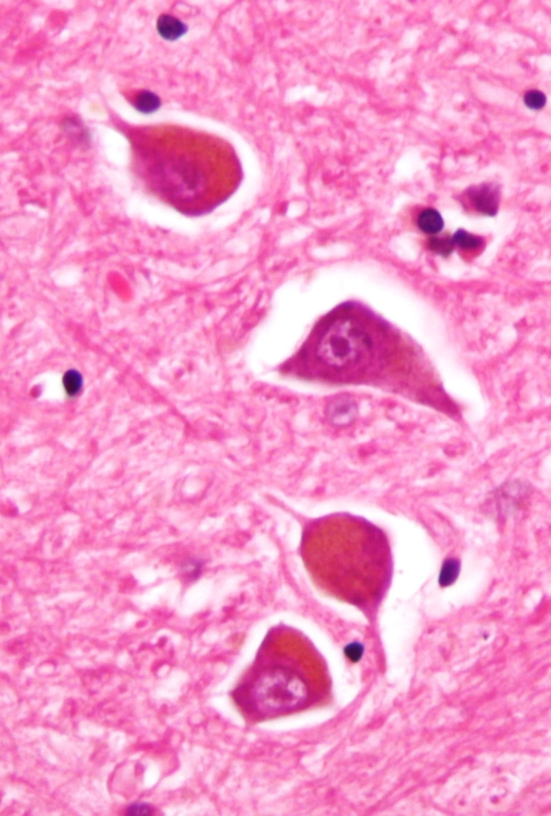
Additional contributors include systemic inflammation and infection (which act synergistically with ammonia), altered gut microbiota, increased GABAergic tone with endogenous benzodiazepine-like compounds, manganese deposition in the basal ganglia, hyponatraemia, and oxidative stress.
Precipitants
| Category | Examples |
|---|---|
| Infection | Spontaneous bacterial peritonitis (always tap the ascites), urinary tract infection, pneumonia, cellulitis, any sepsis |
| Gastrointestinal bleeding | Variceal or ulcer bleeding - blood in the gut is a large protein load that is metabolised to ammonia |
| Constipation | Increases colonic transit time and ammonia absorption - a very common and easily treated cause |
| Drugs | Opioids, benzodiazepines and other sedatives, and diuretics causing electrolyte disturbance |
| Electrolyte and metabolic | Hypokalaemia (increases renal ammonia production), hyponatraemia, dehydration, alkalosis, uraemia, hypoglycaemia |
| Renal impairment | Reduced ammonia clearance; often from over-diuresis, NSAIDs or hepatorenal syndrome |
| Dietary and other | Excess dietary protein load (rare in practice), alcohol binge, surgery, TIPS insertion, portal vein thrombosis, hepatocellular carcinoma |
Clinical features
The presentation ranges from subtle personality change noticed only by family, to deep coma. Features include cognitive impairment, altered consciousness, personality and mood change, a reversed sleep-wake cycle (daytime somnolence with nocturnal insomnia, often an early feature), impaired attention and short-term memory, and slurred speech.
Neurological signs
- Asterixis - the flapping tremor elicited by asking the patient to extend the arms with wrists dorsiflexed and fingers spread. It is a negative myoclonus, a brief loss of postural tone, and is characteristic of grades II-III. It disappears in deep coma and is not specific to liver disease, also occurring in uraemia and carbon dioxide retention
- Constructional apraxia - inability to copy a five-pointed star or draw a clock face, a useful bedside test
- Fetor hepaticus - a sweet, musty breath odour
- Hyperreflexia and increased tone, with extensor plantar responses in advanced grades
- Absence of focal neurological signs - their presence should prompt a search for an alternative diagnosis such as stroke or subdural haematoma
West Haven grading
| Grade | Clinical features |
|---|---|
| Minimal (covert) | Normal examination; abnormalities only on psychometric or neurophysiological testing. Impairs driving and quality of life |
| Grade I | Trivial lack of awareness, euphoria or anxiety, shortened attention span, impaired addition or subtraction, altered sleep rhythm |
| Grade II | Lethargy or apathy, disorientation in time, obvious personality change, inappropriate behaviour, asterixis present |
| Grade III | Somnolence to semi-stupor, responsive to stimuli, gross disorientation, confusion, bizarre behaviour |
| Grade IV | Coma, unresponsive to verbal or painful stimuli |
Grades I and above are collectively termed overt encephalopathy. Grade III-IV encephalopathy indicates the need for critical care assessment and airway protection, since these patients cannot protect their airway and are at high risk of aspiration.
Investigations
Hepatic encephalopathy is a clinical diagnosis of exclusion - there is no confirmatory test. Investigation therefore serves two purposes: excluding alternative causes of confusion, and identifying the precipitant.1
Finding the precipitant
- Full septic screen: blood cultures, urine dipstick and culture, chest radiograph, and diagnostic ascitic tap in anyone with ascites - a neutrophil count above 250 cells/mm³ diagnoses SBP
- Full blood count: infection, and anaemia suggesting GI blood loss
- Urea and electrolytes: hypokalaemia, hyponatraemia, renal impairment, and a raised urea suggesting GI bleeding
- Liver function tests, albumin and INR: to assess synthetic function and severity
- Glucose: hypoglycaemia is both a mimic and a complication
- CRP and, where sepsis is suspected, lactate
- Digital rectal examination and assessment of bowel habit for constipation and melaena
- Medication review for opioids, benzodiazepines and diuretics
Excluding alternatives
- CT head if there are focal neurological signs, a history of head injury or falls (subdural haematoma is easily missed in patients with coagulopathy and alcohol use), or if there is failure to improve with treatment
- Blood alcohol level and consideration of alcohol withdrawal or Wernicke encephalopathy - give IV thiamine (Pabrinex) if there is any doubt
- Toxicology screen and drug levels
- Thyroid function, calcium and B12, and blood gas for CO₂ retention
- EEG occasionally, showing characteristic triphasic waves, though this is rarely needed in practice
Differential diagnosis
- Alcohol withdrawal and delirium tremens - tremor, agitation, autonomic overactivity and visual hallucinations, typically 48-72 hours after the last drink
- Wernicke encephalopathy - confusion, ataxia and ophthalmoplegia; treat empirically with IV thiamine as the two frequently coexist
- Intracranial pathology - subdural haematoma (common in this group given falls and coagulopathy), stroke, intracerebral haemorrhage
- Sepsis and delirium from any cause
- Hypoglycaemia and other metabolic derangement including hyponatraemia and uraemia
- Drug toxicity - opioids, benzodiazepines, alcohol
- Meningitis or encephalitis
- Non-convulsive status epilepticus
- Wilson disease with neuropsychiatric features, in younger patients
Management
Treat the precipitant
This is the most important step and usually the one that resolves the episode: antibiotics and albumin for SBP or other infection, control of variceal bleeding, laxatives for constipation, correction of hypokalaemia and hyponatraemia, rehydration, and withdrawal of sedatives, opioids and excessive diuretics.
Lactulose
Lactulose is first-line, given orally or by nasogastric tube, and titrated to achieve 2-3 soft stools per day - the dose is guided by stool frequency, not by a fixed prescription.3 It works by three mechanisms: it is a non-absorbable disaccharide fermented by colonic bacteria to short-chain fatty acids, which acidify the colon and convert absorbable ammonia (NH₃) into non-absorbable ammonium (NH₄⁺) which is trapped in the lumen; it acts as an osmotic laxative, reducing transit time and ammonia absorption; and it favourably alters the gut flora towards non-ammonia-producing organisms.
Phosphate enemas or lactulose enemas are used where the patient cannot take oral medication or is comatose. Excessive lactulose causes dehydration, hypernatraemia and electrolyte disturbance, which can paradoxically worsen encephalopathy - so more is not better.
Rifaximin
Rifaximin 550 mg twice daily is a poorly absorbed oral antibiotic that reduces ammonia-producing gut bacteria. NICE recommends it in addition to lactulose for the secondary prevention of overt hepatic encephalopathy in patients who have had a second episode, where it significantly reduces recurrence and hospital admission.4 It is well tolerated, with minimal systemic absorption.
Supportive and general measures
- Nutrition: do NOT restrict protein. Historical practice restricted dietary protein, but this is now firmly contraindicated - patients with cirrhosis are catabolic and sarcopenic, and protein restriction worsens muscle wasting and outcomes. Aim for 1.2-1.5 g/kg/day of protein and 35-40 kcal/kg/day, with late-evening snacks to limit overnight catabolism. Vegetable and dairy protein may be better tolerated than animal protein
- Airway protection and critical care for grade III-IV encephalopathy
- Nurse in a quiet, well-lit environment with orientation cues; avoid sedation where possible
- Avoid benzodiazepines, opioids and other sedatives entirely
- Treat coexisting alcohol withdrawal with lorazepam (safer in liver impairment) and give thiamine
- Falls and pressure area care, and catheterisation only if necessary
Other and longer-term measures
- L-ornithine L-aspartate (LOLA) and oral branched-chain amino acids are second-line options with modest evidence
- Neomycin and metronidazole were historically used but are now largely superseded by rifaximin because of ototoxicity, nephrotoxicity and neuropathy
- Embolisation of large spontaneous portosystemic shunts in selected patients with recurrent, refractory encephalopathy
- TIPS reduction or occlusion if encephalopathy followed TIPS insertion
- Liver transplantation - recurrent or refractory hepatic encephalopathy is an indication for transplant assessment, and is the definitive treatment
- Driving advice: patients with overt encephalopathy must be advised not to drive and to inform the DVLA; even minimal encephalopathy impairs driving ability
Complications
- Aspiration pneumonia from a compromised airway in grade III-IV encephalopathy
- Cerebral oedema and raised intracranial pressure - a feature of type A (acute liver failure) encephalopathy, and the leading cause of death in that setting
- Falls, fractures and road traffic accidents, including from minimal encephalopathy
- Malnutrition and sarcopenia, compounded by inappropriate protein restriction
- Recurrence, which is common - around 40% within a year without secondary prophylaxis
- Persistent cognitive impairment, which may not fully reverse after repeated episodes
- Dehydration and electrolyte disturbance from excessive lactulose
- Loss of independence, carer burden and reduced quality of life
Red flags
Prognosis
An episode of overt hepatic encephalopathy is a significant adverse prognostic event in cirrhosis, marking decompensation. Survival after a first episode is roughly 40% at one year and 20% at three years without transplantation, and recurrence affects around 40% within a year unless secondary prophylaxis is used.1
The encouraging counterpoint is that individual episodes are usually fully reversible when the precipitant is identified and treated, and patients frequently return to their previous cognitive baseline. Adding rifaximin to lactulose after a second episode substantially reduces both recurrence and hospitalisation.
Because encephalopathy signals advanced liver disease rather than a primary brain problem, its appearance should trigger assessment for liver transplantation, which resolves the encephalopathy along with the underlying liver failure and offers the only definitive change in prognosis. Repeated episodes may leave a degree of persistent cognitive impairment, which is a further argument for effective secondary prevention.
References
- European Association for the Study of the Liver and American Association for the Study of Liver Diseases. Hepatic encephalopathy in chronic liver disease: practice guideline. J Hepatol. 2014. Available here
- Rose CF et al. Hepatic encephalopathy: novel insights into classification, pathophysiology and therapy. J Hepatol. 2020. Available here
- NICE NG50. Cirrhosis in over 16s: assessment and management. 2016. Available here
- NICE TA337. Rifaximin for preventing episodes of overt hepatic encephalopathy. 2015. Available here
- NICE Clinical Knowledge Summaries (CKS). Cirrhosis. 2023. Available here
- BNF. Lactulose. Available here
- Nephron, CC BY-SA 3.0, via Wikimedia Commons. Available here
- British Liver Trust. Hepatic encephalopathy. Available here
This article is written for revision and education. It is not clinical guidance and must not be used to make decisions about the care of a patient. Always check current NICE guidance and local protocols.