
Hereditary Haemochromatosis: Diagnosis and Management
Key points
- Haemochromatosis: an autosomal recessive disorder of iron metabolism causing excessive intestinal iron absorption and progressive deposition in the liver, pancreas, heart, joints and pituitary.
- Genetics: most cases result from homozygosity for the C282Y mutation in the HFE gene on chromosome 6; the H63D variant is milder and usually only relevant as a compound heterozygote.
- Mechanism: HFE mutations reduce hepcidin, the master negative regulator of iron absorption, so ferroportin remains active and duodenal iron absorption continues unchecked.
- Classic triad: bronze skin pigmentation, diabetes mellitus and hepatomegaly - historically 'bronze diabetes', though most patients now present far earlier.
- Earliest symptoms: fatigue, arthralgia (classically the 2nd and 3rd metacarpophalangeal joints) and erectile dysfunction - all non-specific, causing diagnostic delay.
- First-line tests: transferrin saturation (the most sensitive early marker, raised above 45%) and ferritin; confirm with HFE genotyping.
- Treatment: regular venesection until ferritin falls below 50 µg/L, then lifelong maintenance; iron chelation only if venesection is not tolerated.
- Key principle: venesection reverses or prevents most complications except established cirrhosis, arthropathy and hypogonadism - so early diagnosis is everything.
Introduction
Hereditary haemochromatosis is an inherited disorder of iron metabolism in which inappropriately increased intestinal iron absorption leads to progressive iron accumulation in parenchymal tissues, causing organ damage through oxidative injury and fibrosis.1
It is one of the commonest genetic disorders in people of Northern European descent, with around 1 in 200 to 1 in 400 being homozygous for the C282Y mutation, and roughly 1 in 10 being carriers. Crucially, however, penetrance is incomplete and variable: many homozygotes never develop clinically significant iron overload, while others progress to cirrhosis. Genotype therefore identifies risk rather than disease.
It matters clinically because it is common, easily tested for, and eminently treatable - yet frequently diagnosed late, after irreversible damage such as cirrhosis has occurred. Symptoms typically emerge in the fourth to sixth decades, and considerably later in women, who are protected by menstrual and pregnancy-related iron losses.
Genetics and pathophysiology
Genetics
The condition is autosomal recessive, and over 90% of UK cases are due to homozygosity for the C282Y missense mutation in the HFE gene, located on the short arm of chromosome 6 near the HLA locus.2
| Genotype | Significance |
|---|---|
| C282Y homozygote | The classic genotype, accounting for the large majority of clinically significant cases. Penetrance is incomplete |
| C282Y/H63D compound heterozygote | May develop mild iron overload, usually only with a cofactor such as alcohol excess, MASLD or another liver disease |
| H63D homozygote | Rarely causes clinically significant iron overload on its own |
| Simple heterozygote (carrier) | Not at risk of iron overload; may have marginally raised iron indices |
Rarer non-HFE forms exist, including juvenile haemochromatosis (hemojuvelin or hepcidin mutations, presenting in the teens and twenties with cardiac and endocrine failure) and ferroportin disease, but these are uncommon.
Pathophysiology
The body has no regulated mechanism for excreting iron - balance is maintained entirely by controlling absorption. The central regulator is hepcidin, a peptide hormone made by the liver, which binds the iron export channel ferroportin on duodenal enterocytes and macrophages and causes its internalisation and degradation, thereby shutting off iron entry into the circulation.
The HFE protein normally acts as a sensor of body iron stores and upregulates hepcidin when iron is plentiful. In haemochromatosis, mutated HFE fails to do this, so hepcidin levels are inappropriately low. Ferroportin remains active, duodenal iron absorption continues unchecked at roughly 3-4 mg per day instead of the normal 1-2 mg, and iron accumulates at around 0.5-1 g per year over decades.
Once transferrin becomes saturated, iron circulates as non-transferrin-bound iron, which is taken up avidly by parenchymal cells of the liver, pancreas, heart, anterior pituitary, joints and skin. There it catalyses Fenton chemistry, generating hydroxyl free radicals that cause lipid peroxidation, mitochondrial and lysosomal damage, and activation of hepatic stellate cells, producing fibrosis and ultimately cirrhosis.
Clinical features
The classic teaching triad is bronze skin pigmentation, diabetes mellitus and hepatomegaly - historically termed "bronze diabetes". In practice this represents advanced, late-stage disease, and most patients today present with far more subtle features or are detected on screening.3
Early and non-specific features
- Chronic fatigue and lethargy - the commonest presenting symptom, and easily dismissed
- Arthralgia - classically affecting the 2nd and 3rd metacarpophalangeal joints, producing a characteristic painful handshake. This distribution is highly suggestive and distinguishes it from typical osteoarthritis
- Erectile dysfunction and loss of libido - from pituitary iron deposition causing hypogonadotrophic hypogonadism
- Non-specific abdominal pain and weight loss
Organ-specific manifestations
| Organ | Manifestations | Reversible with venesection? |
|---|---|---|
| Liver | Hepatomegaly, deranged LFTs, fibrosis, cirrhosis, portal hypertension and hepatocellular carcinoma | Fibrosis may improve; established cirrhosis does not reverse |
| Pancreas | Diabetes mellitus from islet beta-cell iron deposition | Glycaemic control may improve; established diabetes usually persists |
| Skin | Slate-grey or bronze hyperpigmentation, from both melanin and iron deposition; most marked in sun-exposed areas | Usually improves |
| Heart | Dilated or restrictive cardiomyopathy, heart failure, arrhythmias (especially atrial fibrillation), conduction defects | Often improves substantially |
| Joints | Arthropathy of the 2nd/3rd MCPs, wrists, hips and knees; chondrocalcinosis and pseudogout from calcium pyrophosphate deposition | Does not reverse, and may progress despite treatment |
| Pituitary | Hypogonadotrophic hypogonadism - erectile dysfunction, amenorrhoea, loss of libido, testicular atrophy, osteoporosis | Usually irreversible |
| Other endocrine | Hypothyroidism, hypoparathyroidism, adrenal insufficiency | Variable |
Investigations
Iron studies - the first-line tests
- Transferrin saturation (serum iron divided by total iron binding capacity) is the most sensitive early marker, rising before ferritin. A fasting value above 45% warrants further investigation, and levels above 55% in men or 50% in women are strongly suggestive1
- Serum ferritin is raised, and correlates with total body iron stores. However, ferritin is an acute phase reactant and is also raised in inflammation, infection, malignancy, alcohol excess, MASLD and cell necrosis - so a raised ferritin alone is not sufficient to diagnose haemochromatosis. The combination of a raised ferritin with a raised transferrin saturation is what points to true iron overload
- Serum iron raised, total iron binding capacity (TIBC) low - the mirror image of iron deficiency anaemia, which is a useful memory aid
Confirmatory and staging tests
- HFE genotyping for C282Y and H63D confirms hereditary haemochromatosis in a patient with raised iron indices, and is used for family screening
- Liver function tests - transaminases are often mildly raised
- MRI liver with T2* or FerriScan sequences quantifies hepatic iron concentration non-invasively and has largely replaced biopsy for assessing iron burden. Cardiac T2* MRI assesses myocardial iron
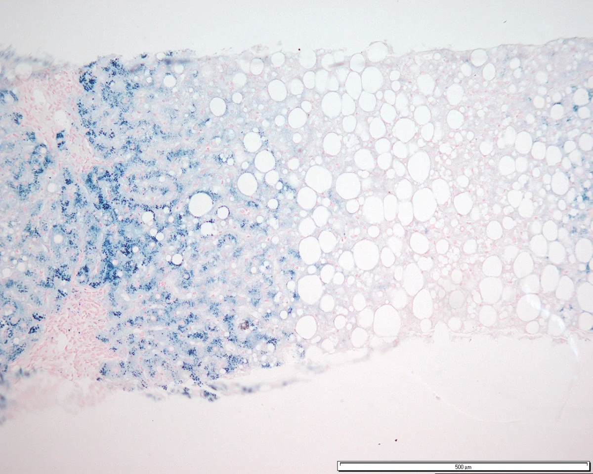
- Liver biopsy is no longer routinely needed for diagnosis, but is used to stage fibrosis where cirrhosis is suspected - typically where ferritin exceeds 1000 µg/L, transaminases are raised, or hepatomegaly is present, as these predict advanced fibrosis. Perls Prussian blue staining demonstrates iron and allows calculation of the hepatic iron index
- Transient elastography (FibroScan) as a non-invasive alternative for fibrosis assessment
- HbA1c or fasting glucose for diabetes, testosterone, LH and FSH for hypogonadism, and thyroid function
- ECG and echocardiogram for cardiomyopathy
- DEXA scan for osteoporosis, given hypogonadism
- Six-monthly ultrasound and AFP surveillance for hepatocellular carcinoma once cirrhosis is established
Differential diagnosis
The main task is distinguishing hereditary haemochromatosis from the far more common causes of a raised ferritin without true iron overload:
- Inflammation, infection or malignancy - ferritin raised as an acute phase reactant, with a normal or low transferrin saturation. This is by far the commonest explanation for a raised ferritin in practice
- Alcohol-related liver disease - raised ferritin with macrocytosis and a raised AST:ALT ratio
- MASLD/metabolic syndrome - the dysmetabolic iron overload syndrome, with modest ferritin elevation and normal transferrin saturation
- Secondary iron overload (haemosiderosis) - from repeated blood transfusions in thalassaemia, sickle cell disease or myelodysplasia, or from ineffective erythropoiesis. Treated with chelation, not venesection, since these patients are anaemic
- Hepatitis C and other chronic liver disease
- Porphyria cutanea tarda, which is associated with iron overload and often coexists
- Wilson disease - the other major inherited metal storage disorder; always consider in unexplained liver disease under 40
- Rare non-HFE haemochromatosis and aceruloplasminaemia
Management
Venesection
Therapeutic venesection (phlebotomy) is the mainstay of treatment and is simple, cheap and highly effective. Each 500 mL of blood removed contains roughly 200-250 mg of iron, forcing mobilisation of stored iron for erythropoiesis.3
- Induction phase: typically weekly venesection of 500 mL, continued until iron stores are depleted. Monitor ferritin regularly, and haemoglobin before each session - hold the session if haemoglobin falls too low. Depletion often takes 1-2 years in established overload
- Target: ferritin below 50 µg/L (some use under 50-100 µg/L) with a transferrin saturation below 50%
- Maintenance phase: lifelong venesection, typically every 2-4 months, titrated to keep ferritin in the target range
- Iron chelation with desferrioxamine or oral deferasirox is reserved for patients who cannot tolerate venesection - for example those with significant anaemia, cardiac disease or poor venous access - and is the treatment of choice in secondary iron overload from transfusion
Lifestyle and general measures
- Avoid iron supplements and iron-fortified foods, and avoid vitamin C supplements around meals, since ascorbate substantially increases iron absorption
- Avoid alcohol, which acts synergistically with iron to accelerate hepatic fibrosis - this is one of the most important interventions
- Avoid uncooked shellfish: patients with iron overload are at markedly increased risk of fatal Vibrio vulnificus septicaemia, and also of Listeria and Yersinia infection, since these organisms thrive in iron-rich environments
- A normal balanced diet is otherwise appropriate; dietary iron restriction is not required because venesection is far more effective. Tea with meals modestly reduces iron absorption
- Vaccinate against hepatitis A and B
- Manage the complications: insulin or oral agents for diabetes, testosterone replacement for hypogonadism, heart failure therapy for cardiomyopathy, and analgesia and rheumatology input for arthropathy
Surveillance and family screening
- Hepatocellular carcinoma surveillance with six-monthly ultrasound and AFP in all patients with cirrhosis. Importantly, the HCC risk persists despite successful venesection, so surveillance is lifelong once cirrhosis has developed
- Screen first-degree relatives with iron studies and HFE genotyping. This is a key intervention, as it identifies relatives before organ damage occurs. Since the condition is autosomal recessive, siblings have a 1 in 4 chance of being homozygous
- Children of an affected individual are all obligate carriers; testing the unaffected parent's genotype clarifies their risk
- Blood donation: patients with haemochromatosis may in many cases donate blood as their means of venesection, which is convenient and altruistic
Complications
- Cirrhosis and portal hypertension - the major determinant of prognosis
- Hepatocellular carcinoma - a roughly 20-fold increased risk once cirrhotic, and the leading cause of death; risk persists after iron depletion
- Diabetes mellitus, with its own microvascular and macrovascular complications
- Cardiomyopathy, heart failure and arrhythmias including atrial fibrillation - a major cause of death in juvenile haemochromatosis
- Hypogonadotrophic hypogonadism, infertility and osteoporosis
- Arthropathy and chondrocalcinosis, which often persist or progress despite treatment and are a leading cause of reduced quality of life
- Increased susceptibility to specific infections - Vibrio vulnificus, Listeria monocytogenes and Yersinia enterocolitica
- Hypothyroidism and other endocrinopathies
- Complications of venesection: anaemia, venous access difficulty and vasovagal reactions
Red flags
Prognosis
The prognosis of hereditary haemochromatosis is determined almost entirely by whether cirrhosis has developed before treatment begins. Patients diagnosed and venesected before cirrhosis have a normal life expectancy - a genuinely excellent outcome and one of the strongest arguments in medicine for early detection and family screening.1
Once cirrhosis is established, it does not reverse, and the patient carries a lifelong risk of decompensation and of hepatocellular carcinoma, which is around 20-fold that of the general population and remains the commonest cause of death. This risk is not abolished by successful iron depletion, which is why surveillance continues indefinitely.
Most other complications respond well: fatigue, skin pigmentation and cardiomyopathy typically improve substantially with venesection, and hepatic fibrosis short of cirrhosis can regress. Arthropathy, hypogonadism and established diabetes generally persist, and arthropathy in particular may continue to progress despite normalised iron stores, which patients should be counselled about so that treatment expectations are realistic.
References
- European Association for the Study of the Liver. EASL Clinical Practice Guidelines on haemochromatosis. J Hepatol. 2022. Available here
- Feder JN et al. A novel MHC class I-like gene is mutated in patients with hereditary haemochromatosis. Nat Genet. 1996. Available here
- Fitzsimons EJ et al. Diagnosis and therapy of genetic haemochromatosis: British Society for Haematology guideline. Br J Haematol. 2018. Available here
- Joseph Mathew, CC BY 2.0, via Wikimedia Commons. Available here
- NICE Clinical Knowledge Summaries (CKS). Haemochromatosis. 2023. Available here
- British Society of Gastroenterology. Guidelines on the management of abnormal liver blood tests. Gut. 2018. Available here
- NHS. Haemochromatosis. 2023. Available here
This article is written for revision and education. It is not clinical guidance and must not be used to make decisions about the care of a patient. Always check current NICE guidance and local protocols.