
Sickle Cell Disease
Key points
- Sickle cell disease: an autosomal recessive haemoglobinopathy caused by a point mutation in the beta-globin gene, producing abnormal haemoglobin S (HbS) that polymerises when deoxygenated, distorting red cells into a rigid sickle shape.
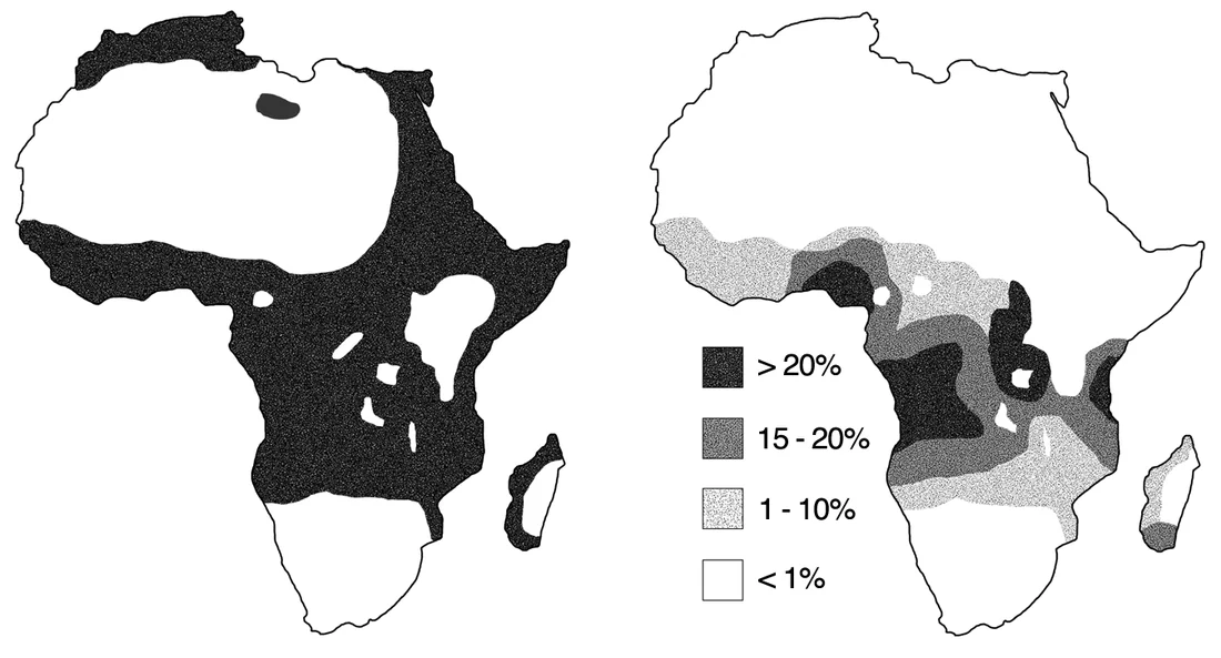
- Genetics: HbSS (sickle cell anaemia) is the most severe homozygous form. Compound heterozygous states (HbSC, HbS/beta-thalassaemia) also cause disease of variable severity. Sickle cell trait (HbAS) is usually asymptomatic and confers relative protection against malaria - the reason the allele persists at high frequency in malaria-endemic regions.
- Pathophysiology: sickled cells cause two parallel problems: chronic haemolytic anaemia (fragile cells are destroyed early) and vaso-occlusion (rigid cells block the microvasculature), producing ischaemia and infarction in almost any organ.
- Acute painful (vaso-occlusive) crisis: the commonest acute presentation - severe bone pain from microvascular occlusion and infarction, often precipitated by cold, dehydration, infection, hypoxia or stress.
- Acute chest syndrome: the leading cause of death in sickle cell disease - a new pulmonary infiltrate with respiratory symptoms and/or hypoxia. It is a medical emergency requiring urgent senior and haematology input.
- Functional hyposplenism: repeated splenic infarction causes autosplenectomy by early adulthood, leaving patients highly susceptible to encapsulated organism infection, particularly Streptococcus pneumoniae.
- Management: acute crises need prompt analgesia, fluids, oxygen if hypoxic, and treatment of any trigger. Long term: hydroxycarbamide to raise HbF, penicillin prophylaxis and vaccination, and curative allogeneic stem cell transplant in selected patients.
- Screening: part of the UK newborn blood spot screening programme, allowing prophylactic penicillin and vaccination to start before life-threatening infection occurs.
Introduction
Sickle cell disease is an inherited haemoglobinopathy and the commonest severe genetic condition in the UK, disproportionately affecting people of African, Caribbean, Middle Eastern, Indian and Mediterranean descent. It is caused by a single point mutation in the beta-globin gene that produces structurally abnormal haemoglobin, haemoglobin S (HbS).1
The disease produces two intertwined problems that explain almost every clinical feature: a chronic haemolytic anaemia, because sickled cells are fragile and destroyed prematurely, and recurrent vaso-occlusion, because rigid sickled cells obstruct small blood vessels and cause ischaemia. Care therefore spans lifelong preventive management punctuated by acute, sometimes life-threatening, crises.
Genetics and pathophysiology
Genetics
Sickle cell disease is autosomal recessive, caused by a single amino acid substitution (glutamic acid to valine at position 6) in the beta-globin chain. Several genotypes exist:
| Genotype | Name | Severity |
|---|---|---|
| HbSS | Sickle cell anaemia | Most severe homozygous form |
| HbSC | Compound heterozygote | Generally milder than HbSS, but with a higher rate of retinopathy and avascular necrosis |
| HbS/beta-thalassaemia | Compound heterozygote | Variable, from mild to as severe as HbSS depending on the beta-thalassaemia mutation |
| HbAS | Sickle cell trait (carrier) | Usually asymptomatic; confers relative protection against malaria |
Pathophysiology
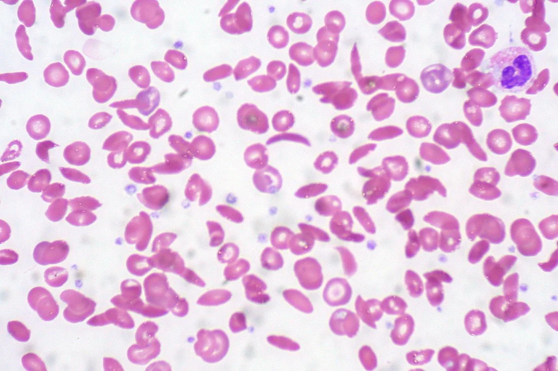
Under conditions of low oxygen tension, HbS molecules polymerise into rigid rod-like fibres, distorting the red cell into the characteristic sickle shape. Initially this is reversible with re-oxygenation, but repeated cycles of sickling and unsickling damage the cell membrane, producing irreversibly sickled cells.
Sickled cells cause disease through two mechanisms:
- Chronic haemolysis - sickled cells are fragile and rigid, and are destroyed both intravascularly and by the reticuloendothelial system, shortening red cell lifespan to around 10-20 days (versus a normal 120)
- Vaso-occlusion - rigid, sickled cells obstruct capillaries and venules, particularly in conditions that promote sickling (hypoxia, dehydration, acidosis, cold, infection), causing tissue ischaemia and infarction. This underlies the painful crisis and most of the disease's chronic organ damage
Sickle cell trait (HbAS) carriers have both normal HbA and HbS, and enough normal haemoglobin that sickling is rare except under extreme hypoxia (e.g. unpressurised high-altitude flight, general anaesthesia). Because sickled cells are hostile to the malaria parasite within them, carriers have a survival advantage in malaria-endemic regions - the classic example of heterozygote advantage, and the reason the allele has persisted despite the severity of homozygous disease.
Clinical features
Presentation typically begins from around 6 months of age, once fetal haemoglobin (HbF), which does not carry the sickle mutation, falls and HbS predominates.
Chronic features
- Chronic haemolytic anaemia - fatigue, pallor, jaundice, gallstones
- Functional hyposplenism from recurrent splenic infarction, progressing to autosplenectomy by early adulthood in HbSS - leaving patients vulnerable to infection with encapsulated organisms
- Growth restriction and delayed puberty
- Leg ulcers, particularly around the malleoli
- Avascular necrosis, especially of the femoral head
- Priapism - from vaso-occlusion within the corpora cavernosa, which is itself a urological emergency if prolonged
- Chronic kidney disease and proteinuria from papillary infarction
- Proliferative retinopathy (particularly in HbSC disease)
Acute crises
- Vaso-occlusive (painful) crisis - the commonest presentation. Severe pain, typically in the long bones, spine, chest or abdomen, often triggered by cold, dehydration, infection, hypoxia, or emotional/physical stress. In children, dactylitis (painful swelling of the hands/feet from small bone infarction) is often the first presentation
- Acute chest syndrome - a new pulmonary infiltrate on chest X-ray with fever, chest pain, cough or hypoxia. May follow a painful crisis, infection, fat embolism from bone marrow infarction, or opioid-related hypoventilation and atelectasis
- Aplastic crisis - a sudden fall in haemoglobin with a low reticulocyte count, classically triggered by parvovirus B19 infection, which transiently halts erythropoiesis
- Splenic sequestration crisis - sudden pooling of blood in the spleen, causing rapid splenic enlargement, a sharp fall in haemoglobin, and circulatory collapse; a paediatric emergency and a leading cause of death in young children with HbSS
- Haemolytic crisis - an acute exacerbation of haemolysis, with a further fall in haemoglobin and rise in jaundice
- Stroke - both ischaemic and haemorrhagic stroke occur at markedly increased rates, including in children, and are a leading cause of long-term disability
Investigations
- Newborn screening - sickle cell disease is included in the UK newborn blood spot screening programme, allowing early diagnosis before life-threatening infection or splenic sequestration occurs
- Haemoglobin electrophoresis / high-performance liquid chromatography (HPLC) confirms the diagnosis and genotype in older children and adults
- Sickle solubility test - a rapid bedside screening test, but does not distinguish disease from trait and does not replace electrophoresis
- FBC and blood film - anaemia with a raised reticulocyte count (or low, in aplastic crisis); the film shows sickle cells, target cells, and Howell-Jolly bodies once hyposplenic
- LDH and bilirubin raised, consistent with chronic haemolysis
- During an acute crisis: FBC, reticulocytes, blood cultures, chest X-ray (if respiratory symptoms - to look for acute chest syndrome), and comparison with the patient's known baseline haemoglobin
- Transcranial Doppler ultrasound - annual screening in children with HbSS to identify those at high stroke risk who benefit from a transfusion programme
Management of the acute crisis
A vaso-occlusive crisis is managed with the mnemonic-friendly bundle of analgesia, hydration, oxygen and treating the trigger:2
- Analgesia - rapid, adequate pain relief using the WHO analgesic ladder, escalating quickly to strong opioids (e.g. IV morphine) for severe pain, given via patient-controlled analgesia where appropriate. Pain is often underestimated and undertreated - individualised, prompt analgesia is a key quality standard
- IV fluids to correct dehydration, which itself promotes sickling
- Oxygen if the patient is hypoxic (saturations below their normal baseline) - oxygen is not required if saturations are normal
- Keep warm and treat any precipitant, including empirical antibiotics if infection is suspected
- Thromboprophylaxis as appropriate for admitted patients
- Escalate early - senior review and haematology involvement for severe, atypical or non-resolving crises
Splenic sequestration requires urgent fluid/blood resuscitation and same-day paediatric/haematology involvement. Priapism lasting beyond around 4 hours is a urological emergency, treated with analgesia, hydration and aspiration/irrigation if it does not settle with conservative measures.
Long-term management
- Hydroxycarbamide (hydroxyurea) - increases fetal haemoglobin (HbF) production, which interferes with HbS polymerisation, reducing the frequency of painful crises and acute chest syndrome. Offered to patients with recurrent crises
- Penicillin V prophylaxis for life, given functional hyposplenism
- Vaccination - full UK schedule plus additional vaccines needed for hyposplenism (pneumococcal, meningococcal, Hib, annual influenza)
- Folic acid supplementation, given chronically increased erythropoietic demand
- Regular transfusion programmes for selected high-risk patients (e.g. abnormal transcranial Doppler, history of stroke), with monitoring for iron overload and need for chelation
- Allogeneic haematopoietic stem cell transplant - the only established curative option, generally reserved for severe disease in children/young adults with a suitable matched donor, balancing cure against transplant-related risk
- Newer disease-modifying agents (e.g. crizanlizumab, voxelotor) and gene therapy are increasingly available for selected patients, reflecting a rapidly evolving treatment landscape
- Patient education on avoiding triggers (dehydration, cold, hypoxia, excessive alcohol, smoking), and genetic counselling for affected families
Complications
- Acute chest syndrome and chronic sickle lung disease/pulmonary hypertension
- Stroke, both in childhood and adulthood
- Avascular necrosis, particularly of the femoral head
- Chronic kidney disease
- Proliferative retinopathy and visual loss, especially in HbSC disease
- Priapism, with a risk of erectile dysfunction if not promptly treated
- Leg ulcers
- Gallstones from chronic haemolysis
- Increased susceptibility to infection, especially encapsulated organisms (Streptococcus pneumoniae, Haemophilus influenzae) due to hyposplenism
- Iron overload in patients on regular transfusion programmes
- Reduced fertility and increased risk of pregnancy complications
Red flags
Prognosis
Outcomes in sickle cell disease have improved dramatically with newborn screening, penicillin prophylaxis, vaccination and hydroxycarbamide, and many patients in the UK now survive well into adulthood, though life expectancy remains reduced compared to the general population.1
Disease severity is highly variable, influenced by genotype (HbSS generally more severe than HbSC or HbS/beta-thalassaemia), HbF level, and access to preventive care. The leading causes of death are acute chest syndrome, infection (particularly in early childhood, before hyposplenism is recognised and treated) and stroke/multi-organ failure. Allogeneic stem cell transplant, where suitable, remains the only route to cure, and emerging gene therapies are beginning to expand curative options further.
References
- NICE. Sickle cell disease: managing acute painful episodes in hospital (CG143). Available here
- British Society for Haematology. Guideline for the management of acute painful sickle cell crisis. Available here
- British Society for Haematology. Guideline for the management of acute chest syndrome in sickle cell disease. Available here
- Anthony Allison, CC0, via Wikimedia Commons. Available here
- Ed Uthman, CC BY 2.0, via Wikimedia Commons. Available here
- NHS. Sickle cell disease. 2023. Available here
- Public Health England. Newborn blood spot screening programme handbook. Available here
This article is written for revision and education. It is not clinical guidance and must not be used to make decisions about the care of a patient. Always check current NICE guidance and local protocols.