
Iron Deficiency Anaemia
Key points
- Iron deficiency anaemia: a microcytic, hypochromic anaemia caused by insufficient iron to support haemoglobin synthesis. It is the commonest cause of anaemia worldwide.
- Mechanism: iron stores are exhausted faster than they are replaced, through blood loss, reduced intake, malabsorption, or increased demand (pregnancy, growth).
- The critical question in adults: where is the blood loss coming from? Iron deficiency in a man or a postmenopausal woman is colorectal or gastric cancer until proven otherwise, and mandates bidirectional endoscopy.
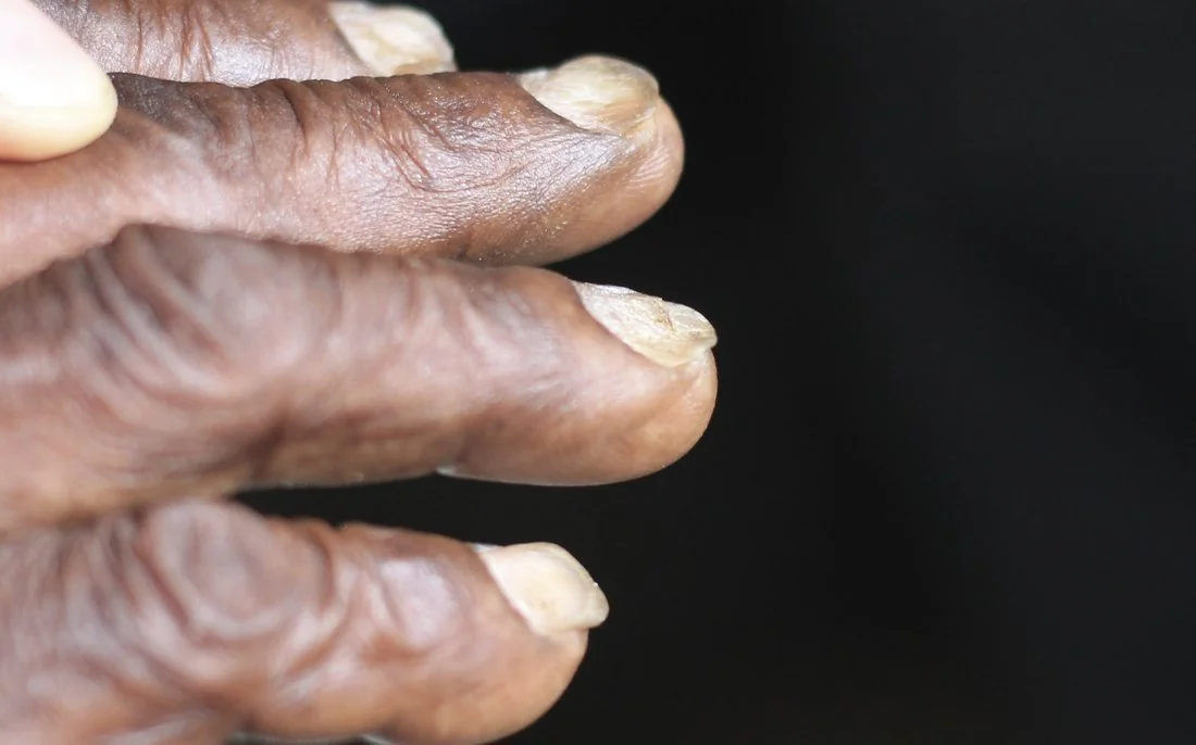
- Clinical features: fatigue, dyspnoea and pallor common to all anaemias, plus iron-specific signs: koilonychia, angular stomatitis, atrophic glossitis and pica.
- Diagnosis: low ferritin is diagnostic, but ferritin is an acute phase reactant and can be normal or raised with concurrent inflammation - interpret alongside transferrin saturation and CRP.
- Key differential: distinguishing IDA from thalassaemia trait and anaemia of chronic disease - ferritin and TIBC point the way, and the Mentzer index helps separate IDA from thalassaemia.
- Management: treat the underlying cause and replace iron, usually oral ferrous sulfate, continued for 3 months after normalisation to replenish stores. IV iron or transfusion for malabsorption, intolerance or severe/symptomatic anaemia.
- Prognosis: excellent once the cause is treated - Hb rises by roughly 10 g/L per week. Failure to respond means reconsider adherence, ongoing loss, malabsorption or the diagnosis itself.
Introduction
Iron deficiency anaemia (IDA) is a microcytic, hypochromic anaemia resulting from an inadequate supply of iron to sustain normal erythropoiesis.1 It is the most common cause of anaemia globally, affecting over a billion people, and one of the commonest reasons for referral to both primary care and gastroenterology.
Iron is essential for the synthesis of haem, the oxygen-carrying component of haemoglobin. When iron stores are depleted, haemoglobin synthesis fails before cell division does, producing red cells that are both small (microcytic) and pale (hypochromic).
IDA itself is a sign, not a final diagnosis - the priority in every adult patient is to identify why iron stores have been depleted, since the cause (particularly occult gastrointestinal blood loss) is frequently more important than the anaemia itself.
Aetiology
Causes fall into four mechanisms: blood loss, reduced intake, malabsorption, and increased demand. In adults in resource-rich settings, chronic blood loss is by far the commonest cause; worldwide, hookworm infection is the leading cause.
Blood loss
- Gastrointestinal: peptic ulcer disease, gastritis (including NSAID- and aspirin-induced), oesophagitis, angiodysplasia, colorectal or gastric cancer, inflammatory bowel disease, diverticular disease, and hookworm (the leading global cause)
- Menorrhagia: the commonest cause in premenopausal women
- Recurrent blood donation
- Haematuria (rarely a significant cause on its own)
Reduced intake
- Poor dietary intake - vegans and vegetarians are at higher risk, as haem iron from meat is far more bioavailable than non-haem plant iron
- Infants weaned onto unmodified cow's milk, which is iron-poor
- Poverty and food insecurity
Malabsorption
- Coeliac disease - always consider, especially with other features of malabsorption
- Previous gastrectomy or bariatric surgery, which bypasses the duodenum where iron is absorbed
- Helicobacter pylori infection - impairs iron absorption independent of any bleeding ulcer
- PPI therapy - reduces gastric acid needed to solubilise dietary iron
- Inflammatory bowel disease affecting the small bowel
Increased demand
- Pregnancy - from expansion of maternal red cell mass and fetal iron requirements
- Prematurity and rapid growth in infancy and adolescence
Clinical features
Many features are generic to any anaemia: fatigue, lethargy, dyspnoea on exertion, palpitations, headache and pallor. Onset is usually gradual, allowing considerable physiological compensation, so patients can present with a surprisingly low haemoglobin.
Features more specific to iron deficiency
- Koilonychia - spoon-shaped nails
- Angular stomatitis (cheilosis) - cracking at the corners of the mouth
- Atrophic glossitis - a smooth, sore tongue from papillary atrophy
- Pica - craving and eating non-food substances, classically ice (pagophagia), but also earth or starch
- Brittle hair and nails, and hair thinning
- Restless legs syndrome
Investigations
Full blood count and blood film
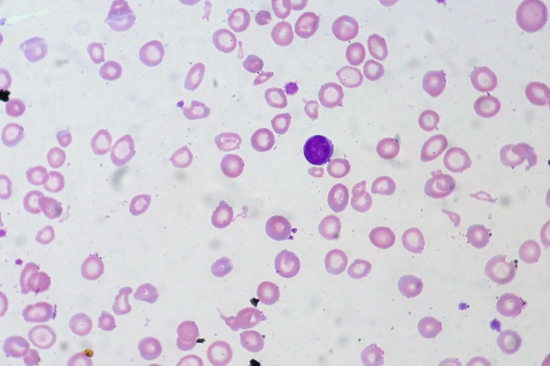
The full blood count shows a low haemoglobin with a low mean corpuscular volume (MCV) and low mean corpuscular haemoglobin (MCH) - a microcytic, hypochromic picture. Platelet count is often mildly raised (reactive thrombocytosis).
The blood film shows microcytic, hypochromic red cells with anisocytosis (variation in size) and poikilocytosis (variation in shape), including pencil cells (elongated, cigar-shaped cells) and occasional target cells.
Iron studies
- Serum ferritin low - this is the most specific test and, when unambiguously low, is diagnostic. However, ferritin is an acute phase reactant and rises with inflammation, infection or malignancy, so it can be falsely normal or elevated in a genuinely iron-deficient patient with concurrent illness. Check CRP alongside, and treat a ferritin at the lower end of normal in an unwell patient with suspicion
- Serum iron low
- Total iron binding capacity (TIBC) / transferrin high - the body upregulates transferrin to maximise capture of scarce iron
- Transferrin saturation low (typically below 16-20%)
Finding the cause
In any man, or any postmenopausal woman, unexplained iron deficiency anaemia should be assumed to represent occult gastrointestinal blood loss until proven otherwise, and requires bidirectional investigation:2
- Upper GI endoscopy (OGD) and colonoscopy to look for peptic ulceration, gastritis, and upper and lower GI malignancy
- Coeliac serology (anti-tissue transglutaminase, IgA) in all patients as part of the standard work-up
- In premenopausal women, a clear history of menorrhagia or a good dietary explanation may make invasive investigation unnecessary, but red flag symptoms should always prompt referral regardless of age or sex
- Faecal occult blood testing is not used in this context - a normal result does not exclude a colonic cancer and should not delay endoscopic investigation
Differential diagnosis
The main task is distinguishing IDA from the other microcytic anaemias, principally thalassaemia trait and, less commonly, anaemia of chronic disease and sideroblastic anaemia.
| Feature | Iron deficiency anaemia | Thalassaemia trait | Anaemia of chronic disease |
|---|---|---|---|
| Ferritin | Low | Normal or raised | Normal or raised |
| Serum iron | Low | Normal | Low |
| TIBC/transferrin | High | Normal | Low or normal |
| Red cell count | Low or normal | Often normal or raised despite low Hb | Low |
| Mentzer index (MCV/RBC) | >13 | <13 | Variable |
| Key extra test | - | Haemoglobin electrophoresis (raised HbA2) | CRP/ESR raised |
Management
Management has two parts: treating the underlying cause (e.g. endoscopic treatment of a bleeding ulcer, resection of a colorectal cancer, gluten-free diet in coeliac disease) and replacing iron stores.
Oral iron replacement
- First line: oral ferrous sulfate 200 mg two to three times daily (ferrous fumarate or gluconate are alternatives with fewer GI side effects at equivalent elemental iron dose)3
- Continue for around 3 months after haemoglobin normalises to fully replenish iron stores, not just correct the haemoglobin
- Side effects are common and include nausea, epigastric discomfort, constipation or diarrhoea, and black stools (a harmless but worth-warning-about finding). Reducing to once-daily or alternate-day dosing improves tolerability and, counter-intuitively, can improve overall absorption - each dose of oral iron transiently raises hepcidin, which blocks absorption from a dose taken too soon afterwards
- Vitamin C (ascorbic acid) enhances absorption when taken together with iron. Tea, coffee, calcium and antacids inhibit absorption and should be avoided around dosing
- Check FBC at 2-4 weeks - haemoglobin should be rising by around 10 g/L per week; a reticulocytosis appears within 7-10 days as the earliest marker of response
Parenteral iron and transfusion
- IV iron (e.g. ferric carboxymaltose, iron isomaltoside) is used where oral iron is not tolerated, where malabsorption limits enteral uptake (e.g. IBD, coeliac disease, post-gastrectomy), where ongoing losses outstrip what oral replacement can achieve, or where rapid correction is needed (e.g. before surgery)
- Blood transfusion is reserved for severe, symptomatic anaemia or haemodynamic instability, and is not a substitute for correcting the underlying iron deficit
Complications
- High-output cardiac failure in severe, long-standing anaemia
- Impaired growth, cognitive development and educational performance in children
- Adverse pregnancy outcomes, including preterm birth and low birth weight
- Plummer-Vinson syndrome - dysphagia from an oesophageal web, with a small increased oesophageal cancer risk
- Reduced quality of life and exercise tolerance, and worsening of any underlying cardiorespiratory disease
Red flags
Prognosis
The prognosis of iron deficiency anaemia itself is excellent once the underlying cause is identified and treated, and iron stores are replaced - haemoglobin typically normalises within 6-8 weeks, with full store repletion by around 3 months of continued treatment.3
The prognosis of the underlying cause, however, is what really matters clinically: an ulcer or coeliac disease responds well to treatment, whereas the outlook for an underlying gastrointestinal malignancy depends entirely on the stage at diagnosis - which is precisely why the diagnostic work-up should never be skipped.
References
- British Society of Gastroenterology. Guidelines for the management of iron deficiency anaemia in adults. Gut. 2021. Available here
- NICE. Suspected cancer: recognition and referral (NG12). 2015 (updated 2023). Available here
- BNF. Iron deficiency anaemia - treatment summary. Available here
- CHeitz, CC BY 2.0, via Wikimedia Commons. Available here
- Ed Uthman, CC BY 2.0, via Wikimedia Commons. Available here
- NICE Clinical Knowledge Summaries (CKS). Anaemia - iron deficiency. 2023. Available here
- World Health Organization. Iron deficiency anaemia: assessment, prevention and control. Available here
This article is written for revision and education. It is not clinical guidance and must not be used to make decisions about the care of a patient. Always check current NICE guidance and local protocols.