
Liver Abscess: Pyogenic and Amoebic
Key points
- Liver abscess: a localised collection of pus within the hepatic parenchyma, most commonly pyogenic (bacterial) or amoebic in origin.
- Classic triad: fever, right upper quadrant pain and jaundice - though all three are present in only a minority; fever with deranged LFTs is the more usual presentation.
- Pyogenic source: most often ascending biliary infection (cholangitis) or portal spread from intra-abdominal sepsis such as appendicitis or diverticulitis.
- Common organisms: Escherichia coli, Klebsiella pneumoniae (notably in diabetes and East Asian patients), Streptococcus milleri group and anaerobes; often polymicrobial.
- Amoebic abscess: Entamoeba histolytica, following travel to endemic areas; classically a single right lobe abscess with 'anchovy paste' contents.
- Key management difference: pyogenic abscesses usually need drainage plus antibiotics; amoebic abscesses usually respond to metronidazole alone without drainage.
- Investigation: ultrasound or contrast CT to identify the collection, blood cultures, and amoebic serology where travel history is relevant.
- Essential step: always search for the underlying source - an undiagnosed biliary obstruction, diverticulitis or colonic malignancy.
Introduction
A liver abscess is a localised collection of pus within the hepatic parenchyma. It is uncommon but important, because it is readily treatable when recognised and carries substantial mortality when missed - and because it is frequently the first sign of an undiagnosed underlying condition such as biliary obstruction, diverticulitis or a colonic malignancy.1
The liver is particularly susceptible to abscess formation because it receives blood from two sources, the hepatic artery and the portal vein, the latter draining the entire gastrointestinal tract and therefore delivering organisms from any intra-abdominal infection. The right lobe is affected in around two thirds of cases, reflecting its larger size and greater portal blood flow.
| Type | Cause | Relative frequency in the UK |
|---|---|---|
| Pyogenic (bacterial) | Bacterial infection, usually biliary or portal in origin | The commonest type in the UK, around 80% |
| Amoebic | Entamoeba histolytica, following travel to endemic areas | The commonest type worldwide; in the UK, associated with travel or migration |
| Fungal | Candida species, usually in immunocompromised or neutropenic patients | Uncommon |
| Hydatid cyst | Echinococcus granulosus - a cyst rather than a true abscess, but an important differential | Rare in the UK; associated with sheep farming areas |
Aetiology
Routes of infection in pyogenic abscess
- Biliary tract (the commonest route): ascending cholangitis from gallstones, biliary strictures, malignant obstruction, or following ERCP or biliary stenting
- Portal vein (pylephlebitis): spread from intra-abdominal sepsis - appendicitis, diverticulitis, inflammatory bowel disease, perforated viscus or pancreatitis
- Hepatic artery: haematogenous seeding in systemic bacteraemia, endocarditis or dental sepsis
- Direct extension: from an adjacent structure such as an empyema of the gallbladder, or a perinephric or subphrenic collection
- Trauma: penetrating or blunt liver injury, or following surgery, liver biopsy, radiofrequency ablation or transarterial chemoembolisation
- Cryptogenic: no source identified in up to a quarter of cases - though this should prompt a determined search rather than acceptance
Organisms
Pyogenic abscesses are frequently polymicrobial, particularly those of biliary or portal origin. Common isolates are:
- Escherichia coli and other Gram-negative enteric organisms
- Klebsiella pneumoniae - increasingly recognised, particularly in patients with diabetes and in those of East Asian origin, where a hypervirulent strain causes a distinctive invasive syndrome with metastatic infection to the eye (endophthalmitis), meninges and lungs
- Streptococcus milleri (anginosus) group - a classic abscess-forming organism
- Anaerobes including Bacteroides fragilis
- Enterococcus, Staphylococcus aureus (usually haematogenous), and Candida in the immunocompromised
Risk factors
- Diabetes mellitus - the strongest single risk factor, and particularly associated with Klebsiella
- Biliary disease - gallstones, strictures, malignancy, and previous biliary intervention
- Immunosuppression, chemotherapy, corticosteroids and HIV
- Underlying malignancy, especially colorectal and pancreaticobiliary
- Cirrhosis and chronic liver disease
- Liver transplantation, and proton pump inhibitor use
- For amoebic abscess: travel to or residence in endemic areas (South Asia, Africa, Central and South America), male sex, alcohol use and immunosuppression
Clinical features
Presentation is often insidious and non-specific, and liver abscess is a recognised cause of pyrexia of unknown origin. The classic triad of fever, right upper quadrant pain and jaundice is present in only a minority.2
- Fever with rigors and night sweats - the most consistent feature, present in around 90%
- Right upper quadrant or epigastric pain, which may be dull and poorly localised, and can radiate to the right shoulder tip through diaphragmatic irritation
- Malaise, anorexia, weight loss and fatigue, often over weeks
- Nausea and vomiting
- Tender hepatomegaly, and tenderness on percussion over the lower right ribs
- Jaundice - present in only around a quarter, and more common with biliary causes or multiple abscesses
- Right-sided pleuritic chest pain, cough or breathlessness from diaphragmatic irritation, a sympathetic pleural effusion or basal atelectasis
- Sepsis and shock in severe cases
Features suggesting an amoebic abscess
Amoebic liver abscess follows intestinal infection with Entamoeba histolytica, with trophozoites reaching the liver through the portal vein. Distinguishing features include:
- Travel to or origin from an endemic area, often weeks to months previously
- Younger patients, and a marked male predominance
- Usually a single, large abscess in the right lobe
- Preceding dysentery in only a minority - most patients have no history of diarrhoea at the time of presentation, which is a common source of diagnostic error
- Aspirate classically resembles 'anchovy paste' or anchovy sauce - reddish-brown, odourless liquefied hepatic tissue, which is usually sterile on bacterial culture
Investigations
Blood tests
- Full blood count: neutrophil leucocytosis, and normocytic anaemia of chronic disease
- CRP and ESR: markedly raised
- Liver function tests: typically a cholestatic picture with raised alkaline phosphatase - the most consistently abnormal enzyme - and raised gamma-GT. Transaminases are mildly raised and bilirubin is often normal
- Albumin: low, reflecting the chronic inflammatory state
- Blood cultures: positive in around half of pyogenic cases; take before starting antibiotics
- Urea, electrolytes, glucose and HbA1c - screen for undiagnosed diabetes, which is a common association
- Amoebic serology (anti-E. histolytica antibodies): highly sensitive for amoebic liver abscess and the key discriminating test; note it remains positive for years after infection, limiting its value in endemic populations
- Stool microscopy and PCR for E. histolytica, though often negative in hepatic disease
Imaging
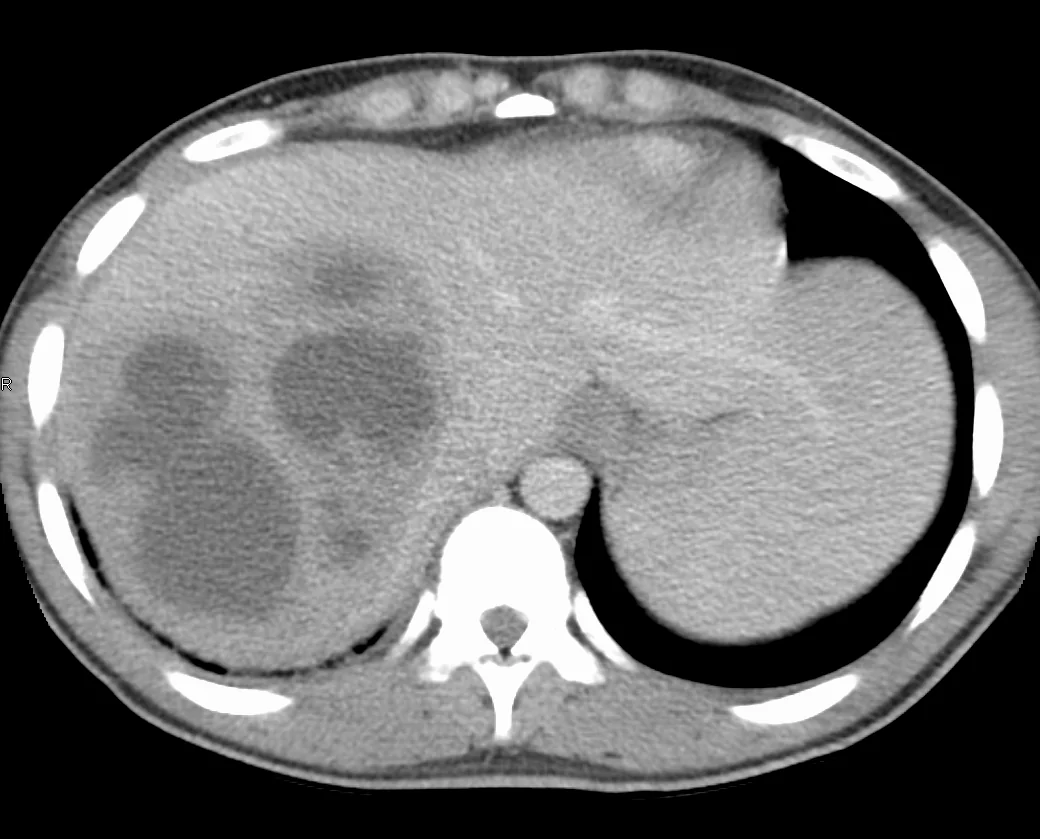
- Abdominal ultrasound: first-line, quick and sensitive, showing a hypoechoic or complex fluid collection. Also assesses the biliary tree for obstruction or stones
- Contrast-enhanced CT abdomen: the investigation of choice for characterising the abscess, defining the number, size and location of collections, planning drainage, and - crucially - identifying the underlying source such as diverticulitis, appendicitis or a colonic tumour. A rim-enhancing lesion with central low attenuation is typical, sometimes with a 'double target' sign
- Chest radiograph: may show a raised right hemidiaphragm, right basal atelectasis or a pleural effusion
- MRI/MRCP: where biliary pathology needs further definition
Aspiration
Image-guided aspiration provides pus for urgent Gram stain, culture and sensitivity, which guides definitive antibiotic therapy, and is frequently therapeutic at the same time. Pyogenic pus is typically foul-smelling and yields organisms; amoebic aspirate is the sterile reddish-brown 'anchovy paste'. Aspiration should be avoided if a hydatid cyst is suspected, since spillage can cause anaphylaxis and peritoneal seeding - check Echinococcus serology first if the imaging shows a cyst with daughter cysts or calcification.
Differential diagnosis
- Acute cholecystitis and ascending cholangitis - which may also be the underlying cause
- Hepatocellular carcinoma or liver metastases, particularly necrotic metastases which can mimic an abscess radiologically
- Hydatid cyst - avoid aspirating; look for calcification and daughter cysts, and check serology
- Simple hepatic cyst or a complicated (haemorrhagic or infected) cyst
- Subphrenic or subhepatic collection, and right lower lobe pneumonia or empyema
- Acute hepatitis, though this gives a hepatitic rather than cholestatic pattern
- Pyrexia of unknown origin from another source - liver abscess should always feature in that differential
- Right-sided renal pathology, including perinephric abscess
Management
Pyogenic liver abscess
Treatment rests on drainage plus antibiotics, together with correction of the underlying cause.1
- Resuscitate using an ABCDE approach; treat sepsis per the sepsis pathway with IV fluids and oxygen
- Take blood cultures, then start empirical broad-spectrum IV antibiotics covering Gram-negatives, streptococci and anaerobes - for example piperacillin-tazobactam, or a cephalosporin with metronidazole, per local policy
- Image-guided percutaneous drainage for abscesses above roughly 3-5 cm - either needle aspiration or, for larger collections, insertion of a percutaneous catheter drain. This is both diagnostic and therapeutic and is the standard of care
- Rationalise antibiotics once culture and sensitivity results are available
- Prolonged antibiotic course: typically 2-3 weeks intravenously followed by oral therapy to complete 4-6 weeks in total, guided by clinical response, inflammatory markers and repeat imaging
- Surgical drainage is reserved for abscesses not amenable to or failing percutaneous drainage, multiloculated collections, rupture, or where laparotomy is needed for the underlying source
- Treat the underlying cause: ERCP for biliary obstruction, cholecystectomy for gallstone disease, and management of diverticulitis or appendicitis
Amoebic liver abscess
The management contrast with pyogenic abscess is a classic examination point: amoebic abscesses usually respond to medical therapy alone and do not require drainage.
- Metronidazole (or tinidazole) for 7-10 days is the mainstay, and produces rapid clinical improvement, often within 72 hours
- Followed by a luminal amoebicide - paromomycin or diloxanide furoate - to eradicate intestinal cysts and prevent relapse and onward transmission. Omitting this step is a common error
- Aspiration is reserved for: diagnostic uncertainty (particularly to exclude a pyogenic abscess), very large abscesses, left lobe abscesses at risk of rupture into the pericardium, failure to respond to metronidazole within 5-7 days, or imminent rupture
- Radiological resolution lags well behind clinical improvement and may take months, so persistent imaging findings in a well patient do not indicate treatment failure
Complications
- Sepsis, septic shock and multi-organ failure
- Rupture - into the peritoneum causing peritonitis, into the pleural space causing empyema, or into the pericardium causing tamponade. Pericardial rupture from a left lobe amoebic abscess is the most feared complication
- Metastatic infection, particularly with the hypervirulent Klebsiella pneumoniae invasive syndrome, causing endophthalmitis (which can rapidly blind), meningitis, brain abscess and septic pulmonary emboli - examine the eyes and ask about visual symptoms
- Pleuropulmonary complications: sympathetic effusion, empyema, basal atelectasis, hepatobronchial fistula
- Portal vein thrombosis and pylephlebitis
- Biliary fistula and haemobilia
- Recurrence, particularly if the underlying source is untreated
- Complications of drainage: bleeding, secondary infection, and seeding along the track
Red flags
Prognosis
With modern imaging, percutaneous drainage and effective antibiotics, the prognosis of pyogenic liver abscess has improved dramatically - mortality has fallen from over 70% in the pre-antibiotic era to around 5-15% today.2 Most patients treated promptly recover fully, though the course is prolonged and antibiotic therapy typically continues for 4-6 weeks with radiological follow-up.
Adverse prognostic factors include multiple abscesses, an underlying malignant biliary obstruction, immunosuppression, delayed diagnosis, septic shock at presentation, and rupture. Recurrence is uncommon provided the underlying source is identified and treated, which is the central determinant of long-term outcome.
Amoebic liver abscess has an excellent prognosis, with mortality under 1% when treated promptly with metronidazole, and most patients improve within days. The main causes of death are rupture - especially into the pericardium - and delayed diagnosis. Because radiological resolution takes months, follow-up imaging should not be over-interpreted in a patient who is clinically well.
References
- Lardière-Deguelte S et al. Hepatic abscess: diagnosis and management. J Visc Surg. 2015. Available here
- Mavilia MG et al. Differentiating pyogenic from amebic liver abscess. J Clin Transl Hepatol. 2016. Available here
- James Heilman, MD, CC BY-SA 4.0, via Wikimedia Commons. Available here
- NICE Clinical Knowledge Summaries (CKS). Gallstones. 2023. Available here
- NICE NG51. Sepsis: recognition, diagnosis and early management. 2016 (updated 2024). Available here
- BNF. Metronidazole. Available here
- UK Health Security Agency. Amoebiasis: guidance and data. Available here
This article is written for revision and education. It is not clinical guidance and must not be used to make decisions about the care of a patient. Always check current NICE guidance and local protocols.