
Graves' Disease and Thyroid Eye Disease
Key points
- Graves' disease: autoimmune hyperthyroidism caused by IgG antibodies that stimulate the TSH receptor, driving unregulated hormone synthesis and diffuse goitre.
- Scale: the commonest cause of hyperthyroidism, accounting for around 70% of cases, with a female to male ratio of about 5 to 10 to 1 and a peak between 30 and 50 years.
- The four extrathyroidal signs: diffuse goitre with a bruit, thyroid eye disease, pretibial myxoedema and thyroid acropachy. Their presence makes the diagnosis without further testing.
- Diagnosis: suppressed TSH with raised free T4 or T3, and positive TSH receptor antibodies (TRAb), which are present in around 95%.
- Eye disease is independent of thyroid status: it can occur while the patient is euthyroid or hypothyroid, and does not necessarily improve when the thyroid is treated.
- Smoking: the strongest modifiable risk factor - it roughly doubles the risk of eye disease, worsens its severity and reduces the response to treatment.
- Sight-threatening features: reduced acuity, loss of colour vision, a relative afferent pupillary defect, disc swelling or corneal breakdown. These require same-day ophthalmology assessment.
- Radioiodine can worsen orbitopathy: so in active moderate to severe eye disease it is avoided, or given under corticosteroid cover.
Introduction
Graves' disease is an autoimmune disorder in which IgG autoantibodies bind and stimulate the TSH receptor on thyroid follicular cells. Unlike TSH itself, these antibodies are not subject to negative feedback, so the gland is driven continuously - producing diffuse hyperplasia, a smooth goitre and unregulated thyroid hormone synthesis.
It is the commonest cause of hyperthyroidism, responsible for around 70% of cases. It is far more common in women, peaks between 30 and 50 years, and clusters with other organ-specific autoimmune disease.
Why the eyes and shins are involved
This is the elegant part of the pathophysiology and explains features that otherwise seem unconnected. The TSH receptor is also expressed on orbital fibroblasts and on dermal fibroblasts of the pretibial skin. The same autoantibodies therefore stimulate these cells, and in concert with the IGF-1 receptor they trigger:
- Secretion of glycosaminoglycans, which are strongly hydrophilic and draw water into the tissue
- Adipogenesis and expansion of orbital fat
- Inflammatory cell infiltration and, later, fibrosis of the extraocular muscles
Because the bony orbit cannot expand, the increase in orbital contents pushes the globe forwards (proptosis), congests venous drainage, restricts eye movement, and at the crowded orbital apex may compress the optic nerve. The same process in pretibial skin produces pretibial myxoedema.
Risk factors
- Female sex and a family history of autoimmune thyroid disease
- Other autoimmune conditions - type 1 diabetes, coeliac disease, pernicious anaemia, vitiligo, Addison's disease, rheumatoid arthritis
- Smoking - the strongest modifiable factor, and particularly for eye disease
- The postpartum period
- Iodine excess, including from amiodarone and contrast media
- Drugs - interferon alfa, immune checkpoint inhibitors, and immune reconstitution after alemtuzumab or antiretroviral therapy
- Stress and major life events, which are consistently reported although the mechanism is unclear
Clinical features
Thyrotoxic features
The general features of thyrotoxicosis are present - weight loss with increased appetite, heat intolerance, sweating, palpitations, tremor, anxiety, diarrhoea, oligomenorrhoea and proximal myopathy, with tachycardia or atrial fibrillation, warm moist skin, brisk reflexes and lid retraction and lid lag (which occur in thyrotoxicosis of any cause).
Features specific to Graves' disease
| Sign | Features | Frequency |
|---|---|---|
| Diffuse goitre with a bruit | Smooth, symmetrical, non-tender enlargement. A bruit reflects the markedly increased vascularity and is not heard in other causes of goitre. | Most patients |
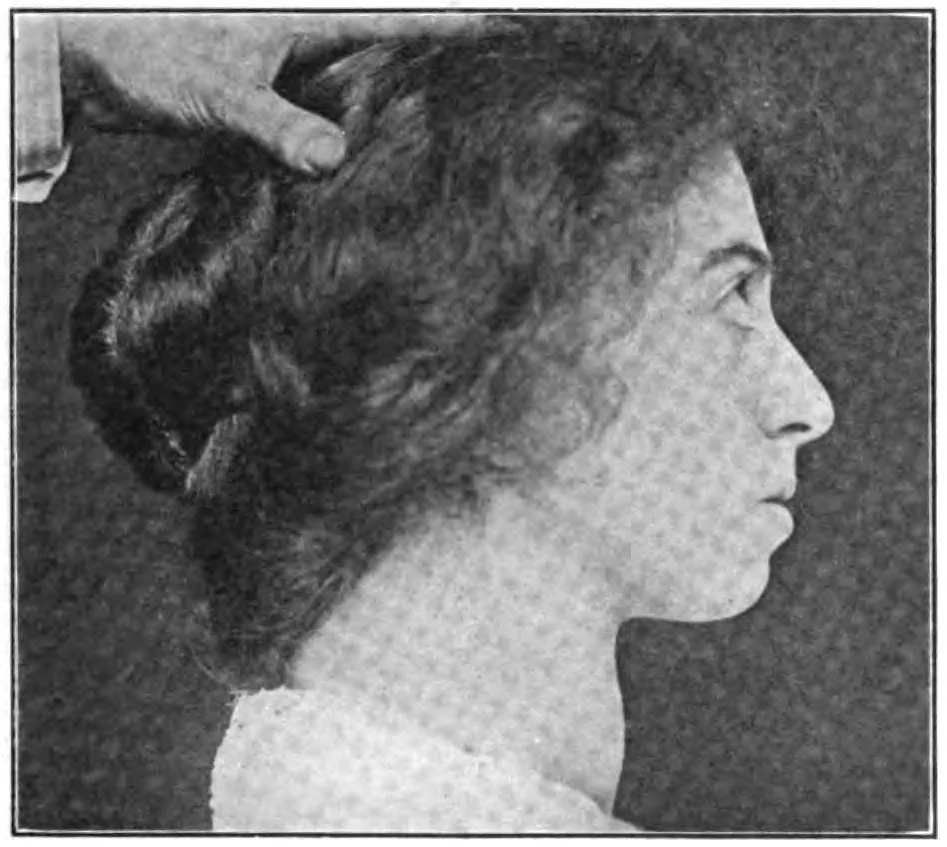
| Thyroid eye disease (Graves' orbitopathy) | Proptosis, periorbital swelling, conjunctival injection, restrictive ophthalmoplegia and, in severe cases, optic neuropathy | Clinically apparent in 25 to 50%, with subclinical changes on imaging in up to 90% |
| Pretibial myxoedema (thyroid dermopathy) | Non-pitting, waxy, raised plaques over the shins and dorsum of the feet, with a peau d'orange texture. Almost always accompanies eye disease. | Around 1 to 5% |
| Thyroid acropachy | Digital clubbing, soft tissue swelling of the hands and feet, and periosteal new bone formation - a triad that is rare but virtually pathognomonic | Under 1% |
Thyroid eye disease
Symptoms and signs
- Grittiness, dryness, watering and photophobia - from corneal exposure
- Retro-orbital ache, and pain on eye movement, which is a marker of active inflammation
- Diplopia - initially on extreme gaze, later in the primary position
- Change in appearance - a staring look, swollen lids, and asymmetry. The psychological impact of this is substantial and consistently under-acknowledged.
- Reduced vision or colour desaturation - the alarming symptom, indicating optic neuropathy
- Signs - lid retraction, lid lag, proptosis (measured with an exophthalmometer), periorbital and lid oedema, conjunctival injection and chemosis, caruncle swelling, incomplete lid closure (lagophthalmos), corneal punctate staining or ulceration, and restrictive ophthalmoplegia
- The extraocular muscles are affected in a characteristic order - inferior rectus first, restricting upgaze, then medial rectus, restricting abduction. This is muscle restriction, not weakness, so forced duction testing is positive.
Assessing activity and severity
- Clinical Activity Score (CAS) - one point each for spontaneous retrobulbar pain, pain on eye movement, eyelid erythema, eyelid swelling, conjunctival redness, chemosis and caruncle swelling. A score of 3 or more out of 7 indicates active disease likely to respond to immunosuppression.
- Severity (EUGOGO) - mild (minor impact on daily life), moderate to severe (sufficient impact to justify immunosuppression or surgery), and sight-threatening
- CT or MRI of the orbits - shows enlarged extraocular muscle bellies with sparing of the tendon insertions (which distinguishes it from orbital myositis and from an orbital mass) and demonstrates apical crowding when optic neuropathy is suspected
Managing eye disease
- Smoking cessation - the single most effective intervention available. Smoking doubles the risk of eye disease, worsens its severity, accelerates progression and blunts the response to steroids and radiotherapy. This should be stated in those terms rather than as generic advice.
- Restore and maintain euthyroidism - and specifically avoid hypothyroidism, which worsens orbitopathy. Monitor closely after radioiodine or thyroidectomy.
- Selenium supplementation - a 6-month course improves outcomes in mild disease of recent onset in selenium-deficient populations
- Supportive measures - artificial tears and ocular lubricants, ointment at night, taping the lids if closure is incomplete, sleeping with the head elevated to reduce periorbital oedema, sunglasses for photophobia, and prisms for stable diplopia
- Moderate to severe active disease - pulsed intravenous methylprednisolone is first line, often with mycophenolate. Second-line options include teprotumumab (an IGF-1 receptor antibody), tocilizumab, rituximab and orbital radiotherapy.
- Inactive (burnt-out) disease - rehabilitative surgery, performed in a strict order: orbital decompression, then strabismus surgery, then eyelid surgery. Doing them out of sequence undoes the previous operation.
- Radioiodine - avoid in active moderate to severe eye disease, or give with oral prednisolone cover. It is acceptable in mild or inactive disease, particularly in non-smokers.
Investigations
- Thyroid function tests - suppressed TSH with raised free T4 and/or free T3
- TSH receptor antibodies (TRAb) - positive in around 95% and effectively confirms the diagnosis. NICE recommends measuring TRAb rather than proceeding to scintigraphy in most patients with suspected Graves' disease.1
- Anti-TPO antibodies - positive in around 75%, but also positive in Hashimoto's, so less useful diagnostically
- Technetium uptake scan - diffuse increased uptake throughout an enlarged gland. Required only where TRAb is negative or the picture is atypical.
- Full blood count and liver function tests - baseline before starting a thionamide
- ECG - for atrial fibrillation
- Bone profile - calcium and alkaline phosphatase are often raised
- Orbital CT or MRI - where eye disease is moderate to severe, atypical, unilateral, or where optic neuropathy is suspected
Managing the hyperthyroidism
- Beta-blockade - propranolol for symptom control while awaiting the effect of definitive treatment
- Carbimazole - first-line antithyroid drug, given as a titration or block-and-replace regimen for 12 to 18 months. Around half of patients relapse after stopping, most within the first year.
- Warn about agranulocytosis at every initiation - stop the drug and obtain an urgent full blood count for sore throat, fever or mouth ulcers
- Radioactive iodine - NICE recommends it as first-line definitive treatment for Graves' disease unless contraindicated. It is simple and highly effective, and hypothyroidism afterwards is expected rather than a failure. Contraindicated in pregnancy and breastfeeding, and in active moderate to severe eye disease without steroid cover.
- Total thyroidectomy - for a large goitre with compressive symptoms, suspected malignancy, severe eye disease, patient preference, or where radioiodine is unsuitable. Render the patient euthyroid first, with potassium iodide for 10 days preoperatively to reduce vascularity.
- Predictors of relapse after drug treatment - large goitre, very high initial T4 and T3, persistently high TRAb, young age, and smoking
Graves' disease in pregnancy
- Distinguish from gestational thyrotoxicosis - the hCG-mediated, self-limiting thyrotoxicosis of early pregnancy, which is associated with hyperemesis, has no goitre, no eye signs and negative TRAb, and needs no antithyroid treatment
- Use propylthiouracil in the first trimester because carbimazole is teratogenic (aplasia cutis, choanal and oesophageal atresia), then switch to carbimazole for the remainder to limit propylthiouracil hepatotoxicity
- Use the lowest effective dose - antithyroid drugs cross the placenta and can cause fetal goitre and hypothyroidism
- TRAb crosses the placenta and can stimulate the fetal thyroid. Measure TRAb, including in women previously treated with radioiodine or surgery who are now euthyroid or hypothyroid, because the antibodies persist. High titres warrant fetal monitoring for goitre, tachycardia and growth restriction, and neonatal thyrotoxicosis can appear in the first days of life.
- Graves' disease often improves in the second and third trimesters as immune tolerance develops, allowing dose reduction, and relapses postpartum
Complications
- Sight loss from dysthyroid optic neuropathy or corneal ulceration
- Permanent diplopia requiring prisms or strabismus surgery
- Disfigurement from proptosis and lid retraction, with substantial psychological morbidity - anxiety, depression and social withdrawal are common and should be asked about
- Atrial fibrillation and thromboembolism
- Heart failure and thyrotoxic cardiomyopathy
- Osteoporosis and fragility fracture
- Thyroid storm
- Relapse after antithyroid drug treatment, in around half
- Permanent hypothyroidism after radioiodine or surgery - an expected outcome requiring lifelong levothyroxine
- Surgical complications - recurrent laryngeal nerve palsy, hypoparathyroidism and postoperative haematoma
- Pregnancy complications - miscarriage, pre-eclampsia, preterm birth, fetal and neonatal thyroid dysfunction
Red flags
Prognosis
The hyperthyroidism of Graves' disease is entirely controllable. Around half of patients remit after a 12 to 18 month course of antithyroid drugs, and those who relapse can be cured with radioiodine or surgery at the cost of lifelong levothyroxine. Cardiovascular and skeletal risks reverse once the patient is euthyroid, and atrial fibrillation frequently returns to sinus rhythm.
Thyroid eye disease follows a different and less satisfying course. It typically runs through an active inflammatory phase lasting 6 to 24 months, followed by a plateau and then an inactive fibrotic phase - the so-called Rundle's curve. Most patients improve spontaneously from the peak, but the improvement is rarely complete: proptosis, lid retraction and diplopia often persist and require rehabilitative surgery years later. Only around 3 to 5% develop sight-threatening disease.
This is why the timing of intervention matters so much. Immunosuppression works only during the active phase, and surgery is reserved for the inactive one - treating in the wrong phase wastes the opportunity in the first case and invites recurrence in the second.
Finally, the psychological burden deserves explicit attention. Thyroid eye disease alters a person's appearance in a way that is visible to everyone they meet, is frequently dismissed as cosmetic by clinicians, and has measurable effects on quality of life comparable to that of serious systemic illness. Acknowledging that, and referring to a specialist service rather than reassuring the patient that their thyroid results are now normal, is a substantial part of good care.
References
- NICE NG145. Thyroid disease: assessment and management. 2019, updated 2023. Available here
- Bartalena L, Kahaly GJ, Baldeschi L et al. The 2021 European Group on Graves' Orbitopathy (EUGOGO) clinical practice guidelines for the medical management of Graves' orbitopathy. European Journal of Endocrinology. Available here
- British Thyroid Association. Current guidelines and statements. Available here
- NICE Clinical Knowledge Summaries. Hyperthyroidism. Available here
- Royal College of Ophthalmologists. Thyroid eye disease guidance. Available here
- MHRA Drug Safety Update. Carbimazole: increased risk of congenital malformations. Available here
- BNF. Carbimazole, propylthiouracil and prednisolone. Available here
- William Seaman Bainbridge, public domain, via Wikimedia Commons. Available here
This article is written for revision and education. It is not clinical guidance and must not be used to make decisions about the care of a patient. Always check current NICE guidance and local protocols.